Ghyle`ne Goudet, François Belin, Jacqueline Be´zard and Nadine Ge´rard1 ..... change much in bovine (Wu et al., 1997; Lévesque and Sirard,. 1996), porcine ...
Molecular Human Reproduction vol.4 no.6 pp. 563–570, 1998
Maturation-promoting factor (MPF) and mitogen activated protein kinase (MAPK) expression in relation to oocyte competence for invitro maturation in the mare Ghyle`ne Goudet, Franc¸ois Belin, Jacqueline Be´zard and Nadine Ge´rard1 I.N.R.A.-Haras Nationaux, Equipe de Reproduction Equine, Station P.R.M.D., 37380 Nouzilly, France 1To
whom correspondence should be addressed
In the equine species, a large proportion of oocytes fail to complete meiosis during in-vitro culture. The biochemical and molecular basis of this failure is unknown. The meiotic cell cycle is controlled in part by the maturation-promoting factor (MPF) and the mitogen-activated protein kinase (MAPK). In this study, we evaluated the oocyte competence for in-vitro maturation and the expression of MPF components (p34cdc2 and cyclin B) and MAPK after in-vitro culture. The maturation rate was influenced by the culture medium and the physiological stage of the mare at the time of oocyte recovery. We showed that MAPK and the two subunits of MPF were present in equine oocytes whatever the nuclear stage they reached after in-vitro culture and whatever the culture medium used. In incompetent oocytes, MAPK remained in its non-phosphorylated form, supposed to be inactive. In conclusion, the incompetence of equine oocytes to resume and complete meiosis is not due to the absence of p34cdc2, cyclin B or MAPK. Our results suggest that it is more probably due to a deficiency of regulators of MPF and/or to an inability to phosphorylate MAPK. Key words: IVM/MAPK/mare/MPF/oocyte
Introduction In the equine species, the conditions for in-vivo oocyte maturation are partly different from those in other domestic mammals; the ovulatory luteinizing hormone (LH) surge is a progressive increase lasting many days, with a maximum concentration occuring 1 day after ovulation (Whitmore et al., 1973; Irvine and Alexander, 1994). Moreover, the in-vitro maturation rate of equine oocytes remains low whatever the culture conditions used: ,70% of the oocytes are in metaphase II at the end of the culture period (Squires, 1996; for review, see Goudet et al., 1997; Hinrichs and Williams, 1997), whereas in the goat (De Smedt et al., 1994), the sow (Singh et al., 1997) and the cow (Sirard, 1989) .90% of the oocytes are mature. We showed recently that the mare’s reproductive status and the follicular diameter have an influence on the oocyte competence for in-vitro maturation (Goudet et al., 1997). At the present time, the reasons of the failure of equine oocytes to complete meiosis during in-vitro culture are unknown, but could be linked to an alteration of the biochemical cascade involved in the meiotic process. The meiotic cell cycle is mainly controlled by a phosphorylation–dephosphorylation regulatory cascade (Maller et al., 1977; Crosby et al., 1984; Kastrop et al., 1990). Among the kinases involved in oocyte maturation, maturation-promoting factor (MPF) (Masui and Markert, 1971) has been found to be a universal cell cycle regulator of both mitosis and meiosis (for review, see Nurse, 1990). It is a heterodimeric complex of two subunits: a serine-threonine protein kinase, p34cdc2 (Dunphy et al., 1988) and a regulatory subunit, cyclin B (Draetta et al., 1989). Activation of MPF depends upon both the association of p34cdc2 and cyclin B, and a subsequent change in the © European Society for Human Reproduction and Embryology
phosphorylated state of key tyrosine and threonine residues on the p34cdc2 subunit (Nurse, 1990). During oocyte meiotic maturation of mammalian species studied so far, MPF activity is very low in the germinal vesicle stage, and peaks at metaphase I and II stages (mouse: Hashimoto and Kishimoto, 1988; Choi et al., 1991; rabbit: Naito and Toyoda, 1991; goat: Jelinkova et al., 1994; pig: Dedieu et al., 1996; bovine: Wu et al., 1997). Other kinases are also involved in the regulation of meiotic events, such as mitogen-activated protein kinase (MAPK, also referred to as extracellular signal-regulated protein kinase, or ERK). MAPK is a serine-threonine kinase activated by phosphorylation at the onset of oocyte maturation in several species, including Xenopus (Ferrell et al., 1991; Gotoh et al., 1991), mouse (Sobajima et al., 1993; Verlhac et al., 1993; Harrouk and Clarke, 1995), rat (Goren et al., 1994), pig (Inoue et al., 1995), goat (Dedieu et al., 1996) and bovine (Fissore et al., 1996). The presence of two isoforms of 42 kDa (ERK2) and 44 kDa (ERK1) of MAPK has been shown in all mammals cited above, except in the goat. The kinase activity in the equine oocyte has never been studied. Our aim was to investigate whether a lack of MAPK, p34cdc2 or cyclin B was associated with the failure of equine oocytes to resume and complete meiosis during in-vitro culture.
Materials and methods Experimental design Adult cyclic pony mares (n 5 56) in good body condition, aged 3– 19 years, kept indoors and fed with concentrates were studied from March to May. They received an initial synchronization treatment (Palmer, 1984) with an intravaginal sponge containing 0.5g altrenogest
563
G.Goudet et al. (Regumate; Roussel UCLAF, Romainville, France) plus 50 mg of oestradiol benzoate (β-oestradiol 3-benzoate; Sigma, La Verpille`re, France) for 1 week. The day of sponge removal, mares received a prostaglandin F2α analogue injection (Cloprostenol, 250 µg/mare i.m.; Estrumate, Pitman-Moore, Meaux, France) to induce luteolysis, and all follicles .5 mm were punctured, in order to make the ovaries free of atretic follicles. New healthy follicles developed afterwards. Ovarian activity was assessed by routine rectal ultrasound scanning (Palmer and Driancourt, 1980) using an Aloka 210 (Socie´te´ Bernard, Nantes, France) with a 5 MHz linear probe.
Follicular puncture and oocyte recovery For each mare, a transvaginal ultrasound-guided follicular puncture was performed at different times during the follicular phase according to the group: group 1 (n 5 12 mares): all follicles .5 mm were punctured at the emergence of the dominant follicle, i.e. 24 h after the largest follicle reached 16 mm; group 2 (n 5 12 mares): all follicles larger than 5mm were punctured at the end of the dominant follicle growth, i.e. 24 h after the largest follicle reached 30 mm; group 3 (n 5 20 mares): an injection (i.v.) of 25 mg of crude equine gonadotrophin (CEG; Duchamp et al., 1987) was performed when the largest follicle reached 33 mm, to induce ovulation; all follicles .5 mm were punctured 34 h after induction of ovulation, i.e. just before ovulation; group 4 (n 5 12 mares): the animals received a daily injection (i.m.) of 0.75 mg of a follicle stimulating hormone (FSH)-enriched fraction of equine pituitary extract containing 12.5% pure FSH and 3% LH as superovulation treatment (Hofferer et al., 1993); the treatment started 2 days after the prostaglandin analogue injection, until the day when the largest follicle reached 33 mm; an injection (i.v.) of 25 mg of CEG was then performed to induce ovulation; all follicles .5 mm were punctured 34 h after induction of ovulation, i.e. just before ovulation. Ovulation in the mare occurs 34–40 h after CEG injection (Duchamp et al., 1987). During the follicular punctures, mares were sedated with Detomidine (0.6 mg/100 kg body weight i.v. Domosedan; Smithkline & French, Courbevoie, France) and the rectum was relaxed with atropine sulphate (4 mg/100 kg body weight i.v. Atropine; Chaix et du Marais, Paris, France). The follicles were punctured using a transvaginal ultrasound-guided follicular aspiration technique (Duchamp et al., 1995; Goudet et al., 1997). All aspirated fluids were individually examined under a stereomicroscope for oocyte recovery. After puncture, the mares received an antibiotic injection (Intramicine: 1 600 000 IU penicillin/100 kg body weight, and 1.3 g dihydrostreptomycine/ 100 kg body weight i.m.; Rhoˆne Me´rieux, Lyon, France). Oocyte culture and nuclear examination At recovery, oocytes were individually classified according to cumulus aspect, as expanded cumulus–oocyte complexes (COCs) or compact COCs (Goudet et al., 1997). In groups 1 and 2, all follicles were non-preovulatory ones. In groups 3 and 4, follicles ,30mm were considered to be non-preovulatory ones. All COCs from mares in groups 1 and 2, and COCs from follicles ,30 mm from mares in groups 3 and 4 were cultured individually in humidified atmosphere (95% air and 5% CO2) at 38.5°C for 30 h in 500 µl of one of the two following maturation media. Medium A: TCM199 with Earle’s salt, 2.2g/l NaHCO3 and L-glutamine (Gibco, Eragny, France) supplemented with 20% inactivated fetal calf serum (FCS; Gibco), antibiotics (100 IU/ml penicillin, 100 µg/ml streptomycin and 0.25 µg/ml fungizone; Gibco), oestradiol-17β (1 µg/ml; Sigma) and CEG (9.5 µg/ml eFSH and 15 µg/ml eLH; Duchamp et al., 1987); medium B: medium A without CEG but supplemented with epidermal growth factor (EGF, 50 ng/ml; Sigma). After culture, the COCs were stripped with small glass pipettes in 500 µl of
564
phosphate-buffered saline (PBS) solution supplemented with 87.5 IU/ ml hyaluronidase (type III, 875 IU/mg, Sigma) at 37°C. Totally denuded oocytes were rinsed in PBS containing 1% FCS at 37°C, stained with 1 µg/ml bis-benzimide (Hoechst 33342; Sigma) in PBS for 5 min at 37°C for DNA detection, and observed in a drop on a slide under an epifluorescent microscope. They were classified according to chromatin configuration as ‘germinal vesicle’, ‘dense chromatin’, ‘metaphase I’, ‘metaphase II’ and ‘degenerated’ (Goudet et al., 1997). They were then rinsed in PBS, frozen in liquid nitrogen and stored at –80°C. In order to have a mature control, 10 COCs collected from preovulatory follicles in group 3 and that reached metaphase II after in-vivo maturation were processed and stored as described above. In order to have an immature control, 10 COCs from follicles ,30mm in group 3 were stripped of their cumulus cells at recovery, stained and kept as described above without any culture time.
Gel electrophoresis and immunoblotting All products were purchased from Sigma, unless otherwise specified. Each oocyte was added to 2 µl of buffer containing 6% w/v sodium dodecyl sulphate (SDS), 62.5 mM Tris–HCl pH 6.8, 10% v/v glycerol, 15% v/v 2-mercaptoethanol and Bromophenol Blue, and was boiled for 4 min. Polypeptides were separated on 10% SDS–polyacrylamide gel electrophoresis (SDS–PAGE) according to Laemmli (1970). Acrylamide–bisacrylamide solution was purchased from Bio-Rad (Hercules, CA, USA). Ten oocytes with the same nuclear stage and from the same culture medium were pooled and loaded onto each lane. At the end of migration, the proteins were submitted to electroblotting on a nitrocellulose sheet (Schleicher & Schuell, Dassel, Germany) overnight at 4°C. The membrane was cut in three strips containing either the 34 kDa proteins, the 42–44 kDa proteins or the 65 kDa proteins. The former and the latter were washed with PBS 0.1% Tween 20 and incubated for 1 h in PBS 0.1% Tween 20 containing 5% dry milk; after a second wash in PBS 0.1% Tween 20, they were incubated for 3 h with a mouse monoclonal antibody raised against recombinant Xenopus p34cdc2 (Tebu, Le Peray-enYvelines, France) and a mouse monoclonal antibody raised against goldfish cyclin B1 (B63; provided by Dr. Yamashita, Sapporo, Japan; Hirai et al., 1992) respectively. After three rinses in PBS 0.1% Tween 20, the membranes were incubated for 1 h with a peroxidaseconjugated rabbit anti-mouse immunoglobulin (Ig)G (Vector Laboratories, Burlingame, CA, USA). The membrane strip containing the 42–44 kDa proteins was washed with TBS (10 mM Tris, 150 mM NaCl) containing 0.1% Tween 20, incubated for 1 h in the blocking solution containing 5% dry milk and 0.2% Nonidet P40 in TBS and incubated for 3 h with a rabbit polyclonal antibody raised against a 16 amino-acid peptide of ERK2 (provided by Dr. Lenormand, Nice, France). This antibody recognizes the two forms of MAPK, p42ERK2 and p44ERK1. After two rinses in TBS 0.1% Tween 20 and a 30 min incubation time in the blocking solution, the membrane was incubated for 1 h with a peroxidase-conjugated goat anti rabbit IgG (Institut Pasteur, Paris, France). The enhanced chemiluminescence detection system (ECL; Amersham Life Science, Buckinghamshire, UK) was used to detect immunoreactive polypeptides. The nitrocellulose membranes were exposed to Hyperfilm MP (Amersham) that were digitalized with an Eikonix 1412 scanner camera (Eastman Kodak, Rochester, NY, USA). Patterns were quantified using the Kepler software (Large Scale Biology Corporation, Rockville, MD, USA). Statistical analysis The χ2 test was used to compare oocyte recovery, cumulus aspect and maturation rate between the four groups and the two maturation media. Analysis of oocyte maturation after in-vitro culture according
MPF and MAPK expression in equine oocyte
Figure 1. Chromatin configuration after in-vitro culture of cumulus–oocyte complexes (COCs) from non-preovulatory follicles, according to the group and the maturation medium (medium A or medium B). For description of the groups and the media, see Materials and methods. The maturation rate of the oocytes from group 1 was lower than the maturation rate of the oocytes from groups 2, 3 and 4 (P , 0.01). The maturation rate was higher in medium B than in medium A (P , 0.01). to follicle diameter was performed by logistical regression analysis. A non-parametric test (G-test 5 2I-test) was used for comparison of oocyte recovery, cumulus aspect and maturation rate between the different classes of follicle diameter. No statistical analyses of the p34cdc2, cyclin B1 and MAPK amounts were performed.
Results Oocyte recovery From the 56 puncture attempts (12 in each of groups 1, 2 and 4, and 20 in group 3), 305 non-preovulatory follicles were flushed and 143 COCs were recovered. Averages of 2.5 COCs per attempt and 0.5 COCs per follicle were obtained. The recovery rate per follicle was not significantly different between the four groups or between the follicular diameters. In the non-preovulatory follicles, 2% (three out of 143) of the COCs had an expanded cumulus. This percentage was not different between the groups or the follicular diameters. Nuclear stage of oocytes All the 143 COCs from non-preovulatory follicles were cultured for 30 h, either in medium A containing crude gonadotrophins (n 5 70) or in medium B containing EGF (n 5 73). One oocyte was lost during analysis, and 142 were analysed for nuclear maturation. After in-vitro culture, 100% of the COCs had an expanded cumulus and 44% (63/142) reached metaphase II. The maturation rate ( percentage of metaphase II oocytes) tended to increase with an increase in follicular diameter (P 5 0.10). The percentage of degenerated oocytes did not vary significantly with the diameter of the follicle of origin. The oocyte competence for in-vitro maturation was influenced by the stage of the follicular growth at recovery (Figure 1): the maturation rate was lower for the oocytes from group 1 than for the oocytes from groups 2, 3 and 4, irrespective of the maturation medium (17 compared with 55, 56 and 54% respectively; P , 0.01) and within each maturation medium (P 5 0.09 for medium A, P , 0.05 for medium B). The
Figure 2. p34cdc2 amounts in equine oocytes. (A) Representative profiles of p34cdc2 amounts in equine oocytes. Lane 1: immature oocytes that were not cultured in vitro (immature control); lane 2: oocytes that remained in germinal vesicle after in-vitro culture in medium A; lane 3: oocytes in metaphase I after in-vitro culture in medium A; lane 4 and 5: oocytes in metaphase II after in-vitro culture in medium A; lane 6: degenerated oocytes after in-vitro culture in medium A; lane 7: preovulatory oocytes that reached metaphase II in vivo (mature control). Data after in-vitro culture in medium B are not shown. (B) Quantitative analysis of p34cdc2 amounts (n 5 number of oocytes). GV 5 oocytes in germinal vesicle after in-vitro culture. MI 5 oocytes in metaphase I after invitro culture. MII 5 oocytes in metaphase II after in-vitro culture. DEG 5 degenerated oocytes after in-vitro culture. IC 5 immature control. MC 5 mature control.
percentage of degenerated oocytes was not significantly different between the four groups, irrespective of the maturation medium, nor within each maturation medium. Irrespective of the groups, the maturation rate was significantly higher in medium B containing EGF (56%; 41/73) than in medium A containing crude gonadotrophins (32%; 22/69) (P , 0.01). The rate of degenerated oocytes was significantly lower in medium B (12%; 9/73) than in medium A (35%; 24/ 69) (P , 0.05). Within each group, the trends were the same, though not significant.
Cell cycle components in oocytes A total of 120 oocytes were analysed by immunoblotting for the presence of p34cdc2, cyclin B and MAPK: 100 oocytes after in-vitro culture, 10 immature oocytes that were not cultured in vitro and 10 preovulatory oocytes that reached metaphase II in vivo. Figure 2A shows the results of a typical immunoblot probed with antibody raised against p34cdc2. A 34kDa signal was observed in the oocytes that were cultured in vitro, whatever their nuclear stage and whatever the maturation medium used. Moreover, this signal was also present in immature oocytes that were not cultured in vitro (immature control), and in metaphase II preovulatory oocytes (mature control). The densitometric analysis (Figure 2B) showed that the intensity of the p34cdc2 signal varied between the different nuclear stages reached after in-vitro culture, and tended to increase during meiotic progression. 565
G.Goudet et al.
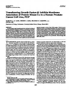
Figure 4. Representative profiles of maturation-promoting factor (MPF) amounts in equine oocytes. Lane 1: immature oocytes that were not cultured in vitro (immature control); lane 2: oocytes that remained in germinal vesicle after in-vitro culture in medium A; lane 3: oocytes in metaphase I after in-vitro culture in medium A; lane 4 and 5: oocytes in metaphase II after in-vitro culture in medium A; lane 6: degenerated oocytes after in-vitro culture in medium A; lane 7: preovulatory oocytes that reached metaphase II in vivo (mature control). Data after in-vitro culture in medium B are not shown.
Figure 3. Cyclin B1 amounts in equine oocytes. (A) Representative profiles of cyclin B1 amounts in equine oocytes. Lane 1: immature oocytes that were not cultured in vitro (immature control); lane 2: oocytes that remained in germinal vesicle after in-vitro culture in medium A; lane 3: oocytes in metaphase I after in-vitro culture in medium A; lane 4: oocytes in metaphase II after in-vitro culture in medium A; lane 5: preovulatory oocytes that reached metaphase II in vivo (mature control); lane 6: degenerated oocytes after in-vitro culture in medium A. Data after in-vitro culture in medium B are not shown. (B) Quantitative analysis of cyclin B1 amounts (n 5 number of oocytes). GV 5 oocytes in germinal vesicle after invitro culture. MI 5 oocytes in metaphase I after in-vitro culture. MII 5 oocytes in metaphase II after in-vitro culture. DEG: degenerated oocytes after in-vitro culture. IC 5 immature control. MC 5 mature control.
A typical immunoblot probed with antibody raised against cyclin B1 is presented in Figure 3A. A 65 kDa signal was observed in the oocytes that were cultured in vitro, whatever their nuclear stage and the maturation medium, in immature oocytes that were not cultured in vitro (immature control), and in metaphase II preovulatory oocytes (mature control). As illustrated in Figure 3B, the intensity of the cyclin B1 signal varied between the different nuclear stages reached after invitro culture, and tended to increase during in-vitro meiotic progression. MAPK was studied by immunoblotting, using a polyclonal anti-ERK2 antibody that recognizes the two forms of MAPK, ERK1 and ERK2. As shown in Figure 4, a lower band, close to 42 kDa, was detected in oocytes at the germinal vesicle stage after in-vitro culture in each maturation medium tested, as well as in immature oocytes that were not cultured in vitro (immature control). Metaphase I oocytes after culture in medium A or medium B exhibited an upper band, higher than 42 kDa. The retarded electrophoretic mobility, due to an increase in molecular mass, is certainly due to modifications by phosphorylation. Lanes containing metaphase II oocytes exhibited various patterns: only the upper band was detected in one lane containing metaphase II oocytes from medium A (n 5 10 oocytes) and two lanes containing metaphase II 566
oocytes from medium B (n 5 20); both the lower and upper bands were detected in two lanes containing metaphase II oocytes, one from medium A (n 5 10 oocytes) and one from medium B (n 5 10). Preovulatory oocytes that reached metaphase II in vivo (mature control) exhibited the upper band. Both the lower and upper bands were detected in the lane containing degenerated oocytes. Because of the various patterns exhibited, those were not quantified.
Discussion The aim of the present study was to investigate the relationship between the incompetence of equine oocytes for in-vitro maturation and the presence or absence of p34cdc2, cyclin B and MAPK. In order to collect oocytes from follicles with various diameters in different hormonal environments, we used invivo ultrasound-guided follicular puncture. This technique is useful for collecting well-characterized populations of oocytes. In the present study, 0.5 COCs per follicle were collected. We observed no significant influence of the follicular diameter and of the hormonal environment on the recovery rate, in agreement with our previous results (Goudet et al., 1997). During in-vitro culture, all the compact COCs underwent cumulus expansion, but only half of the oocytes completed meiosis. Therefore, cumulus expansion during in-vitro culture is not necessarily related to nuclear maturation in equine oocytes, as reported in mouse (Boland and Gosden, 1994), pig (Singh et al., 1993) and cattle (Lorenzo et al., 1994). The maturation rate tended to increase with an increase in follicular diameter, this observation reinforces our previous findings (Goudet et al., 1997). The oocyte competence for in-vitro maturation was influenced by the stage of the follicular phase at recovery; the maturation rate was lower for the oocytes from follicles punctured at the emergence of the dominant follicle (group 1) than for the oocytes from follicles punctured at the end of the dominant follicle development (groups 2, 3 and 4). One could postulate that atresia in subordinate follicles, induced by the emergence of the dominant one, increased the competence of oocytes. A higher percentage of competent oocytes in follicles in early atresia has been previously suggested in cattle (Blondin and Sirard, 1995; Sirard and Blondin, 1996) and has been
MPF and MAPK expression in equine oocyte
recently shown in the horse (Hinrichs and Williams, 1997). The competence of the oocytes was similar between the group with superovulation treatment (group 4) and the group without treatment (group 3). One can assume that, at least in the mare, high concentrations of circulating FSH during follicular phase has no effect on oocyte competence. Finally, the oocyte maturation rate was similar before (group 2) and after (group 3) the CEG injection. This injection induced the maturation of the dominant follicle but had no effect on the competence of the oocytes from subordinate follicles, in contrast with its use during the luteal phase (Goudet et al., 1997). Our results demonstrate the beneficial effect of EGF on invitro maturation of equine oocytes, as well as the deleterious effect of the crude gonadotrophins used as pituitary extract (Duchamp et al., 1987). Similar results were obtained for bovine and porcine oocytes cultured in a medium where gonadotrophins were substituted for EGF (bovine: Harper and Brackett, 1993; porcine: Coskun and Lin, 1995; Singh et al., 1997). Moreover, EGF has been shown to have a positive effect during in-vitro maturation in a variety of species (rodents: Downs, 1989; human: Das et al., 1991; Gomez et al., 1993; cattle: Lorenzo et al., 1994; Lonergan et al., 1996; pig: Ding and Foxcroft, 1994; Singh et al., 1997; rabbit: Lorenzo et al., 1996). The mechanism by which EGF stimulates oocyte maturation in mammalian species is poorly understood. EGF may act as an intraovarian regulator of oocyte maturation in response to gonadotrophin stimulation: EGF expression in hamster ovarian cells is controlled by gonadotrophins, especially FSH (Roy and Greenwald, 1990); FSH action on hamster follicular DNA synthesis is mediated by EGF (Roy and Greenwald, 1991); and the number of EGF binding sites has been shown to be influenced by both gonadotrophin and steroids (rat: St-Arnaud et al., 1983; Feng et al., 1987; pig: Fujinaga et al., 1992). Moreover, EGF may play a role in the biochemical cascade involved in the meiotic cell cycle: the EGF receptor is a transmembrane protein tyrosine kinase; binding of EGF to the receptor activates different intracellular cascades of serine/threonine kinases, one of the most important leading to activation of MAPK (for review, see Boonstra et al., 1995). We investigated the relationship between the failure of equine oocytes to resume meiosis in vitro and the presence or absence of MAPK and MPF, as well as changes in the phosphorylation state of MAPK. MPF plays a crucial role in meiotic maturation since MPF activity has been suggested to cause nuclear lamina disassembly (Peter et al., 1990a), nucleolar disassembly (Peter et al., 1990b), chromosome condensation (Moreno and Nurse, 1990), microfilament rearrangement (Morgan et al., 1989) and reorganization of the intermediate filament network (Chou et al., 1990). The present study demonstrates that the two subunits of MPF, p34cdc2 and cyclin B, were present in equine oocytes whatever the nuclear stage they reached after in-vitro culture, in immature oocytes that were not cultured in vitro, and in preovulatory oocytes that reached metaphase II after in-vivo maturation. Thus, the incompetence for in-vitro maturation in equine oocytes does not result from a lack of p34cdc2 protein, in agreement with findings in pig (Christmann et al., 1994;
Hirao et al., 1995) and rat (Goren et al., 1994). Moreover, during in vitro maturation, basal levels of p34cdc2 do not change much in bovine (Wu et al., 1997; Le´vesque and Sirard, 1996), porcine (Naito et al., 1995; Kubelka et al., 1995) and fish (Hirai et al., 1992) oocytes. Our data are consistent with these findings, since p34cdc2 is present in immature oocytes before in-vitro maturation as well as in mature oocytes after invitro or in-vivo maturation. Incompetent as well as competent equine oocytes contained the cyclin B protein, in agreement with findings in porcine (Christmann et al., 1994), goat (Hue et al., 1997) and mouse (Chesnel and Eppig, 1995; de Vante´ry et al., 1996) oocytes. The amounts of cyclin B in equine oocytes tended to increase between immature oocytes before invitro maturation and mature oocytes after in-vitro maturation, in agreement with findings in bovine oocytes (Le´vesque and Sirard, 1996; Wu et al., 1997), but tended to decrease during in-vivo maturation. Until now, no data on cyclin B amounts after in-vivo maturation of oocytes from mammals are available. Taken together, our data demonstrate that incompetent equine oocytes have the key cell cycle components for the formation of MPF. However, activation of MPF depends not only upon the presence of p34cdc2 and cyclin B, but also upon their association and a subsequent change in the phosphorylated state of key tyrosine and threonine residues on the p34cdc2 subunit (Nurse, 1990). Moreover, low levels of MPF activity can prevent oocyte maturation (Fulka et al., 1997). The incompetence of equine oocytes to resume and complete meiosis may then be due to a deficiency of such regulators of MPF. In the present study, MAPK was present in competent (in metaphase II after in-vitro culture), partially competent (in metaphase I after in-vitro culture) and incompetent (in germinal vesicle after in-vitro culture) equine oocytes, as observed in the goat (Dedieu et al., 1996), the rat (Goren et al., 1994) and the mouse (Harrouk and Clarke, 1995). MAPK is markedly involved in the resumption of meiosis: it is known to activate the MPF and induce germinal vesicle breakdown in Xenopus (Kosako et al., 1994; Haccard et al., 1995; Gotoh and Nishida, 1995 for review); it is associated with microtubules (Xenopus: Fellous et al., 1994); it is a potential regulator of microtubular activity (Xenopus: Gotoh et al., 1991; mouse: Verlhac et al., 1993; Araki et al., 1996; pig: Inoue et al., 1996; goat: Dedieu et al., 1996); and it regulates events that lead to nuclear envelope assembly and disassembly (mouse: Moos et al., 1996; chicken: Peter et al., 1992). In all mammals studied so far, the two forms of MAPK (ERK1 and ERK2) were detected in immature oocytes (pig: Inoue et al., 1995; cattle: Fissore et al., 1996; mouse: Verlhac et al., 1993, 1994). After meiotic maturation, both forms displayed a retarded electrophoretic mobility due to an increase in molecular mass, consistent with modification by phosphorylation. Phosphorylation of MAPK is associated with a marked increase in its kinase activity (Anderson et al., 1990). In equine oocytes, only the form of 42 kDa (ERK2) was detected at the germinal vesicle stage whereas in metaphase I and metaphase II, ERK2 exhibited a decreased mobility through SDS–PAGE, certainly because of its phosphorylation. ERK1 was not detected in equine oocytes either because the band was considerably less intense than the 567
G.Goudet et al.
band representing ERK2, as reported in bovine oocytes (Fissore et al., 1996) or because only ERK2 is present, as reported in Xenopus (Gotoh et al., 1991; Polverino et al., 1995). Thus, in incompetent equine oocytes as well as in immature oocytes before in-vitro maturation, MAPK remained non-phosphorylated and probably inactive, whereas in competent, partially competent and preovulatory equine oocytes, MAPK became phosphorylated and probably with a high kinase activity. These results suggest that: (i) the failure of incompetent oocytes to reach metaphase I may be linked to an inability to phosphorylate MAPK; and (ii) the failure of partially competent oocytes to complete meiosis I reflects a defect acting downstream or independently of MAPK phosphorylation. One must notice that, in the present study, the oocytes in germinal vesicle stage could not be differentiated from the oocytes after germinal vesicle breakdown. In goat (Dedieu et al., 1996), pig (Inoue et al., 1995) and mouse oocyte (Sobajima et al., 1993; Verlhac et al., 1994), MAPK activation occurs beyond MPF activation and germinal vesicle breakdown (GVBD), and may thus not be involved in the early events of oocyte maturation, but rather in the regulation of post-GVBD events such as spindle formation. Further studies are required to specify MAPK activity in the early events of equine oocyte maturation, and the reasons for the inability to phosphorylate MAPK in incompetent equine oocytes. Finally, both the lower and upper bands of ERK2 were detected in degenerated oocytes as well as in some of the oocytes that reached metaphase II after invitro maturation. The presence of the inactive form in the latters could be a sign of degeneration after in-vitro maturation and could be linked to the meiotic spindle disorganization observed in some metaphase II equine oocytes after in-vitro maturation (Goudet et al., 1997). No obvious difference in the presence and phosphorylation state was observed between the two culture media tested in the present study. Actually, as noticed above, EGF has been shown to have an effect on MAPK activity (for review, see Kosako et al., 1994; Boonstra et al., 1995). Further studies are required to specify this field in equine oocyte. In conclusion, the acquisition of meiotic competence in equine oocytes occurred progressively during antral follicle growth and is influenced by the estrus cycle stage. Since this study has shown the presence of p34cdc2, cyclin B and MAPK in partially competent and incompetent equine oocytes, the incompetence to resume meiosis is not due to the absence of these proteins. It may be due to a deficiency of regulators of MPF and/or to an inability to phosphorylate MAPK.
Acknowledgements This work was supported by grants from the Institut National de la Recherche Agronomique, France, and the Haras Nationaux, France. Ghyle`ne Goudet was supported by a fellowship from the Institut National de la Recherche Agronomique, France, and the Re´gion Centre, France. We wish to thank Dr Laurence Gall (INRA, Jouy en Josas, France) for her kind advice during Western blot experiments, Dr Masakane Yamashita (Zoological Institute, Sapporo, Japan) and Dr Fergus R.McKenzie (CNRS, Nice, France) for the gift of the antibodies against cyclin B and MAPK respectively, Guy Duchamp for critical discussion and technical assistance, and Eric Palmer for being at the origin of our work. We are grateful to Isabelle Couty,
568
Monique Ottogalli and the staff of the experimental stud for technical assistance and Alain Beguey and Odile Moulin for photographic work.
References Anderson, N.G., Maller, J.L., Tonks, N.K. and Sturgill, T.W. (1990) Requirement for integration of signals from two distinct phosphorylation pathways for activation of MAP kinase. Nature, 343, 651–653. Araki, K., Naito, K., Haraguchi, S. et al. (1996) Meiotic abnormalities of cmos knockout mouse oocytes: activation after first meiosis or entrance into third meiotic metaphase. Biol. Reprod., 55, 1315–1324. Blondin, P. and Sirard, M.A. (1995) Oocyte and follicular morphology as determining characteristics for developmental competence in bovine oocytes. Mol. Reprod. Dev., 41, 54–62. Boland, N.I. and Gosden, R.G. (1994) Effects of epidermal growth factor on the growth and differentiation of cultured mouse ovarian follicles. J. Reprod. Fertil., 101, 369–374. Boonstra, J., Rijken, P., Humbel, B. et al. (1995) The epidermal growth factor. Cell Biol. Int., 19, 413–430. Chesnel, F. and Eppig, J.J. (1995) Synthesis and accumulation of p34cdc2 and cyclin B in mouse oocytes during acquisition of competence to resume meiosis. Mol. Reprod. Dev., 40, 503–508. Choi, T., Aoki, F., Mori, M. et al. (1991) Activation of P34cdc2 protein kinase activity in meiotic and mitotic cell cycles in mouse oocytes and embryos. Development, 113, 789–795. Chou, Y.-H., Bischoff, J.R., Beach, D. and Goldman, R.D. (1990) Intermediate filament reorganization during mitosis is mediated by p34cdc2 phosphorylation of vimentin. Cell, 62, 1063–1071. Christmann, L., Jung, T. and Moor, R.M. (1994) MPF components and meiotic competence in growing pig oocytes. Mol. Reprod. Dev., 38, 85–90. Coskun, S. and Lin, Y.C. (1995) Mechanism of action of epidermal growth factor-induced porcine oocyte maturation. Mol. Reprod. Dev., 42, 311–317. Crosby, I.M., Osborn, J.C. and Moor, R.M. (1984) Changes in protein phosphorylation during the maturation of mammalian oocytes in vitro. J. Exp. Zool., 229, 459–466. Das, K., Stout, L.E., Hensleigh, H.C. et al. (1991) Direct positive effect of epidermal growth factor on the cytoplasmic maturation of mouse and human oocytes. Fertil. Steril., 55, 1000–1004. Dedieu, T., Gall, L., Crozet, N. et al. (1996) Mitogen-activated protein kinase activity during goat oocyte maturation and the acquisition of meiotic competence. Mol. Reprod. Dev., 45, 351–358. De Smedt, V., Crozet, N. and Gall, L. (1994) Morphological and functional changes accompanying the acquisition of meiotic competence in ovarian goat oocyte. J. Exp. Zool., 269, 128–139. De Vante´ry, C., Gavin, A.C., Vassalli, J.D. and Schorderet-Slatkine, S. (1996) An accumulation of p34cdc2 at the end of mouse oocyte growth correlates with the acquisition of meiotic competence. Dev. Biol., 174, 335–344. Ding, J. and Foxcroft, G.R. (1994) Epidermal growth factor enhances oocyte maturation in pigs. Mol. Reprod. Dev., 39, 30–40. Downs, S.M. (1989) Specificity of epidermal growth factor action on maturation of the murine oocyte and cumulus oophorus in vitro. Biol. Reprod., 41, 371–379. Draetta, G., Luca, F., Westendorf, J. et al. (1989) cdc2 protein kinase is complexed with both cyclin A and B: evidence for proteolytic inactivation of MPF. Cell, 56, 829–838. Duchamp, G., Bour, B., Combarnous, Y. and Palmer, E. (1987) Alternative solutions to hCG induction of ovulation in the mare. J. Reprod. Fertil., 35 (Suppl.), 221–228. Duchamp, G., Be´zard, J. and Palmer, E. (1995) Oocyte yield and the consequences of puncture of all follicles larger than 8 millimeters in mares. In Sharp, D.C. and Bazer, F.W. (eds), Equine Reproduction VI. Society for the Study of Reproduction, Madison, WI, USA, pp. 233–241. Dunphy, W.G., Brizuela, L., Beach, D. and Newport, J. (1988) The Xenopus cdc2 protein is a component of MPF, a cytoplasmic regulator of mitosis. Cell, 54, 423–431. Fellous, A., Kubelka, M., Thibier, C. et al. (1994) Association of p34cdc2 kinase and MAP kinase with microtubules during the meiotic maturation of Xenopus oocytes. Int. J. Dev. Biol., 38, 651–659. Feng, P., Knecht, M. and Catt, K. (1987) Hormonal control of epidermal growth factor receptors by gonadotropins during granulosa cell differentiation. Endocrinology, 120, 1121–1126.
MPF and MAPK expression in equine oocyte Ferrell, J.E.Jr., Wu, M., Gerhart, J.C. and Martin, G.S. (1991) Cell cycle tyrosine phosphorylation of p34cdc2 and a microtubule-associated protein kinase homolog in Xenopus oocytes and eggs. Mol. Cell. Biol., 11, 1965–1971. Fissore, R.A., He, C.L. and Vande Woude, G.F. (1996) Potential role of mitogen-activated protein kinase during meiosis resumption in bovine oocytes. Biol. Reprod., 55, 1261–1270. Fujinaga, H., Yamoto, M., Nakano, R. and Shima, K. (1992) Epidermal growth factor binding sites in porcine granulosa cells and their regulation by follicle-stimulating hormone. Biol. Reprod., 46, 705–709. Fulka Jr, J., Kalab, P., First, N.L. and Moor, R.M. (1997) Damaged chromatin does not prevent the exit from metaphase I in fused mouse oocytes. Hum. Reprod., 12, 2473–2476. Gomez, E., Tarin, J.J. and Pellicer, A. (1993) Oocyte maturation in humans: the role of gonadotropins and growth factors. Fertil. Steril., 60, 40–46. Goren, S., Piontkewitz, Y. and Dekel, N. (1994) Meiotic arrest in incompetent rat oocytes is not regulated by cAMP. Dev. Biol., 166, 11–17. Gotoh, Y. and Nishida, E. (1995) Activation mechanism and function of the MAP kinase cascade. Mol. Reprod. Dev., 42, 486–492. Gotoh, Y., Nishida, E., Matsuda, S. et al. (1991) In vitro effects on microtubule dynamics of purified Xenopus M phase-activated MAP kinase. Nature, 349, 251–254. Goudet, G., Be´zard, J., Duchamp, G. et al. (1997) Equine oocyte competence for nuclear and cytoplasmic in vitro maturation: effect of follicle size and hormonal environment. Biol. Reprod., 57, 232–245. Haccard, O., Lewellyn, A., Hartley, R.S., et al. (1995) Induction of Xenopus oocyte meiotic maturation by MAP kinase. Dev. Biol., 168, 677–682. Harper, K.M. and Brackett, B.G. (1993) Bovine blastocyst development after in vitro maturation in a defined medium with epidermal growth factor and low concentrations of gonadotropins. Biol. Reprod., 48, 409–416. Harrouk, W. and Clarke, H.J. (1995) Mitogen-activated protein (MAP) kinase during the acquisition of meiotic competence by growing oocytes of the mouse. Mol. Reprod. Dev., 41, 29–36. Hashimoto, N. and Kishimoto, T. (1988) Regulation of meiotic metaphase by a cytoplasmic maturation-promoting factor during mouse oocyte maturation. Dev. Biol., 126, 242–252. Hinrichs, K. and Williams, K.A. (1997) Relationships among oocyte–cumulus morphology, follicular atresia, initial chromatin configuration, and oocyte meiotic competence in the horse. Biol. Reprod., 57, 377–384. Hirai, T., Yamashita, M., Yoshikuni, M. et al. (1992) Cyclin B in fish oocytes: its cDNA and amino acid sequences, appearance during maturation, and induction of p34cdc2 activation. Mol. Reprod. Dev., 33, 131–140. Hirao, Y., Tsuji, Y., Miyano, T. et al. (1995) Association between p34cdc2 levels and meiotic arrest in pig oocytes during early growth. Zygote, 3, 325–332. Hofferer, S., Lecompte, F., Magallon, T. et al. (1993) Induction of ovulation and superovulation in mares using equine LH and FSH separated by hydrophobic interaction chromatography. J. Reprod. Fertil., 98, 597–602. Hue, I., Dedieu, T., Huneau, D. et al. (1997) Cyclin B1 expression in meiotically competent and incompetent goat oocytes. Mol. Reprod. Dev., 47, 222–228. Inoue, M., Naito, K., Aoki, F., et al. (1995) Activation of mitogen-activated protein kinase during meiotic maturation in porcine oocytes. Zygote, 3, 265–271. Inoue, M., Naito, K., Nakayama, T. and Sato, E. (1996) Mitogen-activated protein kinase activity and microtubule organisation are altered by protein synthesis inhibition in maturing porcine oocytes. Zygote, 4, 191–198. Irvine, C.H.G. and Alexander, S.L. (1994) The dynamics of gonadotrophinreleasing hormone, LH and FSH secretion during the spontaneous ovulatory surge of the mare as revealed by intensive sampling of pituitary venous blood. J. Endocrinol., 140, 283–295. Jelinkova, L., Kubelka, M., Motlik, J. and Guerrier, P. (1994) Chromatin condensation and histone H1 kinase activity during growth and maturation of rabbit oocytes. Mol. Reprod. Dev., 37, 210–215. Kastrop, P.M.M., Bevers, M.M., Destre´e, O.H.J. and Kruip, Th.A.M. (1990) Changes in protein synthesis and phosphorylation patterns during bovine oocyte maturation in vitro. J. Reprod. Fertil., 90, 305–310. Kosako, H., Gotoh, Y. and Nishida, E. (1994) Regulation and function of the MAP kinase cascade in Xenopus oocytes. J. Cell Sci., 18 (Suppl.), 115–119. Kubelka, M., Rimkevicova, Z., Guerrier, P. and Motlik, J. (1995) Inhibition of protein synthesis affects histone H1 kinase, but not chromosome condensation activity, during the first meiotic division of pig oocytes. Mol. Reprod. Dev., 41, 63–69. Laemmli, U.K. (1970) Cleavage of structural proteins during the assembly of the head of bacteriophage T4. Nature, 227, 680–685.
Le´vesque, J.T. and Sirard, M.-A. (1996) Resumption of meiosis is initiated by the accumulation of cyclin B in bovine oocytes. Biol. Reprod., 55, 1427–1436. Lonergan, P., Carolan, C., Van Langendonckt, A. et al. (1996) Role of epidermal growth factor in bovine oocyte maturation and preimplantation embryo development in vitro. Biol. Reprod., 54, 1420–1429. Lorenzo, P.L., Illera, M.J., Illera, J.C. and Illera, M. (1994) Enhancement of cumulus expansion and nuclear maturation during bovine oocyte maturation in vitro by the addition of epidermal growth factor and insulin-like growth factor I. J. Reprod. Fertil., 101, 697–701. Lorenzo, P.L., Rebollar, P.G., Illera, M.J. et al. (1996) Stimulatory effect of insulin-like growth factor I and epidermal growth factor on the maturation of rabbit oocytes in vitro. J. Reprod. Fertil., 107, 109–117. Maller, J., Wu, M. and Gerhart, J.C. (1977) Changes in protein phosphorylation accompanying maturation of Xenopus laevis oocytes. Dev. Biol., 58, 295–312. Masui, Y. and Markert, C.L. (1971) Cytoplasmic control of nuclear behavior during meiotic maturation of frog oocytes. J. Exp. Zool., 177, 129–146. Moos, J., Xu, Z., Schultz, R.M. and Kopf, G.S. (1996) Regulation of nuclear envelope assembly/disassembly by MAP kinase. Dev. Biol., 175, 358–361. Moreno, S. and Nurse, P. (1990) Substrates for p34cdc2: In Vivo Veritas? Cell, 61, 549–551. Morgan, D.O., Kaplan, J.M., Bishop, J.M. and Varmus, H.E. (1989) Mitosisspecific phosphorylation of p60c-src by p34cdc2-associated protein kinase. Cell, 57, 775–786. Naito, K. and Toyoda, Y. (1991) Fluctuation of histone H1 kinase activity during meiotic maturation in porcine oocytes. J. Reprod. Fertil., 93, 467–473. Naito, K., Hawkins, C., Yamashita, M. et al. (1995) Association of p34cdc2 and cyclin B1 during meiotic maturation in porcine oocytes. Dev. Biol., 168, 627–634. Nurse, P. (1990) Universal control mechanism regulating onset of M-phase. Nature, 344, 503–508. Palmer E. (1984) Recent attempts to improve synchronisation of ovulation and to induce superovulation in the mare. Equine Vet. J., 3 (Suppl.), 11–18. Palmer, E. and Driancourt, M.A. (1980) Use of ultrasonic echography in equine gynecology. Theriogenology, 13, 203–206. Peter, M., Nakagawa, J., Dore´e, M. et al. (1990a) In vitro disassembly of the nuclear lamina and M phase-specific phosphorylation of lamins by cdc2 kinase. Cell, 61, 591–602. Peter, M., Nakagawa, J., Dore´e, M. et al. (1990b) Identification of major nucleolar proteins as candidate mitotic substrates of cdc2 kinase. Cell, 60, 791–801. Peter, M., Sanghera, J.S., Pelech, S.L. and Nigg, E.A. (1992) Mitogenactivated protein kinases phosphorylate nuclear lamins and display sequence specificity overlapping that of mitotic protein kinase p34cdc2. Eur. J. Biochem., 205, 287–294. Polverino, A., Frost, J., Yang, P. et al. (1995) Activation of mitogen-activated protein kinase cascades by p21-activated protein kinases in cell-free extracts of Xenopus oocytes. J. Biol. Chem., 270, 26067–26070. Roy, S.K. and Greenwald, G.S. (1990) Immunohistochemical localization of epidermal growth factor-like activity in the hamster ovary with a polyclonal antibody. Endocrinology, 126, 1309–1317. Roy, S.K. and Greenwald, G.S. (1991) Mediation of follicle-stimulating hormone action on follicular deoxyribonucleic acid synthesis by epidermal growth factor. Endocrinology, 129, 1903–1908. Singh, B., Barbe, G.J. and Armstrong, D.T. (1993) Factors influencing resumption of meiotic maturation and cumulus expansion of porcine oocyte– cumulus cell complexes in vitro. Mol. Reprod. Dev., 36, 113–119. Singh, B., Meng, L., Rutledge, J.M. and Armstrong D.T. (1997) Effects of epidermal growth factor and follicle-stimulating hormone during in vitro maturation on cytoplasmic maturation of porcine oocytes. Mol. Reprod. Dev., 46, 401–407. Sirard, M.A. (1989) Practical aspects of in vitro fertilization in cattle. J. Reprod. Fertil., 38 (Suppl.), 127–134. Sirard, M.A. and Blondin, P. (1996) Oocyte maturation and IVF in cattle. Anim. Reprod. Sci., 42, 417–426. Sobajima, T., Aoki, F. and Kohmoto, K. (1993) Activation of mitogenactivated protein kinase during meiotic maturation in mouse oocytes. J. Reprod. Fertil., 97, 389–394. Squires, E.L. (1996) Maturation and fertilization of equine oocytes. Vet. Clin. N. Am. (Equine Pract.), 12, 31–45.
569
G.Goudet et al. St.-Arnaud, R., Walker, P., Kelly, P.A. and Labrie, F. (1983) Rat ovarian epidermal growth factor receptors: characterization and hormonal regulation. Mol. Cell. Endocrinol., 31, 43–52. Verlhac, M-H., De Pennart, H., Maro, B. et al. (1993) MAP kinase becomes stably activated at metaphase and is associated with microtubule-organizing centers during meiotic maturation of mouse oocytes. Dev. Biol., 158, 330–340. Verlhac, M.-H., Kubiak, J.Z., Clarke, H.J. and Maro, B. (1994) Microtubule and chromatin behavior follow MAP kinase activity but not MPF activity during meiosis in mouse oocytes. Development, 120, 1017–1025. Whitmore, H.L., Wentworth, B.C. and Ginther, O.J. (1973) Circulating concentrations of luteinizing hormone during estrous cycle of mares as determined by radioimmunoassay. Am. J. Vet. Res., 34, 631–636. Wu, B., Ignotz, G., Currie, W.B. and Yang, X. (1997) Dynamics of maturationpromoting factor and its constituent proteins during in vitro maturation of bovine oocytes. Biol. Reprod., 56, 253–259. Received on December 18, 1997; accepted on March 23, 1998
570