extracardiac activity (17). For scintimammography using. 99mTc..methoxyisobutyl isonitrile, recently published data clearly favor the MLEM and OSEM, with 10% ...
Maximum-Likelihood Reconstruction with Ordered Subsets in Bone SPECT Didier Blocklet, Alain Seret, Niculaie Popa and AndréSchoutens Hôpital Universitaire Erasme, Radioisotopes, UniversitéLibre de Bruxelles, Brussels, Belgium
the lumbar spine. In articulations, This study was aimed at determining whetherthe ordered-subset expectation maximum (OSEM) is more effective than filtered backprojection (FBP) for bone SPECT in the routine clinical
context. Methods. Fifty-seven consecutive bone SPECT studies were analyzed. They included pelvic and lumbar spine, thoraco lumbar spine, head and neck, feet and shoulders. A64-projection SPECT study was acquired over 360°by single-head cameras 2—3h after the injection of 750 MBq @Tc-methyIene diphospho nate. Three observers compared the OSEM and FBP recon structed images. Results. Streak artifacts, always present with FBP, were rarely generated with the OSEM. When present (n = 24), artifacts associated with negative values near hyperactivities
in FBP were not generated with the OSEM in 67% of the cases (n = 16), permitting a satisfactory interpretation ofthese regions. In half of the other cases
(17%, n = 4/24), interpretation was
precluded. In only one case did the three observers agree that more hyperactivities were seen with the OSEM. Ninety-six percent of the OSEM pictures were superior or equal to FBP for anatomic resolution and were clearly better in 12% of the cases. The extent of the lesion with the OSEM seemed better or equally defined in 96% and clearly better in 14% of the cases. The low-activity regions were better or equally visualized in all cases
and were clearly better seen in 23% of the cases. The quality of the pictures was found to be better or superior with the OSEM in 98% of the cases and definitely better in 65% of the cases. Conclusion:Replacementof FBPbytheOSEM inboneSPECT would be beneficial to clinical practice. KeyWords:boneSPECT;filteredbackprojection; iterativerecon struction; maximum likelihood of expectation maximization; or dered-subset expectation maximum
J NucIMed1999;40:1978—1984
rounding
may be partially
masked
the nonpathologic
sur
by the streak artifact
centered on the areas of hyperactivities and the negative values in the vicinity of these hyperactivities. The maximum likelihood of expectation maximization (MLEM) algorithm does not suffer these limitations. No filter must be chosen. The reconstructed
spatial resolution is
better than that achieved with only ramp-FBP for an equivalent noise level (9). The streak artifacts should not appear, and the positivity (absence of negative values) of the reconstruction is guaranteed (3,6, 10). Finally, the algorithm converges (3, 10), but slowly (6, 11). The major drawback of the method is the high number of iterations that are required. Indeed, the higher the number of iterations, the higher the noise level (9, 12). Therefore, stopping rules must be defined either by limitation of the iteration number or by use of an a priori constraint (6,9,13). The MLEM has been widely studied theoretically and on phantoms. To our knowledge, the MLEM has been rarely used in clinical research, probably because of the powerful computer system needed (6). However, present-day workstations are able to perform this kind of processing in a relatively short time. The use of
the ordered subsets (OS) accelerating procedure further shortens the reconstruction time (6). The aim of this study was to determine whether the fast iterative reconstruction method, the ordered-subset expecta tion maximum (OSEM), is more effective, essentially from a qualitative point of view, than classical FBP for bone SPECT in the routine clinical context.
MATERIALSAND METHODS n the routine clinical context, filtered backprojection (FBP) has been for many years the only reconstruction method applied to bone SPECT. In addition to the loss of resolution associated with the filter and the difficulty in choosing the more appropriate filter (1,2), FBP may generate artifacts, mainly streaking artifacts and negative values (3—6) near the border of intense hyperactivities. In SPECT of the pelvis, the artifacts originating from the bladder activity may partially or totally obscure the hips (4, 7,8). Highly active kidneys are a frequent problem in SPECT studies of
Materials From 15 June to 30 September 1997, 57 consecutive bone SPECT studies were analyzed. All patients referred to this depart ment for bone SPECT were included in the study. The regions of interest were the pelvic and lumbar spine (n = 48), the thoracolum bar spine (n = 4), the head and the neck (n = 3), the feet (n = 1)
and the shoulders (n = 1).
Methods For each patient, a whole-body scan was acquired 2—3h after
injection of750 MBq @“Tc-methylene diphosphonate, followed by 64-projection (20 s per projection) tomography over 360°in a 64 X 64 format. This acquisition protocol follows the recommendations ReceivedNov.11,1998;revisionacceptedApr.19,1999. of the Society of Nuclear Medicine (14). Single-head cameras were For correspondencecontact: DidierBlocklet,MD, Hôpital Universitaire used: Sophy DSX (Sopha Medical Vision, Buc, France) and Erasme, Radioisotopes,Route de Lennik808, B-1070Bruxelles,Belgium.
1978
THE JOURNALOF NUCLEAR MEDICINE •Vol. 40 •No. 12 •December
1999
@
3 General Electric AC400 (General Electric Medical Systems, Mil
criteria: presence and extent of streak and negative-value artifacts
waukee, WI). The Sophy camera was equipped with the low energy, ultra-high-resolution collimator, and an electronic zoom of
near hyperactivities, number of lesions, definition (anatomic defini
8/6 provided a pixel size of 6.9 mm. The General Electric camera
quality of the pictures (general impression).
tion, lesion extent and visualization of low-activity regions) and
had a low-energy, high-resolution collimator, and the pixel size was
6.4 mm. The resolution at 10 cm (full width at half maximum for a point source in air) was 6.5 ±0.3 mm for the Sophy system and 8.0 ±0.3 mm for the General Electric camera. Both systems had almost identical sensitivities of5.0 ±0.5 cpmlkBq. FBP reconstruction was performed using Vision software (SMV).
A Hanningfilterwitha cutofffrequency of 0.5 cycle/pixel was
RESULTS Results are presented regardless of the anatomic regions examined because they are not influenced by topography.
applied in the course of the backprojection.
Artifacts
The iterative reconstruction algorithm was the OSEM imple mented as a two-dimensional reconstruction. Neither attenuation
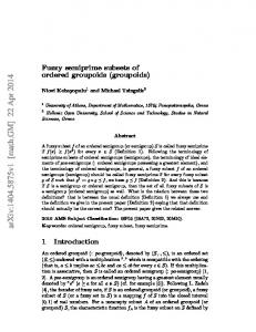
Streak artifacts, which are always present with FBP, were rarely generated with the OSEM (Fig. 1 and Table 1). A paravenous injection in each of two patients who were
correction or resolution recovery was attempted. Two iterations were applied to eight subsets. This subset—iterationcombination gave visually the best images. For the 64 transverse slices, the reconstruction time was 3 s for FBP and 14 s for the OSEM on an RISC 6000 3AT workstation (International Business Machines, White Plains, NY). All acquisitions were reconstructed by both methods (i.e., FBP and OSEM). None of the reconstructions was postfiltered. Trans
verse, coronal and sagittal slices were displayed with a gray scale and printed on black background. Analysis of Results The 57 SPECT studies were submitted to visual analysis by three observers. The paired reconstructions were randomized, and
the method of reconstruction was not indicated. Two of the observers had experience with FBP and OSEM pictures; the other observer had experience with only FBP pictures. The observers compared FBP and OSEM pictures for different
S
13
unable
to raise their arms caused
cases,
FBP streak
permitting
a satisfactory
interpretation
of these re
gions (Figs. 3 and 4). However, in 33% (n = 8) of the cases, OSEM provided unsatisfactory images. In half of these cases (17%, n = 4), interpretation was impossible (Fig. 2).
Number of Lesions The three observers
agreed in only one case on the
presence of an additional hyperactivity
on the OSEM picture
(Fig. 5 and Table 2).
p
14
13
14
FBP
OSEM
30
31
32
30
31
32
FIGURE1.
significant
artifacts that were less intense in the OSEM pictures (Figs. 2 and 3). When present (n = 24), artifacts associated with negative values near hyperactive regions were not generated or only minimally generated by the OSEM in 67% (n = 16) of the
FRP
OSEM
33
34
33
34
The onlyshoulderSPECTstudyperformed in thisserieswasblurredby streakartifactsgeneratedby filtered
backprojection (FBP) reconstruction technique, but these artifacts were not present with ordered-subset
expectation maximum
(OSEM). Resolution seems to be inferiorwith FBP.
OSEM AND BONE SPECT
•Blocklet et al.
1979
@
.@
@,
TABLE1
Definition
Common Artifacts Generated by FBP Reconstruction Method
Ninety-six
Artifactor ofvalue negativeNo. Generation Streak artifacts
lesions
Not generated by OSEM when pre
%
57/57
100
sent in FBP (n = 57)
Stillpresent withOSEM
0/57
Negative values added Not generated or mini near hyperactivities mallygenerated by OSEM (interpret able) when present
16/24
0 66.6
percent of the OSEM pictures
clearly were better seen in 23% of the cases.
GeneralImpression The quality of the pictures obtained was better or identical with the OSEM than with FBP in 98% of the cases (Table 4). The quality was definitely better with the OSEM in 65% of the cases (Figs. 1, 2 and 4).
in FBP (n = 24) Also generated by OSEM
Found uninterpret able by 2/3 of observers
4/24
16.7
Found interpretable by 2/3 of observers
4/24
16.7
DISCUSSION
FBP = filteredbackprojection;OSEM = ordered-subset expecta tion maximum. Allstreak artifacts and most negative values were not observed in OSEM pictures.
In this study, FBP is state of the art in routine clinical bone SPECT. The filter used in FBP remains the user's choice; the Hanning filter is used routinely for bone SPECT at this institution.
Because this filter is popular and is available
subset number
•*..—@--
can be found in previous
studies
,,@@L;
(6, 11) and
as follows. Use the highest number of
@@::z@-..@-@-
.
@_.4#..$T'
in
virtually all commercial software, we used it in this study. The number of subsets and the number of iterations must be set with the OSEM technique. Recommendations for the are summarized
@
were superior or
equal to FBP for anatomic definition (Table 3). Moreover, in 12% of the pictures, all observers agreed that the anatomic definition with the OSEM was clearly better. The lesion area with the OSEM seemed to be inferior or equal to FBP in 96% of the cases. It was obviously inferior in 14% of the cases (Fig. 6). The low-activity regions were better visualized or equally visualized in all cases and
,@
@.
j 0t .;
al
£ FBP
!@:
-,@,
-.@..@
,,1@a•:@
‘e@1@',: I
@OSEM OSEM
—
-
Ii
FIGURE 2.
•
11
4.;
Importantartifactswere generated by filtered backprojection(FBP) after reconstructionof pelvicand lumbar SPECT
images in patient with paravenous injection in left arm, which entailed low statistics counting. Patient was not able to raise the arms and presented with important bladder residual activity. Ordered-subset expectation maximum (OSEM) helped with both artifacts, erasing Li (paravenous injection, transverse slice 11) and erasing both hips (bladder activity, transverse slice 43). Images remained
unsatisfactory, but general outlook improved with OSEM.
1980
THEJOURNAL OFNUCLEAR MEDICINE• Vol. 40 • No. 12 • December 1999
*
FBP d@
@
‘@
,-
.
a
l@
@p
•d/'
.@.
I ---@ @,;---‘@
—
-@
p @
36
*
‘
.il•I@@
:ii
V@
.,j
OSEM
I
.1,
@
@
-
‘
C-
:@
1%
OSEM
I S
p
36
17
‘I
4'—'
‘
ti;
@•
@.
FIGURE 3. Secondpatientwithparavenousinjectionof tracerwas unableto raisethe arms.Withordered-subsetexpectation maximum (OSEM), streak artifact was of lower intensity than with filtered backprojection (FBP), making examination interpretable. subsets while keeping at least 4 projections per subset. The higher the subset number, the more enhanced the reconstruc tion speed.
Below
4 projections
per subset,
significant
differences could arise between MLEM and OSEM recon structions (15).
With eight subsets and 64 projections, all subsets contain 8 projections.
In this way, we are not too close to the limit of
4 projections per subset, and the reconstruction time is divided by 8 in comparison with the MLEM. Once the number of subsets is chosen, the number of iterations
- ‘
4
‘
FBP
a
4$
59
(@0
t, I
V.,-.
59
•.
a
4.
FBP@
...,..‘
-4
S ,
@
,@4
,
34@―@lI@
@
-
@
LOSEM
:1(
:t ,‘
.
@t& :i@@
@1@.
@@IIII@
.*. h
‘1IIIIIIPP@
FIGURE 4. In somepatients,hipswere almosterasedby filteredbackprojection (FBP) technique,whichgeneratesnegative values near high bladder activity.Ordered-subset expectation maximum (OSEM) method leads to interpretable pictures.
OSEM AND BONE SPECT
•Blocklet et al.
1981
A
FBP
@ @
0
A
A
16
17
I
‘2t)
OSEM
,
•@lk
lE)
it
, ‘,
I (;Ø
4i
!
‘
@1_@- OSEM
44
,
p
41
@
B
@
@ @
i:'
FBP-Hann
@
.
S
U
I
•\
FIGURE5.
i:i
FBP-Ramp V
S
7
@‘
20
I
FBP
it @
4'
17
I
•
14
FBP-Butterworth
‘
0 a
f
)
• ;,
OSEM .
. •\
a
@•
5,
4
1@
(A)Suspected borderline lesionwasdetectedwithonlyordered-subset expectation maximum (OSEM)reconstruc
tion method (arrow). Other lesions were also more precise with OSEM than with filtered backprojection (FBP). (B) Borderline lesion
was visualized with FBP using less smoothing filter(ramp or Butterworth filter,order 10 and cutoff 0.5 cycle/pixel) than Hanning (Hann) filter (cutoff 0.5 cycle/pixel). entirely controls the final resolution,
contrast and noise of
the reconstructed images (15). The limiting factor here is the noise. It first decreases with the iteration number. After a few
tens of iterations in the MLEM and only a few iterations in the OSEM (with eight subsets), the noise starts to increase dramatically. A preliminary study of 10 cases (not included in this study) showed that two iterations gave visually the
1982
best compromise. Postfiltering of the reconstructed data or a noise regularization method based on priors could change
the compromise toward more iterations, and therefore better contrast, but at the cost of a loss of resolution (6,9,12,13). The FBP SPECT reconstruction method has some disad vantages. The most important disadvantages in spine SPECT are streak artifacts (Figs.
THE JOURNALOF NUCLEAR MEDICINE •Vol. 40 •No. 12 •December
1—3)and negative
1999
values in the
TABLE2
borderof highlyactiveregions.In pelvicSPECT,negative
Number of Lesions
values, sometimes almost erasing the hips (Figs. 2 and 4), are generated when significant bladder activity is present
No.ofNo.ofReconstruction
method
@ @ @
Same number withboth methods More lesions seen withOSEM
3
48 @l
More lesions seen with FBP
Same number withboth methods More lesions withOSEM More lesions with FBP Same number or one more lesion
(4,7,8). The choiceof a correctfilter is not alwayseasy.
%
observerslesions
Moreover, any filtering operation leads to a global smooth
84.2
. activity on the nearby pixels (1,2,5). 1@
2
0
3
57
ing ofthe reconstructed volume. In particular, the hyperactiv ity becomes less focal because of spreading out of the In contrast, OSEM methods do not use any filter. This study shows that the OSEM is able to eliminate, at least
partially, almost all artifacts observed with FBP. With FBP, 100
24 patients presented with significant
generally observed with both reconstruction methods. In very few cases, one more borderline lesion was detected withOSEM.
TABLE3
paravenous injection,remindusthattheacquisitionquality
Definition
remains the key point of any scintigraphic investigation.
OSEM = ordered-subset expectation maximum; FBP = filtered backprojection. Total number of lesions = 57. Same number of lesions was
No lesion was missed No.ofNo.ofParameter observerslesions
Anatomicdefinition Superior withOSEM Identicalwithboth techniques Inferior with OSEM
@
negative artifacts near
the bladder. Better results were obtained with the OSEM (Fig. 4), but this technique failed to satisfactorily eliminate the negative-value artifacts in 4 patients (Fig. 2). Measure ment of bladder activity in the first and last projections of the SPED' study showed that the bladder contained high activity without further filling (