Biochimica et Biophysica Acta 1838 (2014) 2399–2403
Contents lists available at ScienceDirect
Biochimica et Biophysica Acta journal homepage: www.elsevier.com/locate/bbamem
MCLIP, an effective method to detect interactions of transmembrane proteins of the nuclear envelope in live cells Mohammed Hakim Jafferali a, Balaje Vijayaraghavan a, Ricardo A. Figueroa a, Ellinor Crafoord a, Santhosh Gudise a,b, Veronica J. Larsson a, Einar Hallberg a,⁎ a b
Department of Neurochemistry, Stockholm University, SE10691 Stockholm, Sweden Department of Biosciences and Nutrition, Karolinska Institute (Novum), SE14189 Huddinge, Sweden
a r t i c l e
i n f o
Article history: Received 15 May 2014 Received in revised form 9 June 2014 Accepted 10 June 2014 Available online 17 June 2014 Keywords: Samp1 Nuclear envelope Nuclear membrane Crosslinking CoIP Protein–protein interaction
a b s t r a c t Investigating interactions of proteins in the nuclear envelope (NE) using co-immunoprecipitation (Co-IP) has previously been difficult or even impossible due to their inherent resistance to extraction. We have developed a novel method, MCLIP (Membrane protein Cross-Link ImmunoPrecipitation), which takes advantage of a cell permeable crosslinker to enable effective detection and analysis of specific interactions of NE proteins in live cells using Western blot. Using MCLIP we show that, in U2OS cells, the integral inner nuclear membrane protein Samp1 interacts with Lamin B1, the LINC (Linker of nucleoskeleton and cytoskeleton) complex protein, Sun1 and the soluble small GTPase Ran. The results show that the previously detected in vitro interaction between Samp1 and Emerin also takes place in live cells. In vitro pull down experiments show, that the nucleoplasmic domains of Samp1 and Emerin can bind directly to each other. We also, show that MCLIP is suitable to coprecipitate protein interactions in different stages of the cell cycle. © 2014 The Authors. Published by Elsevier B.V. This is an open access article under the CC BY-NC-ND license (http://creativecommons.org/licenses/by-nc-nd/3.0/).
1. Introduction Co-immunoprecipitation (Co-IP) followed by protein identification using MS analysis or Western blotting is an invaluable approach to identify protein interaction partners [1,2]. Classic immunoprecipitation (IP) is difficult to apply on extraction resistant proteins, like nuclear envelope (NE) proteins which require solubilization conditions that disrupt protein–protein interactions [3–6]. A strategy to stabilize and thereby improve chances to identify labile protein–protein interactions when using harsh solubilization conditions, is to use chemical crosslinkers possessing homobifunctional groups such as N-hydroxysuccinimide esters. The ester groups of the crosslinker react with primary amines on proteins to form covalent amide bonds [7–9]. Dithiobis [succinimidyl propionate] (DSP) is one such crosslinker, which has a spacer arm length of 12 Å containing a disulfide bond in the middle allowing reversal of the crosslink [10–12]. DSP is membrane permeable, it penetrates and crosslinks proteins in live cells [12–14]. The covalent bonds formed by DSP connect interacting proteins during extraction under denaturing conditions, allowing the solubilization of “hard-to-extract” binding partners. The NE consists of the inner and outer nuclear membranes, the nuclear pore complexes and the nuclear lamina [15,16]. More than a hundred unique transmembrane proteins are located in the inner ⁎ Corresponding author. Tel.: +46 8 16 35 98. E-mail address:
[email protected] (E. Hallberg).
nuclear membrane (INM) [17–19]. Here we present a method to detect and study interactions of NE proteins combining crosslinking with DSP, and subsequent solubilization using detergent and chaotropic agents followed by IP, after diluting to non-denaturing conditions. We call this method MCLIP (Membrane protein Cross-Link ImmunoPrecipitation) and show that MCLIP is an efficient tool to detect specific interactions in the NE. 2. Material and methods 2.1. Cell culture and transfection Human osteosarcoma U2OS (ATCC) cells were grown and maintained in 1× Dulbecco's modified Eagle's (DMEM) GlutaMAX™ medium (GIBCO), supplemented with 10% fetal bovine serum (FBS, v/v) and 1% penicillin-streptomycin (v/v) at 37 °C in a humidified atmosphere containing 5% CO2. Stable U2OS cell lines expressing Samp1-YFP or YFP-Samp1 were grown as above in media supplemented with 200 μg/mL of G418. For transfection of cells with plasmids encoding the YFP fusion proteins, X-treme gene HP DNA transfection reagent (Roche) was used and cells were analyzed after incubation for 24 h. 2.2. Crosslinking Cells were grown in 10 cm culture dishes until they were confluent, rinsed with 1 × phosphate-buffered saline (PBS) and subsequently
http://dx.doi.org/10.1016/j.bbamem.2014.06.008 0005-2736/© 2014 The Authors. Published by Elsevier B.V. This is an open access article under the CC BY-NC-ND license (http://creativecommons.org/licenses/by-nc-nd/3.0/).
2400
M.H. Jafferali et al. / Biochimica et Biophysica Acta 1838 (2014) 2399–2403
crosslinked with 1 mM DSP (Pierce/Thermo Scientific # 22585) in standard culture media for 15 min at RT. The reaction was quenched with 15 mM Tris-Cl (pH 7.4) for 10 min at RT. 2.3. Cell synchronization Cells were incubated with 100 ng/mL nocodazole diluted in cell culture media for 16 h. Prometaphase cells were collected by mitotic shake off. To synchronize cells in metaphase, the cultures were washed three times with 1 × PBS and cultured in media containing 25 μM MG132 for an additional 3–4 h before harvest. The synchronization was monitored by viewing the metaphase plate using phase contrast microscopy. 2.4. Nuclear preparation from cross-linked cells Cells were trypsinized or scraped and collected from 10 cm culture dishes were resuspended in 1 mL ice-cold hypotonic buffer (10 mM HEPES pH 7.4, 5 mM MgCl2, 10 mM NaCl) with 1 × protease inhibitor (04693159001, Roche), incubated for 30 min at 4 °C, and then homogenized 50–100 strokes in a Dounce homogenizer using a tight fitting pestle to rupture the plasma membrane. The soluble cytoplasmic fraction was separated from the insoluble nuclear fraction by centrifugation through a 200 μl 40% sucrose cushion at 800 ×g for 20 min at 4 °C. The nuclear pellet was washed twice with 1× PBS and collected. 2.5. Nuclear envelope protein extraction from cross-linked cells The isolated nuclear pellet from interphase cells or the cell pellets from synchronized or asynchronous cells were resuspended in 5 volumes of 7 M urea and 1% Triton X-100 (TX-100) with protease inhibitor and incubated on ice for 20 min. Homogenization by resuspending the nuclei through a 23-gauge needle was performed to rupture the membranes. A part of the lysate was saved as input and combined with equal volume of 2× sample buffer containing 200 mM DTT and boiled for 10 min. The remaining lysate was diluted 8 times with 1 × PBS containing protease inhibitors and sonicated on ice and cleared by centrifugation at 1000 ×g for 10 min. 2.6. Protein extraction from non cross-linked cells Nuclear pellets were isolated from U2OS cells as mentioned above. A part of the nuclear pellet was saved as input. The remaining nuclear pellet was incubated with 0.5 M NaCl and 1% TX-100 or 1 M NaCl and 1% TX-100 for 20 min on ice. The lysates were centrifuged at 23,000 ×g for 25 min at 4 °C. Input, soluble and insoluble fractions were combined with equal volume of 2 × sample buffer containing 200 mM DTT and boiled for 10 min.
2.8. Western blot analysis SDS-PAGE separated proteins were transferred onto PVDF (Bio-Rad) membranes and blocked with 5% milk in PBS-T (blocking solution) for 1 h at RT. The membranes were incubated with primary antibodies in the blocking solution for 1 h or overnight at 4 °C. After 3 × 10 min washes in PBS-T, the membranes were incubated with secondary antibody in the blocking solution for 1 h. After 4 × 10 min washes in PBS-T, the membranes were subjected to ECL detection (SuperSignal West Dura, Thermo Fisher Scientific). The emitted chemiluminescent signal was analyzed by ChemiDoc XRS + imaging system (Bio-Rad). 2.9. Antibodies Primary antibodies used for Western blots were rabbit polyclonal Samp1 (1:500) (Buch et al., 2009), rabbit polyclonal anti-Sun1 (1:500) (HPA008346, Atlas Antibodies), rabbit polyclonal anti-Sun2 (1:500) (HPA001209, Atlas Antibodies), mouse monoclonal anti-Emerin (1:500) (sc-25284, Santa Cruz Biotechnology), rabbit polyclonal antiLamin B1 (1:1000) (ab16048, abcam), mouse monoclonal anti-Lamin A/C (1:200) (ab40567, abcam), mouse monoclonal anti-mAb414 (1:2500) (MMS120P, BioSite), goat polyclonal anti-Ran (c-20) (1:200) (Sc1156, Santa Cruz Biotechnology), rabbit polyclonal anti-Pom121 (1:6000) [21], rabbit polyclonal anti-Nup210 (1:300) [22,23], mouse monoclonal anti-GST (1:100) (P1A12, Developmental Studies Hybridoma Bank) and mouse monoclonal anti-His (1:2500) (MA121315, ThermoScientific). Primary antibodies used for immunofluorescence were rabbit polyclonal anti-GFP (1:2500) (A11122, Invitrogen) and mouse monoclonal anti-GFP (1:1000) (11814460001, Roche), which both recognizes the YFP variant of GFP. The secondary antibodies used for immunofluorescence were Alexa Flour 488 goat anti-rabbit IgG (A11008, Invitrogen) and Alexa Fluor 488 goat anti-mouse IgG (A11001, Invitrogen). As secondary antibodies for western blotting, horseradish-peroxidase-coupled donkey anti-mouse IgG (NA931), or donkey anti-rabbit IgG (NA934) from GE health care or rabbit antigoat IgG (Abcam, ab6741) were used. 2.10. Immunoflorescence Stable U2OS cell lines expressing Samp1-YFP or YFP-Samp1 and transiently transfected U2OS cells over-expressing YFP-Emerin were grown on glass coverslips. The cells were washed twice with PBS, fixed on ice in 3.7% PFA in PBS for 20 min and permeabilized with 0.5% TX-100 in PBS for 5 min. After three washes with PBS the cells were blocked in 2% BSA in PBS-T (blocking buffer) for 1 h and incubated with primary antibodies in blocking buffer for 1 h. After four washes with blocking buffer the samples were incubated with secondary antibodies in blocking buffer for 1 h. After additional washes with PBS-T, the cover slips were carefully mounted in Fluoromount-G (SouthernBiotech) and sealed using nail polish.
2.7. Immunoprecipitation
2.11. Imaging and image processing
Cleared lysates from the nuclear envelope protein extractions were preincubated with 2% BSA blocked protein G sepharose beads (control) for 1 h at 4 °C with end-over-end rotation. The precleared lysates were then added to the agarose beads that are covalently coupled with camel anti-GFP antibodies (GFP-Trap_A, ChromoTek), which also recognize the YFP variant of GFP [20] and incubated for 2 h at 4 °C with endover-end rotation. The protein G sepharose beads (control) and the GFP-Trap_A beads were washed with wash buffer (250 mM NaCl, 10 mM HEPES, 0.5% TX-100, 0.1% Tween-20, pH 7.4). Bound proteins were eluted and the crosslink was reversed with 2 × Sample buffer containing 200 mM DTT. The unbound proteins from GFP-Trap_A were concentrated by TCA precipitation. Samples were loaded on 10% SDS-PAGE precast gels (Bio-Rad) or 8% SDS-PAGE gels.
Imaging was performed using Leica DM/IRBE 2 epi-fluorescence microscope controlled by micro manager [24] with a 63× 1.4 NA oil immersion objective. Emission was collected between 500 and 550 nm (Alexa Fluor 488). Micrographs were collected for cells expressing equal amounts of YFP fusion proteins, assessed by total fluorescence. Cells were manually segmented and region intensities is quantified using Fiji (ImageJ) [25]. 2.12. RNA extraction and cDNA synthesis Chaetomium thermophilum variant: thermophilum La Touche 1950 was obtained from DSMZ, Germany. Total RNA was extracted from C. thermophilum (Ct) using RNeasy plant mini kit (Qiagen). To generate
M.H. Jafferali et al. / Biochimica et Biophysica Acta 1838 (2014) 2399–2403
2401
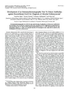
cDNA, RNA was reverse transcribed using Superscript III reverse transcriptase (Invitrogen), according to the manufacturer's instructions. Reactions were performed in a final volume of 20 μl including 1 μM hexamer and 1 μg total RNA. Reverse transcription was performed in a thermocycler under the following reaction conditions: 25 °C for 5 min, 50 °C for 60 min and the enzyme was inactivated at 85 °C for 5 min. 2.13. PCR amplification and bacterial expression A PCR reaction mixture (50 μl) containing 50 ng of cDNA as template, 5 μl of 10× Pfu buffer, 2 mM dNTP, 1 μM of forward and reverse primer, and 2.5 U Pfu DNA polymerase (Thermo Scientific) was used for CtSamp1 and human Emerin PCR amplification. The cDNA encoding fulllength Emerin (IMAGE clone ID: 3505626) was used as a template for amplification. The thermocycler parameters were 34 cycles of 94 °C for 1 min, 63 °C for 1 min, and 72 °C for 1 min. The nucleotide sequences of Ct-Samp1(1–180) were 5′-GCAGGATCCAATGCCCCTCCGTAC-3′ (forward) and 5′-CAGAAGCTTTTACCTCTTTCCCAGCGC-3′ (reverse) and for Emerin(1–222) primers were 5′-GCAGGATCCAGATGGACAACTA CGC-3′ (forward) and 5′-CAGCTCGAGCATTACTGGCGATCCTGG-3′ (reverse). The amplified PCR products were separated on a 1.5% agarose gel. The excised fragments were purified using PCR cleanup system kit (Fermentas) and cloned into pET-33b (+) and pGEX-5X-3 vectors, respectively. The positive plasmids were verified by DNA sequencing and expressed in rosetta DE3 competent cells (Millipore). Protein expression was induced at a cell density of A600 0.6 with 0.5 mM of isopropyl-β-D-thiogalactopyranoside (IPTG, Sigma) and the cells were harvested after 3 h post-induction at 30 °C. 2.14. Pull-down experiment Bacterial cells (5 × 10⁶) were treated with 1 mL of lysis buffer (100 mM HEPES, 200 mM NaCl, 1 mg/mL lysozyme, 0.1% TX-100, 10% glycerol and 1× protease inhibitor, pH 7.4) on ice for 45 min. The cells were sheared by brief sonication and soluble proteins were recovered in the supernatant following centrifugation at 15,500 ×g for 30 min at 4 °C. Nickel or glutathione magnetic agarose beads (25 μl) were incubated with 5% BSA in PBS. To the pre-incubated beads, lysate containing the bait protein (200 μg total protein) was added, the total volume was brought to 500 μl by the addition of lysis buffer and subsequently imidazole concentration was adjusted to 50 mM in order to avoid non-specific binding and incubated at 4 °C for 1 h with end-over-end rotation. The unbound lysate was separated by a magnetic separator and collected in an Eppendorf tube. To the bound beads, lysate containing the prey protein (400 μg total protein) was added along with extra lysis buffer and imidazole as before and incubated at 4 °C for 1 h with end-over-end rotation. The beads were washed 3 × 15 min with wash buffer (100 mM HEPES, 1 M NaCl, 1% TX-100, 60 mM imidazole, pH 7.4). The unbound lysates containing both bait and prey proteins were TCA precipitated. An equivalent amount of lysate, bound and unbound proteins was loaded onto 12% SDS PAGE and analyzed by Western blotting using anti-His and anti-GST antibodies, respectively. 3. Results and discussion 3.1. Extraction and distribution of NE proteins A major challenge when investigating interactions between proteins of the NE is that some of these proteins are known to be difficult to solubilize using non-denaturing conditions. To illustrate this we show that Sun1 completely resisted extraction using 1% TX-100 and 1 M NaCl (Fig. 1A), consistent with its poor solubilization reported earlier [26]. Another problem is that large proportions of overexpressed NE proteins often localize in other compartments compared to their endogenous interacting partners. Endogenous Samp1 localized specifically in the INM [27]. In contrast, in cell lines over-expressing YFP-tagged Samp1,
Fig. 1. Extraction and distribution of NE proteins. A) Extraction of NE protein. Nuclei were isolated from U2OS cells and solubilized with 0.5 M NaCl + 1% Triton X-100 (TX-100) or 1 M NaCl + 1% TX-100. Western blot analysis of Sun1 in nuclear lysate (NL), soluble (S) and insoluble (P) fractions. Note that Sun1 is resistant to extraction. B) Distribution of overexpressed NE proteins. The panels show stable U2OS cell lines expressing Samp1-YFP (a–c), YFP-Samp1 (d–f) or transiently expressing YFP-Emerin (g–i). YFP-fluorescence at normal exposure (a, d, g) or overexposed images (b, e, h) are shown with corresponding phase contrast images (c, f, i). Scale bar, 5 μm.
11% of Samp1-YFP (Fig. 1B a–c) and 21% of YFP-Samp1 (Fig. 1B d–f), respectively, was located in the endoplasmic reticulum (ER). In comparison, as much as 65% of transiently overexpressed YFP-Emerin (Fig. 1B g–i) is located in the ER. This clearly illustrates that when determining protein–protein interactions that actually take place in the NE, isolation of nuclei is required (c.f. Fig. 2A). 3.2. Reversible cross-linking of intact cells enables coprecipitation of hard to extract INM proteins To overcome the limitations in NE protein solubilization we developed a membrane protein crosslinking IP (MCLIP) protocol (c.f., Fig. 2A) and tested it on proteins interacting with the INM protein Samp1. For this, we crosslinked proteins of live U2OS cells stably expressing Samp1-YFP or YFP-Samp1 using the cell permeable reversible crosslinker DSP, untransfected U2OS cells were used as control. We isolated the nuclei, to exclude protein–protein interactions taking place in the ER (c.f. Fig. 1). The isolated nuclei were subsequently solubilized in 1% TX100 and 7 M urea (Fig. 2A). We separated the soluble and insoluble fractions of the nuclear lysate by centrifugation. After extraction the urea concentration was diluted to 0.8 M, which is tolerated by antibodies and
2402
M.H. Jafferali et al. / Biochimica et Biophysica Acta 1838 (2014) 2399–2403
Fig. 2. In vivo cross-linking immunoprecipitation. A) Flowchart. B) Solublilization of different nuclear envelope proteins after DSP crosslinking. U2OS cells were treated with (+) or without (−) DSP for 15 min followed by lysis, isolation of nuclei, solubilization in 7 M urea and 1% Triton X-100. Western blot analysis of proteins in the nuclear lysate (NL), soluble (S) fractions and insoluble (P) fractions probed with antibodies specific for the indicated proteins. C) Analysis of co-precipitation of Samp1 and NE marker proteins. Stable U2OS cell lines expressing Samp1YFP (SY) or YFP-Samp1 (YS) or untransfected (UTR) cells were treated with DSP as described in Fig. 2B. Input (nuclear lysates) and the soluble fractions were subjected to immunoprecipitation (IP) with (+) or without (−) α-GFP antibodies, respectively. The proteins in the different fractions were separated by SDS-PAGE and analyzed by Western blotting using antibodies specific for Sun1, Sun2, Emerin, Lamin B1, Lamin A/C, Pom121, Nup210 or the nuclear pore complex specific antibody mAb414 (Nup214, Nup62). The efficiency of the bait in IP was assessed using anti-Samp1 antibodies. Lysates show that total levels of input remain constant in the three different samples. Note that Sun2, Lamin A/C, Nup214, Nup62, Pom121, and Nup210 did not coprecipitate with SY or with YS.
immunoprecipitation of crosslinked complexes was performed (Fig. 2A). Western blotting using NE marker antibodies shows that after extraction the NE proteins were completely recovered in the soluble fraction in both DSP treated and non crosslinked cells (Fig. 2B). The fact that Lamin A/C, Sun2, Nup62, Nup214, Pom121 or Nup210 did not coprecipitate with either Samp1-YFP or YFP-Samp1 (Fig. 2C), shows that MCLIP identifies specific protein–protein interactions and argues against trapping of proteins in artificial complexes created by DSP-induced over-crosslinking. Previous studies [28] using high resolution confocal microscopy combined with deconvolution in HeLa cells showed that Samp1 partially colocalizes with Sun1. The poor solubilization of Sun1 under nondenaturing conditions prompted us to investigate if an interaction between Sun1 and Samp1 could be detected using MCLIP. Indeed, we were able to show that Samp1-YFP or YFP-Samp1 coprecipitates Sun1 in U2OS cells (Fig. 2C). Samp1-YFP was more efficient than YFP-Samp1 indicating that N-terminally positioned YFP might interfere with the Sun1– Samp1 interaction. We also found that Samp1-YFP and YFP-Samp1 were able to coprecipitate Emerin and Lamin B1 (Fig. 2C). The results show that the previously identified in vitro interaction between Samp1 and Emerin in HeLa cells [28] also occurs in the NE of live U2OS cells. Taken together the results show that our approach is an efficient method to identify specific protein\protein interactions of NE proteins. 3.3. The small GTPase Ran coprecipitates with Samp1 Samp1 is predicted to interact with Ran [29], a small GTPase involved in various cellular functions such as nucleocytoplasmic transport
[30], microtubule assembly [31] and postmitotic nuclear assembly [32, 33]. Here we investigated the extent of this interaction throughout the cell cycle of stable U2OS cell lines expressing Samp1-YFP. The results show that Samp1-YFP interacts with Ran in interphase, prometaphase and metaphase (Fig. 3). Thus we show that MCLIP method can be used to identify protein–protein interactions in different phases of cell cycle.
Fig. 3. Samp1-YFP coprecipitates Ran during different phases of the cell cycle. Stable U2OS cell lines expressing Samp1-YFP were synchronized in either prometaphase or metaphase. Asynchronous cells represent interphase. The cells were treated with the cell permeable reversible crosslinker DSP, followed by lysis, extraction and immunoprecipitation with (+) or without (−) α-GFP antibodies. The fractions were separated by SDS-PAGE and analyzed by Western blotting using antibodies specific for Ran. Lysates show that total levels of Ran remain constant in the different cell populations.
M.H. Jafferali et al. / Biochimica et Biophysica Acta 1838 (2014) 2399–2403
Fig. 4. Direct interaction between Samp1 and Emerin. His6-Ct-Samp1(1–180) and GSTEmerin(1–222) were subjected to pull-down experiment using either nickel agarose beads (His6 pull down) or glutathione agarose beads (GST pull down). The beads were incubated either with GST-Emerin and His6-Ct-Samp1 or GST and His6-Ct-Samp1. Lysate (L), bound (P) and unbound (S) proteins were separated by SDS PAGE and analyzed by Western blotting using anti-GST and anti-His anitibodies as indicated.
3.4. The interaction between Samp1 and Emerin is direct To investigate whether Samp1 and Emerin can directly interact with each other, we turned to the thermophilic fungus C. thermophilum (Ct), which has ideal properties for structural and biochemical studies, compared to other mesophilic counterparts [34]. We found that the hypothetical protein CTHT_0001390 contains an N-terminal domain with conserved CXXC zinc finger motifs, which is homologous to human Samp1 [27,28] and also has a predicted membrane topology similar to that of human Samp1, suggesting that it is the Ct homologue of Samp1. We designed and expressed the recombinant fusion proteins, GST-Emerin(1–222) and His6-Ct-Samp1(1–180) in Escherichia coli. The expressed proteins were subjected to nickel or glutathione agarose beads in pull-down experiments to determine Samp1–Emerin interaction. The results show that Ct-Samp1 and human Emerin are able to bind each other directly (Fig. 4). The hydrophobic segment (13–35) of human Samp1 is not conserved in Ct-Samp1 and is thus not required for Samp1–Emerin interactions. This suggests that the nucleoplasmic part of Ct-Samp1 is responsible for interaction with the nucleoplasmic part of Emerin supporting the idea that the binding involves the zinc finger domains of Samp1 [28]. 4. Conclusion MCLIP (Membrane protein Cross-Link ImmunoPrecipitation) is an efficient method to detect specific protein interactions of extraction resistant proteins in the NE. Crosslinking of intact cells enables the detection of interactions taking place in live cells. Acknowledgments This work was made possible by grants from the Swedish Research Council #621-2010-448, Cancerfonden #110590 and the foundation Olle Engkvists mine. We would like to thank Dr. Katherine Wilson, Johns Hopkins University School of Medicine, Baltimore for sharing plasmid encoding YFP-Emerin. References [1] R. Aebersold, M. Mann, Mass spectrometry-based proteomics, Nature 422 (2003) 198–207. [2] P. Yaciuk, Co-immunoprecipitation of protein complexes, Methods Mol. Med. 131 (2007) 103–111. [3] C.M. Snow, A. Senior, L. Gerace, Monoclonal antibodies identify a group of nuclear pore complex glycoproteins, J. Cell Biol. 104 (1987) 1143–1156. [4] N. Dwyer, G. Blobel, A modified procedure for the isolation of a pore complexlamina fraction from rat liver nuclei, J. Cell Biol. 70 (1976) 581–591. [5] A. Radu, G. Blobel, R.W. Wozniak, Nup155 is a novel nuclear pore complex protein that contains neither repetitive sequence motifs nor reacts with WGA, J. Cell Biol. 121 (1993) 1–9. [6] R. Ashery-Padan, A.M. Weiss, N. Feinstein, Y. Gruenbaum, Distinct regions specify the targeting of otefin to the nucleoplasmic side of the nuclear envelope, J. Biol. Chem. 272 (1997) 2493–2499.
2403
[7] M. Zhou, S. Felder, M. Rubinstein, D.R. Hurwitz, A. Ullrich, I. Lax, J. Schlessinger, Realtime measurements of kinetics of EGF binding to soluble EGF receptor monomers and dimers support the dimerization model for receptor activation, Biochemistry 32 (1993) 8193–8198. [8] L. Zhang, S. Rayner, N. Katoku-Kikyo, L. Romanova, N. Kikyo, Successful coimmunoprecipitation of Oct4 and Nanog using cross-linking, Biochem. Biophys. Res. Commun. 361 (2007) 611–614. [9] C. Guerrero, C. Tagwerker, P. Kaiser, L. Huang, An integrated mass spectrometrybased proteomic approach: quantitative analysis of tandem affinity-purified in vivo cross-linked protein complexes (QTAX) to decipher the 26 S proteasomeinteracting network, Mol. Cell. Proteomics 5 (2006) 366–378. [10] B. Craige, G. Salazar, V. Faundez, Phosphatidylinositol-4-kinase type II alpha contains an AP-3-sorting motif and a kinase domain that are both required for endosome traffic, Mol. Biol. Cell 19 (2008) 1415–1426. [11] A.J. Lomant, G. Fairbanks, Chemical probes of extended biological structures: synthesis and properties of the cleavable protein cross-linking reagent [35S] dithiobis(succinimidyl propionate), J. Mol. Biol. 104 (1976) 243–261. [12] A.L. Smith, D.B. Friedman, H. Yu, R.H. Carnahan, A.B. Reynolds, ReCLIP (reversible cross-link immuno-precipitation): an efficient method for interrogation of labile protein complexes, PLoS ONE 6 (2011) e16206. [13] P. Percipalle, A. Jonsson, D. Nashchekin, C. Karlsson, T. Bergman, A. Guialis, B. Daneholt, Nuclear actin is associated with a specific subset of hnRNP A/B-type proteins, Nucleic Acids Res. 30 (2002) 1725–1734. [14] G. Salazar, S. Zlatic, B. Craige, A.A. Peden, J. Pohl, V. Faundez, Hermansky–Pudlak syndrome protein complexes associate with phosphatidylinositol 4-kinase type II alpha in neuronal and non-neuronal cells, J. Biol. Chem. 284 (2009) 1790–1802. [15] L. Gerace, B. Burke, Functional organization of the nuclear envelope, Annu. Rev. Cell Biol. 4 (1988) 335–374. [16] M.W. Hetzer, The nuclear envelope, Cold Spring Harb. Perspect. Biol. 2 (2010) a000539. [17] E.C. Schirmer, L. Florens, T. Guan, J.R. Yates III, L. Gerace, Nuclear membrane proteins with potential disease links found by subtractive proteomics, Science 301 (2003) 1380–1382. [18] G.S. Wilkie, N. Korfali, S.K. Swanson, P. Malik, V. Srsen, D.G. Batrakou, J. de las Heras, N. Zuleger, A.R. Kerr, L. Florens, E.C. Schirmer, Several novel nuclear envelope transmembrane proteins identified in skeletal muscle have cytoskeletal associations, Mol. Cell. Proteomics 10 (2011) M110 (003129). [19] N. Korfali, G.S. Wilkie, S.K. Swanson, V. Srsen, J. de Las Heras, D.G. Batrakou, P. Malik, N. Zuleger, A.R. Kerr, L. Florens, E.C. Schirmer, The nuclear envelope proteome differs notably between tissues, Nucleus 3 (2012) 552–564. [20] U. Rothbauer, K. Zolghadr, S. Muyldermans, A. Schepers, M.C. Cardoso, H. Leonhardt, A versatile nanotrap for biochemical and functional studies with fluorescent fusion proteins, Mol. Cell. Proteomics 7 (2008) 282–289. [21] M. Kihlmark, G. Imreh, E. Hallberg, Sequential degradation of proteins from the nuclear envelope during apoptosis, J. Cell Sci. 114 (2001) 3643–3653. [22] M. Olsson, S. Scheele, P. Ekblom, Limited expression of nuclear pore membrane glycoprotein 210 in cell lines and tissues suggests cell-type specific nuclear pores in metazoans, Exp. Cell Res. 292 (2004) 359–370. [23] R.W. Wozniak, E. Bartnik, G. Blobel, Primary structure analysis of an integral membrane glycoprotein of the nuclear pore, J. Cell Biol. 108 (1989) 2083–2092. [24] A. Edelstein, N. Amodaj, K. Hoover, R. Vale, N. Stuurman, Computer control of microscopes using microManager, in: Frederick M. Ausubel, et al., (Eds.), Current Protocols in Molecular Biology, 2010, (Chapter 14, Unit14 20). [25] J. Schindelin, I. Arganda-Carreras, E. Frise, V. Kaynig, M. Longair, T. Pietzsch, S. Preibisch, C. Rueden, S. Saalfeld, B. Schmid, J.Y. Tinevez, D.J. White, V. Hartenstein, K. Eliceiri, P. Tomancak, A. Cardona, Fiji: an open-source platform for biologicalimage analysis, Nat. Methods 9 (2012) 676–682. [26] F. Haque, D.J. Lloyd, D.T. Smallwood, C.L. Dent, C.M. Shanahan, A.M. Fry, R.C. Trembath, S. Shackleton, SUN1 interacts with nuclear lamin A and cytoplasmic nesprins to provide a physical connection between the nuclear lamina and the cytoskeleton, Mol. Cell. Biol. 26 (2006) 3738–3751. [27] C. Buch, R. Lindberg, R. Figueroa, S. Gudise, E. Onischenko, E. Hallberg, An integral protein of the inner nuclear membrane localizes to the mitotic spindle in mammalian cells, J. Cell Sci. 122 (2009) 2100–2107. [28] S. Gudise, R.A. Figueroa, R. Lindberg, V. Larsson, E. Hallberg, Samp1 is functionally associated with the LINC complex and A-type lamina networks, J. Cell Sci. 124 (2011) 2077–2085. [29] T. Schmitt, C. Ogris, E.L. Sonnhammer, FunCoup 3.0: database of genome-wide functional coupling networks, Nucleic Acids Res. 42 (2014) D380–D388. [30] M.S. Moore, G. Blobel, The GTP-binding protein Ran/TC4 is required for protein import into the nucleus, Nature 365 (1993) 661–663. [31] M.V. Nachury, T.J. Maresca, W.C. Salmon, C.M. Waterman-Storer, R. Heald, K. Weis, Importin beta is a mitotic target of the small GTPase Ran in spindle assembly, Cell 104 (2001) 95–106. [32] A. Harel, R.C. Chan, A. Lachish-Zalait, E. Zimmerman, M. Elbaum, D.J. Forbes, Importin beta negatively regulates nuclear membrane fusion and nuclear pore complex assembly, Mol. Biol. Cell 14 (2003) 4387–4396. [33] T.C. Walther, P. Askjaer, M. Gentzel, A. Habermann, G. Griffiths, M. Wilm, I.W. Mattaj, M. Hetzer, RanGTP mediates nuclear pore complex assembly, Nature 424 (2003) 689–694. [34] S. Amlacher, P. Sarges, D. Flemming, V. van Noort, R. Kunze, D.P. Devos, M. Arumugam, P. Bork, E. Hurt, Insight into structure and assembly of the nuclear pore complex by utilizing the genome of a eukaryotic thermophile, Cell 146 (2011) 277–289.