Timo Piironen,1 Martti Nurmi,3 Kerttu Irjala,4 Olli Heinonen,5 Hans Lilja,6. Timo Lo¨vgren,2 ...... Corey E, Brown LG, Corey MJ, Buhler KR, Vessella RL. LNCaP.
Clinical Chemistry 47:4 703–711 (2001)
Enzymes and Protein Markers
Measurement of Circulating Forms of Prostate-specific Antigen in Whole Blood Immediately after Venipuncture: Implications for Point-of-Care Testing Timo Piironen,1 Martti Nurmi,3 Kerttu Irjala,4 Olli Heinonen,5 Hans Lilja,6 Timo Lo¨vgren,2 and Kim Pettersson2*
Background: The purpose of this study was to validate the use of whole-blood samples in the determination of circulating forms of prostate-specific antigen (PSA). Methods: Blood samples of hospitalized prostate cancer and benign prostatic hyperplasia patients were collected and processed to generate whole-blood and serum samples. Three different rapid two-site immunoassays were developed to measure the concentrations of total PSA (PSA-T), free PSA (PSA-F), and PSA-␣1-antichymotrypsin complex (PSA-ACT) to detect in vitro changes in whole-blood samples immediately after venipuncture. The possible influence of muscle movement on the release of PSA from prostate gland was studied in healthy men by measuring the rapid in vitro wholeblood kinetics of PSA forms before and after 15 min of physical exercise on a stationary bicycle. Results: Rapid PSA-T, PSA-F, and PSA-ACT assays were designed using a 10-min sample incubation. No significant changes were detected in the concentrations of PSA-T, PSA-F, and PSA-ACT from the earliest time point of 12–16 min compared with measurements performed up to 4 h after venipuncture. Physical exercise did not influence the concentrations of the circulating
forms of PSA. Hematocrit-corrected whole-blood values of PSA-T and PSA-F forms were comparable to the respective serum values. Calculation of the percentage of PSA-F (PSA F/T ratio ⴛ 100) was similar irrespective of the sample format used, i.e., whole blood or serum. Conclusions: We found that immunodetectable PSA forms are likely at steady state immediately after venipuncture, thus enabling the use of anticoagulated whole-blood samples in near-patient settings for pointof-care testing, whereas determinations of PSA (e.g., PSA-T, PSA-F, or PSA-ACT) performed within the time frame of the office visit would provide results equivalent to conventional analyses performed in serum. © 2001 American Association for Clinical Chemistry
During the past 15 years, the immunochemical detection of prostate-specific antigen (PSA)7 has become an indispensable prostate cancer (PCa) marker. In addition to the established use of PSA for monitoring disease progression, increasing interest has been directed toward the potential of PSA for early detection of PCa. Populationbased screening for PCa in the absence of definitive evidence for the clinical usefulness of PSA remains a controversial issue (1 ). Despite this, the use of PSA testing as part of regular physical check-ups has led to an increase in the reported number of cases of PCa in both symptomatic and nonsymptomatic urology patients. PSA is a 28.4-kDa glycoprotein that consists of 237 amino acids (2 ). It is a chymotrypsin-like serine protease and a member of the glandular kallikrein family (3 ).
1 The Finsen Laboratory af.sn. 8621, Strandboulevarden 49, 2100 Copenhagen, Denmark. 2 Department of Biotechnology and 5 Paavo Nurmi Center, University of Turku, 20520 Turku, Finland. 3 Department of Surgery, Turku University Central Hospital, 20520 Turku, Finland. 4 Department of Clinical Chemistry, Turku University Central Hospital, 20520 Turku, Finland. 6 Department of Laboratory Medicine, Division of Clinical Chemistry, Lund University, University Hospital, 20502 Malmo¨, Sweden. *Author for correspondence. Fax 358-2-3338050; e-mail kim.pettersson@ utu.fi. Received in revised form January 3, 2001; accepted January 19, 2001.
7 Nonstandard abbreviations: PSA, prostate-specific antigen; PCa, prostate cancer; ACT, ␣1-antichymotrypsin; AMG, ␣2-macroglobulin; PSA-F, free PSA; BPH, benign prostatic hyperplasia; PSA-T, total immunoreactive PSA; and MAb, monoclonal antibody.
703
704
Piironen et al.: Circulating Forms of PSA in Whole Blood
Reference values for PSA in serum are six orders of magnitude lower than those for seminal fluid (4 – 6 ). In vitro, the active single-chain form of PSA forms stable covalent complexes with several major extracellular protease inhibitors, such as ␣1-antichymotrypsin (ACT), ␣2macroglobulin (AMG), pregnancy-zone protein, protein C inhibitor, and ␣1-antitrypsin (7–12 ). In vivo, enzymatically active serum PSA is regulated mainly by ACT (13 ). The significance of AMG as a complexing ligand for PSA in vivo has been difficult to clarify because the PSA moiety in PSA-AMG complexes is prevented from interacting with antibodies and, therefore, remains unrecognized by conventional immunoassays because of steric shielding resulting from engulfment by the surrounding 720-kDa AMG molecule (14 ). By contrast, many independent antigenic epitopes on PSA remain exposed after complexation with ACT, leading to the loss of one antigenic epitope region mapped to a small portion of the kallikrein loop surrounded by the active site cleft on PSA (15 ). In 1991, two groups reported independently that immunoreactive PSA in serum exists predominantly (65– 95%) as an ⬃90-kDa complex with ACT (PSA-ACT) and to a smaller extent (5–35%) as the ⬃30-kDa noncomplexed free PSA (PSA-F) form (7, 8 ). Stenman et al. (8 ) demonstrated that the proportion of serum PSA complexed to ACT was significantly higher in serum samples collected from patients with PCa compared with subjects with benign prostatic hyperplasia (BPH) (8 ). Christensson et al. (16 ) confirmed and extended this finding by showing that the percentage of PSA-F (or PSA F/T ratio, where T represents total immunoreactive PSA, consisting of PSA-F and PSA-ACT) in serum was substantially lower in PCa compared with BPH. Today, these findings have been widely confirmed by numerous other studies (17–20 ). In general, measurements of the percentage of PSA-F have been found to be most useful in patients with moderately increased total PSA concentrations of 4 –10 g/L. However, recently reported data have suggested that the percentage of PSA-F may also enhance the specificity of PSA testing below the conventional 4 g/L cutoff (21–24 ). The release mechanism(s) and competition for different metabolic pathways that regulate the presence of different PSA forms in the circulation have not been clarified. There are several reports that indicate that PSA-F manifests significant heterogeneity (25 ), although the detailed nature of PSA-F in serum and the exact site at which active PSA is inactivated by forming covalent complexes with the various serpins has not been resolved. Several reports have suggested that the free noncomplexed form(s) are inactive because they are essentially nonreactive with active inhibitors, such as ACT and AMG, which occur in large excess compared with PSA in the blood circulation (26 –31 ). According to another hypothesis, PSA would be enzymatically active when it enters the blood circulation. Therefore, it would be conceivable that the different forms of PSA, assayed immediately after venipuncture, may not
have reached steady state because the kinetics of PSA forming complexes with ACT and AMG are slow (29 ). If so, PSA testing performed ahead of equilibrium may be misleading compared with testing performed on conventionally processed serum samples. The aim of the present study was to investigate whether there are any significant changes in the concentrations and proportions of free and serpin-complexed PSA and to define the magnitude of changes occurring immediately after venipuncture in PCa and BPH patients and in healthy subjects with or without stressed conditions. Therefore, we designed assay protocols by which PSA in whole-blood specimens could be rapidly and quantitatively immobilized to the capture antibody, thus minimizing the time during which complex formation could occur ex vivo.
Materials and Methods specimens Blood samples from hospitalized PCa and BPH patients were obtained from the Department of Surgery, Turku University Central Hospital, Turku, Finland. The procedures followed were in accordance with the Helsinki Declaration of 1975, as revised in 1996. Specimen characteristics of the hospitalized patients and men who volunteered for the bicycle exercise are shown in Table 1. Whole-blood and plasma samples were obtained using heparin (0.015 US Pharmacopeia units/L) and EDTA (0.047 mmol/L) tubes (Venoject). Serum samples were collected in tubes (Vacutainer) that did not contain anticoagulants. Approximately 30 min after venipuncture, serum and plasma samples were generated by centrifugation.
monoclonal antibodies and purified proteins The reactivity of anti-PSA monoclonal antibodies (MAbs) with free and complexed PSA has been described previously (15, 32 ). MAb 5A10 recognizes PSA-F, whereas MAbs H117 and H50 recognize free and complexed forms of PSA. MAb 241 was generated against ACT and used as an Eu3⫹-labeled tracer in the assay for PSA-ACT (33 ). Tracer MAbs were labeled with 4 –7 molecules of Eu3⫹/
Table 1. Specimen characteristics. Mean (SD)
Minimum
A. Hospitalized PCa and BPH patients (n ⴝ 8) Serum PSA-T, g/L 16.7 (14.8) 3.3 Serum PSA-F, g/L 1.5 (1.4) 0.7 Serum PSA-ACT, g/L NDa ND Serum PSA F/T, % 11.8 (8.2) 4.7 Hematocrit, % 44.4 (1.6) 42.0 B. Men who volunteered for the bicycle exercise (n ⴝ 5) Serum PSA-T, g/L 4.0 (3.0) 1.2 Serum PSA-F, g/L 0.9 (0.7) 0.2 Serum PSA-ACT, g/L 3.3 (2.6) 1.0 Serum PSA F/T, % 22.6 (2.8) 19.8 a
ND, not determined.
Maximum
49.0 4.6 ND 30.3 46.0 8.9 1.9 7.6 27.2
Clinical Chemistry 47, No. 4, 2001
IgG according to the instructions of the DELFIA® Eu3⫹labeling product (Perkin-Elmer Wallac Oy). Purified PSA from seminal plasma containing ⬃85% of the catalytically active single-chain form and ⬃15% of the inactive twochain form, purified ACT, and PSA-ACT complexes were generated, purified, and stored as reported previously (9, 32 ).
immunoassay validation Microtitration wells coated with anti-PSA MAb H117, DELFIA ProstatusTM PSA Dual Assay, DELFIA 1234 Plate Fluorometer, DELFIA Buffer, DELFIA Wash Solution, and DELFIA Enhancement Solution for the immunofluorometric assays were from Perkin-Elmer Wallac Oy. We used modified Prostatus immunofluorometric assays for the detection of PSA-F and PSA-T and an in-house investigational assay for the detection of PSA-ACT (33 ). All assays used the same capture MAb, H117, whereas Eu3⫹labeled detection MAbs 5A10, H50, and 241 were used in the PSA-F, PSA-T, and PSA-ACT assays, respectively. Modifications of assay conditions were made to directly accommodate whole-blood samples and, in particular, to rapidly reach an equilibrium with the capture antibody. The subsequent wash step interrupted any complex formation occurring ex vivo and was followed by a 2-h incubation with the detection antibodies. Assay kinetics in the reaction wells were determined by analyzing whole-blood samples from three male individuals. The PSA-T, PSA-F, and PSA-ACT concentrations were 1.2–22.3, 0.7–13.8, and 0.6 –7.1 g/L, respectively. The incubation time of the first assay step was 5–120 min with a sample volume of 25 L and a total reaction volume of 75 L. The time required for the reaction to reach an average of ⱖ90% of the signal obtained after the 2-h incubation was considered sufficient for the rapid assays. The final rapid assay protocols were as follows. Duplicates of calibrators or unknown samples (25 L) together with 50 L of DELFIA Buffer were incubated at room temperature for 10 min in microtitration wells precoated with MAb H117. After a wash step, 150 ng per well of the Eu3⫹-labeled detection MAbs, H50, 5A10, and 241, respectively, was added in 200 L of DELFIA Buffer and incubated for 2 h. The final wash step was followed by the addition of 200 L per well of DELFIA enhancement solution. Analytical detection limits of the rapid assays were determined by analyzing 12 replicates of the zero calibrator and calculating the dose corresponding to 2 SD of the calibration diluent multiplied by the slope of the calibration curve. Between-assay imprecision was studied using 12 whole-blood samples from female individuals after the addition of purified PSA-F (derived from seminal plasma) and in vitro-prepared purified PSA-ACT complex. The PSA-T, PSA-F, and PSA-ACT concentrations were 1.1–
705
35.3, 0.2– 8.2, and 0.8 –27 g/L, respectively. The assays were performed on 3 different days with duplicate measurements. Within-assay imprecision was calculated after measuring the same 12 whole-blood samples with 6 replicates within the same assay run. Linearity studies were performed using serial dilutions (1:2, 1:4, and 1:8) of whole blood from male individuals. For one set of dilutions, DELFIA Assay Buffer was used as diluent, and for other set, pooled whole-blood samples from female individuals not containing measurable PSA concentrations were used as the diluent.
time course studies of psa-t, psa-f, and psa-act Recovery of purified PSA. Purified PSA was incubated at 37 °C with freshly obtained serum, EDTA- and heparinanticoagulated whole blood, and EDTA and heparin plasma from female individuals (n ⫽ 5). At various time points (1 min to 12 h) after venipuncture, aliquots of the reaction mixture were withdrawn and rapidly measured for PSA-T, PSA-F, and PSA-ACT. Endogenous PSA forms in PCa, BPH, and control samples. EDTA-anticoagulated whole-blood and serum samples from healthy controls (n ⫽ 3) and hospitalized BPH (n ⫽ 3) and PCa (n ⫽ 5) patients were collected and processed. PSA-T, PSA-F, and PSA-ACT measurements of wholeblood specimens were initiated starting from as soon as 1– 6 min after venipuncture and monitored until 2– 48 h after sampling. Hematocrit values were measured by centrifugation. Endogenous PSA forms after physical exercise. A person biking on a stationary bicycle develops a special muscle movement that squeezes the prostate gland. We chose the method described previously by Oremek and Seiffert (34 ). Healthy volunteers (n ⫽ 5) were subjected to 15 min of exercise on a stationary bicycle with a 100-W setting. Heparin-anticoagulated whole-blood and serum samples were collected and processed before and after the biking exercise. Any changes in PSA-T, PSA-F, and PSA-ACT concentrations or proportions in samples collected after the exercise were monitored by initiating measurements from 1 min to 2 h after venipuncture.
statistics SPSS 7.5 for Windows was used to perform nonparametric Kruskal–Wallis H-tests and Mann–Whitney U-tests to determine whether the results of the rapid assay measurements followed at various time points were significantly different (P ⬍0.05) from the initial reference values. The change in PSA immunoreactivity with time was determined by the least-squares regression method. The slope of the linear regression was determined with the 95% confidence limits.
706
Piironen et al.: Circulating Forms of PSA in Whole Blood
Results design of rapid immunoassays
The high affinity of MAb H117, 4 ⫻ 1010 L/mol (35 ), enabled the design of rapid PSA capture for the PSA-T, PSA-F, and PSA-ACT immunoassays. After 10 min of incubation, the mean ⫾ SD concentrations of the PSA-T, PSA-F, and PSA-ACT assays were 94% ⫾ 5%, 94% ⫾ 3%, and 90% ⫾ 6% complete compared with the signals obtained after 120 min of incubation. Therefore, a 10-min incubation for the capture phase was considered sufficient to reliably reflect the analyte concentrations measured in the subsequent steps for the measurement of PSA-T, PSA-F, and PSA-ACT. Analytical detection limits of the PSA-T, PSA-F, and PSA-ACT assays were 0.02, 0.01, and 0.11 ng/L, respectively. Within-assay CVs for the PSA-T, PSA-F, and PSAACT assays were 4.2– 8.0%, 1.9 –9.7%, and 4.1–14%, respectively. Corresponding between-assay CVs were 1.5– 21%, 2.6 –16%, and 5.0 –21%, respectively. In the sampledilution studies, the recoveries in the assay buffer (percentage of the value obtained for the undiluted samples) for the PSA-T, PSA-F, and PSA-ACT assays were 96 –107%, 78 –90%, and 119 –136%, respectively. When the sample was diluted in the whole blood of female individuals, corresponding recoveries were 87–135%, 109 –111%, and 90 –118%, respectively. Taken together, the technical quality of rapid-capture immunoassays was considered adequate for the quantitative estimation of rapid changes in concentrations of PSA forms immediately after venipuncture.
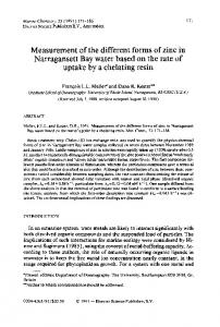
time course studies of psa-t, psa-f, and psa-act Recovery of purified PSA. The recovery of purified PSA-F added to a serum from female individuals as measured by PSA-T, PSA-F, and PSA-ACT assays is shown in Fig. 1. Twelve hours after the addition of purified PSA-F, ⬃5% of the initially added PSA was detected as PSA-ACT, whereas ⬃10% remained as PSA-F. The majority (⬃85% of the initially added PSA) was not immunodetected by
these assays, which we attributed to the loss of immunoreactivity through steric shielding by complex formation with AMG. This was as also evidenced by an investigational in-house assay for PSA-AMG (30 ) (data not shown). Highly similar elimination and complex formation patterns were obtained using EDTA-anticoagulated whole-blood, heparin whole-blood, EDTA plasma, and heparin plasma samples from three individuals (data not shown). Analyses performed at 15 and 30 min after the initial addition of PSA showed that the majority of the enzymatically reactive part of the added PSA (i.e., ⬎50%) had already reacted with AMG after 15 min. Further analyses performed at 1, 2, and 4 h after the initial addition of PSA showed that the PSA-ACT concentrations were 87–91% of those measured after 12 h, whereas PSA-T and PSA-F concentrations were slightly higher (128 –132% and 118 –120%, respectively) than the concentrations detected after 12 h. Endogenous PSA forms in PCa, BPH, and control samples. The established protocols were first validated with EDTA whole blood, heparin whole blood, and serum obtained from healthy male subjects. The in vitro kinetics of one individual (PSA-T, 0.66 g/L; PSA-F, 0.13 g/L) are shown in Fig. 2. No significant changes over a time span of 1 min to 48 h after venipuncture were seen between the different sample matrices and the concentrations of PSA-T and PSA-F. These procedures were then extended to three BPH patients and five PCa samples (PSA-T range, 3.3– 49 g/L) from which EDTA whole-blood samples were obtained. No significant changes in the three measured analytes (PSA-T, PSA-F, PSA-ACT) were observed over a time span of 2 min to 4 h, indicating that the samples were already at steady-state equilibrium before the first measuring point (2– 6 min) after venipuncture (Fig. 3). The slopes (95% confidence intervals) of the linear regression
Fig. 1. Elimination curve of purified PSA-F added to serum from a female individual.
Fig. 2. Rapid in vitro kinetics of PSA forms measured in EDTA whole blood, heparin whole blood, and serum from a male individual (PSA-T, 0.66 g/L; PSA-F, 0.13 g/L).
PSA-T, PSA-F, and PSA-ACT assays were determined in a time scale of 1 min to 12 h after the addition of purified PSA.
PSA-T and PSA-F assays were determined in a time scale of 1 min to 48 h after the venipuncture.
Clinical Chemistry 47, No. 4, 2001
Fig. 3. Rapid in vitro kinetics of PSA forms measured in EDTA whole blood from PCa (n ⫽ 5; filled symbols) and BPH patients (n ⫽ 3; open symbols). PSA-T (A), PSA-F (B), and PSA-ACT (C) concentrations were determined in a time scale of 2 min to 4 h after venipuncture and plotted as concentrations relative to the first time point measured.
for pooled data in a linear scale were ⫺0.045 (⫺0.100 to 0.010) for PSA-T, ⫺0.048 (⫺0.112 to 0.016) for PSA-F, and ⫺0.016 (⫺0.063 to 0.031) for PSA-ACT; thus, none of the slopes were statistically different from a slope of zero. Endogenous PSA forms after physical exercise. In the study of five healthy male volunteers (PSA-T range, 1.2– 8.9 g/L) subjected to 15 min of bicycle exercise, statistically significant differences in PSA-T, PSA-F, or PSA-ACT concentrations were not found when samples taken before and
707
Fig. 4. Rapid in vitro kinetics of PSA forms measured in EDTA whole blood from men (n ⫽ 5) sampled immediately after 15 min of exercise on a stationary bicycle. PSA concentrations were plotted relative to the preexercise sample. (A), PSA-T; (B), PSA-F; and (C), PSA-ACT.
after the physical exercise were compared. Similarly, initiating the measurements with the postexercise sample at various time points after venipuncture revealed no significant changes in the measured concentrations of the various forms of PSA (Fig. 4). As shown in Fig. 5, there was an excellent correlation between PSA-F and PSA-T concentrations in whole-blood specimens compared with those measured in serum samples. Correlation coefficients for PSA-T, PSA-F, and the PSA F/T ratio between whole-blood and serum values
708
Piironen et al.: Circulating Forms of PSA in Whole Blood
Fig. 5. Comparison of PSA-F (⽧; ⫻ 10 g/L), PSA-T (⫹; g/L), and PSA F/T (E; %) values in whole-blood and serum samples (n ⫽ 13). Lines: (⫺ 䡠 ⫺ 䡠 ⫺), y ⫽ x; (⫺ ⫺ ⫺ ⫺), hematocrit-corrected trend (y ⫽ 0.56x).
were 0.998, 0.989, and 0.983, respectively (P ⬍0.001 for all). Corresponding linear-regression fitting functions for PSA-T, PSA-F, and the PSA F/T ratio were: y ⫽ 0.58x ⫹ 0.01; y ⫽ 0.68x ⫹ 0.03; and y ⫽ 0.97x ⫹ 0.05, respectively. The PSA F/T ratio (percentage of PSA-F) had a slope close to 1 (0.97), whereas the slopes for PSA-T and PSA-F closely followed the mean hematocrit-corrected wholeblood values (slope ⫽ 0.56).
Discussion Measurements of the circulating forms of PSA, i.e., mainly PSA-F and PSA complexed to various serpin ligands (mainly ACT), provide useful clinical information by enhancing the separation of PCa from benign prostatic conditions. However, it is not known why PSA-F remains in a noncomplexed form despite the very large excess (⬎104-fold) of potentially complexing agents (mainly AMG and ACT). Internally cleaved PSA molecules or the zymogen form of PSA represent two obvious alternatives that render PSA enzymatically inactive. Furthermore, it is not yet known at which anatomical site the enzymatically active PSA interacts to form PSA-ACT complexes. Because ACT is expressed in the prostate epithelium (36 ), it cannot be excluded that some of the complex formation may occur within the tissue. However, it is generally assumed that complexation occurs extracellularly before the complex enters the circulation after enzymatically active PSA is released from the prostate gland. The aim of the present study was to investigate whether rapid conversion of the circulating forms of PSA can be demonstrated immediately after venipuncture in healthy subjects, as well as in patients with BPH and PCa. Our study clearly showed that there were no significant changes in the concentrations of PSA-T, PSA-F, and PSA-ACT, as can
be seen from the earliest time points, i.e., 12–16 min after venipuncture up to several hours, indicating that these PSA forms are practically at steady state at the time of venipuncture. Addition of purified PSA to fresh serum, plasma, or whole-blood samples from females demonstrated that the proportion of PSA that was rendered “invisible” (⬃85% of the purified PSA) to the PSA-F, PSA-T, and PSA-ACT assays was closely correlated to the enzymatically active fraction estimated to account for ⱖ80% of the purified preparation. This loss of immunoreactivity correlated with the concentration of PSA-AMG as has been suggested previously (9, 29 ). Compared with AMG, a much smaller proportion (ⱕ5%) of the added PSA formed complexes with ACT, which is also in agreement with previous results (12, 27 ). However, compared with many other serine protease/serpin interactions, the rate at which PSA forms complexes with AMG and ACT in vitro is comparatively slow (13 ). Approximately one-fifth of the enzymatically active PSA remained in the free form after 1-h incubations in vitro with sera from females compared with the selected endpoint after a 12-h incubation. Only 10 min after PSA was added in vitro, the noncomplexed proportion was as high as 60 –70%. Therefore, if PSA released from the prostate is enzymatically active when it enters the circulation, one would expect that the time required to reach steady state for total immunodetectable PSA as well as for the PSA F/T ratio would be in the order of several hours after venipuncture. Following this logic, it has been suggested (37 ) that sufficient time after venipuncture should be allowed for such an equilibrium to occur before initiating routine PSA determinations. However, this assumption has not been verified experimentally. Our results showing that the concentrations of the free and complexed forms of PSA in anticoagulated whole blood are at steady state at or very shortly after venipuncture can be interpreted in two ways that are not mutually exclusive. One interpretation would be that the proportion of enzymatically active PSA released extracellularly into the circulation may be close to insignificant. The other interpretation would be that it is likely that enzymatically active PSA released extracellularly has already reached equilibrium regarding its interaction with AMG and ACT before entering the circulation. In this context, it is of interest to compare the present results with those reported by Bjo¨rk et al. (38 ) showing that PSA released into the circulation during radical retropubic prostatectomy did not form complexes with ACT. Similar results were also obtained by Lilja et al. (30 ), where the investigators found that the PSA-F released during radical prostatectomy appeared enzymatically nonreactive because it did not complex with ACT or AMG. These results are in full agreement with the present study, although the release mechanism after radical prostatectomy may be different from the one in the in vivo situation. The rapid assay protocols described here have also been tested with
709
Clinical Chemistry 47, No. 4, 2001
samples collected from patients subjected to transurethal resection of the prostate at the time point when the electroresection was completed (data not shown). In much the same way as the results from the present study show, measurements of PSA-T, PSA-F, and PSA-ACT in rapidly processed plasma samples were at complete steady state from the first initiated analysis at 10 min to the last measurement performed after 24 h (Charlotte Becker, personal communication). Recently, Zhang et al. (37 ) reported on the measurement of PSA-AMG complex in retrospective clinical serum sample specimens from PCa and BPH patients and found that the proportion of this complex relative to PSA-T was higher in BPH (17%) than in PCa (12%) patients. Considering the assumed rapid clearance of PSA-AMG from the circulation, the authors hypothesized that the increased concentrations may be attributable to differences in postsampling complex formation. BPH patients were assumed to contain more clipped PSA, which then slowly reacted with AMG. Our present study does not agree with this hypothesis because we did not find any significant changes in the PSA-T, PSA-F, and PSAACT concentrations from the earliest time point after venipuncture up to several hours. In a previous study (39 ) on the stability of PSA forms at different temperatures over a 1-week period, we showed insignificant changes both in the absolute concentrations and in the percentage of PSA-F when blood samples had been collected with the presence of anticoagulants, such as heparin and EDTA. In the same study, we found that in serum samples studied over the same time interval, there was a substantial decrease in the PSA-F fraction and, to a lesser extent, in PSA-ACT. This may offer an explanation for the surprisingly high concentrations of PSA-AMG found by Zhang et al. (37 ) using frozen serum samples: proteolytic activation of the zymogen form of PSA and/or dissociation of enzymatically active PSA from PSA-ACT may constitute the basis for the increased PSA-AMG concentration. Prostatic massage carried out as an exercise on stationary bicycles and studies of its possible effect on the release of PSA into the circulation have been performed using stationary exercise bicycles. Oremek et al. (34 ) reported that increases in PSA as high as a threefold were found after 15 min of exercise on a bicycle ergometer. The reported increases correlated directly with the preexercise PSA concentration and the patients’ ages; however, increases were observed in all age categories. In disagreement with this, our study, which used five subjects with preexercise PSA concentrations that were within reference values to slightly above the upper limit of normal, did not demonstrate any significant changes in PSA-T, PSA-F, or PSA-ACT. The reason for the discrepancy is not known, but other studies have also been unable to demonstrate any significant changes in concentrations of circulating PSA forms subsequent to physical activity (40 – 42 ). In the absence of definite results from ongoing randomized PCa-screening studies, population-based screen-
ing remains a controversial issue. However, the widespread use of PSA by general practitioners or urologists for the early diagnosis of PCa in nonsymptomatic men is a fact. In our present study, we have shown that rapid quantitative analysis of the different PSA forms performed on whole-blood-based samples are feasible from a technical point of view. The steady state of PSA forms at venipuncture shows that near-patient determinations of PSA forms (PSA-T, PSA-F, or PSA-ACT) performed within the time frame of an office visit with a healthy patient can provide results equivalent to conventional PSA assays where samples are allowed to equilibrate for hours before the measurement. The excellent correlation between whole-blood specimens and the corresponding plasma fractions or serum further illustrate this issue. In performing whole-blood-based determinations of PSA-T or PSA-F, researchers and clinicians need to consider whether a hematocrit correction should be done to preserve the comparability of results to conventionally used cutoff limits or whether new reference values should be provided. However, the PSA F/T ratio or any other ratio of two analytes measured from the same specimen will still be independent of variations in the hematocrit values. In conclusion, although the detection of PCa by no means requires urgent testing, the possibility to provide a quantitative PSA determination directly from a whole-blood sample during the first consultation represents a convenience from both the point of view of the physician and the patient. An increased PSA result and/or a decreased PSA F/T ratio can be followed directly by biopsy. A result within reference values, on the other hand, provides immediate relief from unnecessary psychological stress. Thus, an immediately obtained PSA result is advantageous with regard to both optimal patient care and to providing more rational and efficient use of resources. Further advantages of point-of-care determinations may be achieved by simple sample logistics and by minimizing reported PSA stability problems (39, 43 ).
This study was supported by grants from the Academy of Finland (Project 45252), the Finnish Cultural Foundation, the Swedish Medical Research Council (Project 13X-7903), the Swedish Cancer Society (Project 3555), the Faculty of Medicine at Lund University, the Research Fund, and the Cancer Research Fund at University Hospital, Malmo¨, Crafoord Foundation, and Fundacion Frederico S.A.
References 1. de Koning H, Schroder F. PSA screening for prostate cancer: the current controversy. Ann Oncol 1998;9:1293– 6. 2. Belanger A, van Halbeek H, Graves HC, Grandbois K, Stamey TA, Huang L, et al. Molecular mass and carbohydrate structure of prostate specific antigen: studies for establishment of an international PSA standard. Prostate 1995;27:187–97.
710
Piironen et al.: Circulating Forms of PSA in Whole Blood
3. Lundwall A, Lilja H. Molecular cloning of human prostate specific antigen cDNA. FEBS Lett 1987;214:317–22. 4. Sensabaugh GF. Isolation and characterization of a semen-specific protein from human seminal plasma: a potential new marker for semen identification. J Forensic Sci 1978;23:106 –15. 5. Wang MC, Valenzuela LA, Murphy GP, Chu TM. Purification of a human prostate specific antigen. Invest Urol 1979;17:159 – 63. 6. Lilja H, Abrahamsson PA. Three predominant proteins secreted by the human prostate gland. Prostate 1988;12:29 –38. 7. Lilja H, Christensson A, Dahlen U, Matikainen MT, Nilsson O, Pettersson K, Lovgren T. Prostate-specific antigen in serum occurs predominantly in complex with ␣1-antichymotrypsin. Clin Chem 1991;37:1618 –25. 8. Stenman UH, Leinonen J, Alfthan H, Rannikko S, Tuhkanen K, Alfthan O. A complex between prostate-specific antigen and ␣1-antichymotrypsin is the major form of prostate-specific antigen in serum of patients with prostatic cancer: assay of the complex improves clinical sensitivity for cancer. Cancer Res 1991;51: 222– 6. 9. Christensson A, Laurell CB, Lilja H. Enzymatic activity of prostatespecific antigen and its reactions with extracellular serine proteinase inhibitors. Eur J Biochem 1990;194:755– 63. 10. Espana F, Gilabert J, Estelles A, Romeu A, Aznar J, Cabo A. Functionally active protein C inhibitor/plasminogen activator inhibitor-3 (PCI/PAI-3) is secreted in seminal vesicles, occurs at high concentrations in human seminal plasma and complexes with prostate-specific antigen. Thromb Res 1991;64:309 –20. 11. Christensson A, Lilja H. Complex formation between protein C inhibitor and prostate-specific antigen in vitro and in human semen. Eur J Biochem 1994;220:45–53. 12. Zhang WM, Leinonen J, Kalkkinen N, Stenman UH. Prostatespecific antigen forms a complex with and cleaves ␣(1)-protease inhibitor in vitro. Prostate 1997;33:87–96. 13. Chen Z, Komatsu K, Prestigiacomo A, Stamey TA. Addition of purified prostate specific antigen to serum from female subjects: studies on the relative inhibition by ␣2-macroglobulin and ␣1antichymotrypsin. J Urol 1996;156:1357– 63. 14. Sottrup-Jensen L. ␣-Macroglobulins: structure, shape, and mechanism of proteinase complex formation. J Biol Chem 1989;264: 11539 – 42. 15. Piironen T, Villoutreix BO, Becker C, Hollingsworth K, Vihinen M, Bridon D, et al. Determination and analysis of antigenic epitopes of prostate specific antigen (PSA) and human glandular kallikrein 2 (hK2) using synthetic peptides and computer modeling. Protein Sci 1998;7:259 – 69. 16. Christensson A, Bjork T, Nilsson O, Dahlen U, Matikainen MT, Cockett AT, et al. Serum prostate specific antigen complexed to ␣1-antichymotrypsin as an indicator of prostate cancer. J Urol 1993;150:100 –5. 17. Catalona WJ, Smith DS, Wolfert RL, Wang TJ, Rittenhouse HG, Ratliff TL, Nadler RB. Evaluation of percentage of free serum prostate-specific antigen to improve specificity of prostate cancer screening [see comments]. JAMA 1995;274:1214 –20. 18. Luderer AA, Chen YT, Soriano TF, Kramp WJ, Carlson G, Cuny C, et al. Measurement of the proportion of free to total prostate-specific antigen improves diagnostic performance of prostate-specific antigen in the diagnostic gray zone of total prostate-specific antigen. Urology 1995;46:187–94. 19. Prestigiacomo AF, Lilja H, Pettersson K, Wolfert RL, Stamey TA. A comparison of the free fraction of serum prostate specific antigen in men with benign and cancerous prostates: the best case scenario. J Urol 1996;156:350 – 4. 20. Bjo¨rk T, Piironen T, Pettersson K, Lovgren T, Stenman UH, Oesterling JE, et al. Comparison of analysis of the different
21.
22.
23.
24.
25.
26.
27.
28.
29.
30.
31.
32.
33.
34.
35.
prostate-specific antigen forms in serum for detection of clinically localized prostate cancer. Urology 1996;48:882– 8. Becker C, Piironen T, Pettersson K, Hugosson J, Lilja H. Clinical value of human glandular kallikrein 2 and free and total prostatespecific antigen in serum from a population of men with prostatespecific antigen levels 3.0 ng/mL or greater. Urology 2000;55: 694 –9. Catalona WJ, Partin AW, Finlay JA, Chan DW, Rittenhouse HG, Wolfert RL, Woodrum DL. Use of percentage of free prostatespecific antigen to identify men at high risk of prostate cancer when PSA levels are 2.51 to 4 ng/mL and digital rectal examination is not suspicious for prostate cancer: an alternative model. Urology 1999;54:220 – 4. Catalona WJ, Ramos CG, Carvalhal GF, Yan Y. Lowering PSA cutoffs to enhance detection of curable prostate cancer. Urology 2000;55:791–5. Tornblom M, Norming U, Adolfsson J, Becker C, Abrahamsson PA, Lilja H, Gustafsson O. Diagnostic value of percent free prostatespecific antigen: retrospective analysis of a population-based screening study with emphasis on men with PSA levels less than 3.0 ng/mL. Urology 1999;53:945–50. Hilz H, Noldus J, Hammerer P, Buck F, Luck M, Huland H. Molecular heterogeneity of free PSA in sera of patients with benign and malignant prostate tumors. Eur Urol 1999;36:286 – 92. Charrier JP, Tournel C, Michel S, Dalbon P, Jolivet M. Twodimensional electrophoresis of prostate-specific antigen in sera of men with prostate cancer or benign prostate hyperplasia. Electrophoresis 1999;20:1075– 81. Corey E, Brown LG, Corey MJ, Buhler KR, Vessella RL. LNCaP produces both putative zymogen and inactive, free form of prostate-specific antigen. Prostate 1998;35:135– 43. Kumar A, Mikolajczyk SD, Hill TM, Millar LS, Saedi MS. Different proportions of various prostate-specific antigen (PSA) and human kallikrein 2 (hK2) forms are present in noninduced and androgeninduced LNCaP cells. Prostate 2000;44:248 –54. Leinonen J, Zhang WM, Stenman UH. Complex formation between PSA isoenzymes and protease inhibitors. J Urol 1996;155:1099 – 103. Lilja H, Haese A, Bjo¨rk T, Friedrich MG, Piironen T, Pettersson K, et al. Significance and metabolism of complexed and non-complexed prostate-specific antigen forms and human glandular kallikrein 2 in clinically localized prostate cancer before and after radical prostatectomy. J Urol 1999;162:2029 –34. Mikolajczyk SD, Millar LS, Wang TJ, Rittenhouse HG, Wolfert RL, Marks LS, et al. “BPSA,” a specific molecular form of free prostate-specific antigen, is found predominantly in the transition zone of patients with nodular benign prostatic hyperplasia. Urology 2000;55:41–5. Pettersson K, Piironen T, Seppala M, Liukkonen L, Christensson A, Matikainen MT, et al. Free and complexed prostate-specific antigen (PSA): in vitro stability, epitope map, and development of immunofluorometric assays for specific and sensitive detection of free PSA and PSA-␣1-antichymotrypsin complex. Clin Chem 1995; 41:1480 – 8. Oesterling JE, Jacobsen SJ, Klee GG, Pettersson K, Piironen T, Abrahamsson PA, et al. Free, complexed and total serum prostate specific antigen: the establishment of appropriate reference ranges for their concentrations and ratios. J Urol 1995;154: 1090 –5. Oremek GM, Seiffert UB. Physical activity releases prostatespecific antigen (PSA) from the prostate gland into blood and increases serum PSA concentrations [see comments]. Clin Chem 1996;42:691–5. Lovgren J, Piironen T, Overmo C, Dowell B, Karp M, Pettersson K,
Clinical Chemistry 47, No. 4, 2001
36.
37.
38.
39.
et al. Production of recombinant PSA and HK2 and analysis of their immunologic cross-reactivity. Biochem Biophys Res Commun 1995;213:888 –95. Bjo¨rk T, Bjartell A, Abrahamsson PA, Hulkko S, di-Sant’Agnese A, Lilja H. ␣1-Antichymotrypsin production in PSA-producing cells is common in prostate cancer but rare in benign prostatic hyperplasia. Urology 1994;43:427–34. Zhang WM, Finne P, Leinonen J, Vesalainen S, Nordling S, Rannikko S, Stenman UH. Characterization and immunological determination of the complex between prostate-specific antigen and ␣2-macroglobulin. Clin Chem 1998;44:2471–9. Bjo¨rk T, Ljungberg B, Piironen T, Abrahamsson PA, Pettersson K, Cockett AT, Lilja H. Rapid exponential elimination of free prostatespecific antigen contrasts the slow, capacity-limited elimination of PSA complexed to ␣1-antichymotrypsin from serum. Urology 1998;51:57– 62. Piironen T, Pettersson K, Suonpaa M, Stenman UH, Oesterling JE,
711
Lovgren T, Lilja H. In vitro stability of free prostate-specific antigen (PSA) and prostate-specific antigen (PSA) complexed to ␣1-antichymotrypsin in blood samples. Urology 1996;48:81–7. 40. Leventhal EK, Rozanski TA, Morey AF, Rholl V. The effects of exercise and activity on serum prostate specific antigen levels. J Urol 1993;150:893– 4. 41. Crawford E, Mackenzie S, Safford H, Capriola M. The effect of bicycle riding on serum prostate specific antigen levels. J Urol 1996;156:103–5. 42. Banfi G, Pontillo M, Dolci A, Roi GS. Prostate-specific antigen is not increased in young men by ultraendurance sport performances [Letter; see comment]. Clin Chem 1997;43:1465– 6. 43. Leinonen J, Lovgren T, Vornanen T, Stenman UH. Double-label time-resolved immunofluorometric assay of prostate-specific antigen and of its complex with ␣1-antichymotrypsin. Clin Chem 1993;39:2098 –103.