Materials Research, 321-325, 2003. Vol. 6, No. 3, 2003Vol. 6, No. 3,Mechanical Characterisation of Porous Glass Reinforced Hydroxyapatite Ceramics – Bonelike
© 2003 321
Mechanical Characterisation of Porous Glass Reinforced Hydroxyapatite Ceramics – Bonelike Marcelo Henrique Prado da Silvaa*, Alexandra Fernandes Lemosb, José Maria da Fonte Ferreirab, José Domingos Santosc,d a
DEMP – Universidade Federal do Ceará, Campus PICI, Bl. 714, 60455-760 Fortaleza - CE, Brazil b Dep. Eng. Cer. e do Vidro, CICECO, Univ. de Aveiro, Aveiro, Portugal c Faculdade de Engenharia da Universidade do Porto (FEUP), Rua Dr. Roberto Frias, 4200, Porto Codex, Portugal d INEB – Lab. Biomateriais, Rua do Campo Alegre, 823, 4150-180 Porto, Portugal Received: October 10, 2002; Revised: February 4, 2003 In the present study, mechanical properties of porous glass reinforced hydroxyapatite bioceramics were assessed by microhardness, bending and compression tests and fracture toughness determination. Porous discs were produced by a dry method using wax spheres as pore formers. Green bodies were sintered and the final microstructure of the composites consists of hydroxyapatite, alpha and beta tricalcium phosphate (α and β-Ca3(PO4)2)due to the reaction between the glassy phase and the hydroxyapatite matrix. The results of the mechanical tests showed that the glassy phase yielded higher fracture toughness and bending strength when comparing with literature data for single hydroxyapatite. There is a compromise between mechanical properties and the porosity level for bioceramics: for example, according to Weibull statistics for composites with 65% porosity the maximum bending stress level is 0.2 MPa for 100% survival probability whereas this stress level increases to 2.5 MPa for composites with 40%. However, only the 65% porosity composite samples seem to have the complete adequate morphology for bone ingrowth.
Keywords: CaO-P2O5 glass, hydroxyapatite, porous composites, bending, compression
1. Introduction Porous biomaterials can be used in applications where bone ingrowth is needed, such as bone diseases or bone fracture. Bioactive glasses and ceramics are examples of biomaterials used for bone reconstruction. Hydroxyapatite (HA), (Ca10(PO4)6(OH)2), is the most well characterised bioceramic and there are several studies on HA bioactivity, i. e., the ability to chemically bonding to living bone. However, medical applications of HA are restricted to sites of low-to-medium load-bearing applications1,2. A novel class of biomaterials designed by glass reinforced hydroxyapatite, GR-HA, exhibit bioactivity and higher mechanical properties when compared to single phase HA. GR-HA can be produced by mixing HA and bioactive glasses. When a CaO-P2O5 based glass is added to hydroxyapatite (HA) and sintered, the glassy phase reacts with HA. The phases present *e-mail:
[email protected] Trabalho apresentado no 1º Congresso da Sociedade Brasileira em Materiais, Rio de Janeiro, Julho de 2002.
will depend on the sintering temperature and the glass composition1,2. In applications where bone ingrowth is intended, it is particularly beneficial to have bioresorbable phases like beta tricalcium phosphate (β-TCP) and alpha tricalcium phosphate (α-TCP), as these phases are known to be more soluble than HA. These resorbable phases may be substituted by new bone formation and therefore the bone defect area may be completely regenerated keeping the original morphology. In order that it may happen, the in vivo degradation of the bioceramic should be similar to the new bone formation rate1-3. Porous bioceramics can be produced by several techniques such as the polymeric sponge technique, foaming processes and techniques using organic additives3-5. The main morphological requisites for allowing bone ingrowth are the existence of open and interconnected pores, with
322
Silva et al.
pore diameters larger than 100 µm for proper vascularisation6. The interconnectivity of the pores can be achieved by adding pore formers, for example. However, there is a compromise between interconnectivity and mechanical strength7. In this study, a patented glass reinforced-hydroxyapatite (GR-HA), whose commercial name is Bonelike 8 consisting of HA and a CaO-P2O5 based glass was used as the base material. In previous studies, the authors developed a dry method for the production of porous GR-HA specimens for applications where bone ingrowth is needed9. In the present study, this novel biomaterial was fully mechanically characterised and results interpreted in terms of the level of porosity present in the microstructure.
2. Experimental GR-HA powders were produced using a calcium-phosphate based glass with the composition, in molar fraction, 0.75P2O5 - 0.15CaO - 0.10CaF2. To produce the material, the glass was melted in air for one hour at 1450 °C in a platinum crucible. The produced glass was quenched in cold water and then crushed in an agate mortar. It was then planetary ball milled in ethanol until 90% of the particles had an average particle size less than 14 µm. The glass powder was dried in an oven at 100 °C overnight, then disaggregated in an agate mortar and sieved. GR-HA powders were obtained by wet mixing (ethanol) 4.0 wt.% of glass with HA (Ca10(PO4)6(OH)2) powder (batch P201, Plasma Biotal; Tideswell, U.K.). The suspension was planetary mixed for 12 h, dried and sieved. A detailed method for the preparation of the composite has been described elsewhere8. GR-HA powders were dry mixed with 40 and 65% in volume of wax spheres in a planetary mill for 5 min and then used to produce green bodies by uniaxial pressing at 44 MPa for 30 s. The pressed samples were heat treated to burn out the organic additives at 550 °C for 4 h at a heating rate of 0.5 °C/min and then sintered at 1300 °C during 1 h at a heating rate of 4 °C/min. The porous glass reinforced hydroxyapatite (GR-HA) ceramics consisting of hydroxyapatite and 4.0 wt.% of a phosphate based-glass were characterised by scanning electron microscopy (SEM), mercury porosimetry, bending test with Weibull statistics analysis, compression test, microhardness and fracture toughness determination using Laugier, Evans and Lawn equations modified by Nihara10. Porous discs (20 mm diameter) were used in the compression and in the concentric ring-on-ring bending tests. Bulk density, size distribution of the pore interconnections and pore volume fractions of the samples were determined in a previous study9. Bending tests in dense standard bars were performed for determining the Young’s modulus of the dense material, according to the method described the European standard
Materials Research
for Young’s modulus determination of ceramic materials11. For this purpose, dense standardised bars were produced by uniaxial pressing at 44 MPa and sintering at 1300 °C during 1h at a heating rate of 4 °C/min. The test was carried on in a Shimadzu Universal Testing Machine Autograph, AG-25TA (Japan). Vickers microhardness analysis was performed on polished specimens. For this purpose, specimens were vacuum embedded in epoxy resin (Buehler), ground with SiC papers and polished with diamond paste (Struers), 1 µm particle size. Vickers microhardness tests were carried out in a Shimadzu Micro Hardness Tester Type-M (Shimadzu, Dataletty 150). The indentations diagonals and the length of the crack on the indentations tips were measured using a scanning electron microscope. Compression tests were performed on porous discs (12.4 ± 0.2 mm diameter) with 40 and 65 vol% porosity (n = 7). The test was carried using a Shimadzu Universal Testing Machine Autograph, AG-25TA (Japan) at a crosshead speed of 0.50 mm/min. The flexural bending strength was assessed by the concentric ring-on-ring test method and the rupture modulus was calculated according to the equation proposed by Soltesz12.
3. Calculation procedures Eight standard bars were used for calculating the average Young’s modulus of GR-HA uniaxially pressed with 44 MPa, Young’s modulus was determined according to the equation:
(1) where: E = Young’s modulus expressed in N/m2; P = upper load, expressed in N; L = test jig outer span, expressed in m; a = test piece width, expressed in m; b = test piece thickness, expressed in m; d = displacement of the test specimen, recorded in m. Microhardness and fracture toughness determination Eleven Vickers indentations were produced on 65 vol% porosity specimens. Average values of diagonals of each indentation as well as average values of crack length on the tips of the indentations were obtained by measurement in SEM. Fracture toughness (K1c) was determined according to the formula: (2)
Vol. 6, No. 3, 2003
Mechanical Characterisation of Porous Glass Reinforced Hydroxyapatite Ceramics – Bonelike
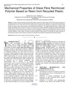
where: K1c = Fracture toughness; E = Young’s modulus expressed in N/m2; Ø = constraint factor (=H/σy ~ 3, where H is hardness and σy is the yield stress); l = crack length; a = half of indentation diagonal length; HV = Vickers microhardness; Figure 1 shows one Vickers indentation as it was measured using SEM technique, where Vickers indentation diagonals and cracks at the tips of the indentation may clearly be seen. Flexural bending strength: The rupture modulus, σ, was calculated according to Soltesz12:
(3)
where: σ = rupture modulus; F = load; υ = Poisson’s ratio = 0.28 (from the literature13); t = thickness; r = radius of the disc; r1 = radius of the loading ring; r2 = radius of the supporting ring; Weibull statistic analysis was performed and the Weibull
modulus was calculated for both compression and bending tests. Weibull statistic is indicated for porous ceramic specimens and indicates variability of the strength of the analysed ceramic14.
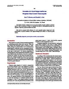
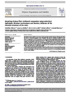
4. Results Table 1 shows the calculated values for Young’s modulus (E), fracture toughness (K1c) and Vickers microhardness (HV). The obtained values represent the mechanical features of the struts of the studied materials since localized techniques were used to perform the mechanical analysis. Figure 2 shows the probability of failure vs. compression strength for the specimens with 40 and 65 vol%. From the curves slopes, it is possible to identify that the two sets of specimens exhibited similar variations in Weibull moduli under compression tests. From this statistical analysis one may observe that no failure will occur when compressive tensions below 2 MPa are acting on specimens with 65 vol% porosity. For specimens with 40 vol% porosity, this value is shifted to 30 MPa. Figure 3 shows the probability of failure vs. bending strength for the specimens with 40 and 65 vol%. The bending tests showed a significant difference in the variability of bending strength for the two sets of specimens. This finding may be observed by the difference in the slopes of the two curves. Figure 3 shows that the minimum bending stress that 40 vol% samples will withstand is
Table 1. Young’s modulus (E), fracture toughness (KIc) and Vickers microhardness (HV) values for 65 vol% porosity GR-HA specimens.
E (GPa) 77 ± 12
Figure 1. Vickers indentation showing the diagonals and crack length as measured for KIc determination.
323
KIc (MPa.m1/2) 1.18 ± 0.1
HV 404
Figure 2. Probability of failure of porous GR-HA discs vs. compressive strength.
324
Silva et al.
Materials Research
Table 2. Weibull moduli for porous GR-HA specimens tested under compression and bending.
Porosity (vol%) 40 65
mc 9.19 8.23
mb 7.24 3.7
Figure 3. Probability of failure of porous GR-HA discs vs. bending strength.
0.2 MPa for the 65 vol% specimens and 2.5 MPa for the 40 vol% samples. Table 2 shows Weibull moduli for compression (mc) and bending (mc). It is noteworthy that although no significant change in mc values were obtained for samples with 40 and 65 vol% porosity, mb values decreased considerably when the porosity of samples increased. The calculated values for m quantitativaly confirm the observation of Figs. 2 and 3.
5. Discussion
Figure 4. SEM picuture of porous GR-HA specimen with 65 vol% porosity.
In a previous study, dense and porous GR-HA specimens were characterised by SEM and XRD with Rietveld refinement and mercury porosimetry9. These analyses showed that both presented a microstructure composed by HA, β-TCP and α-TCP. SEM analysis of the GR-HA composites showed that β-TCP and α-TCP phases were welldispersed in the HA matrix, which confirm previous studies that showed that CaO-P2O5 acted as a sintering aid for hydroxyapatite matrix. This previous work also demonstrated that only composite samples with 65 vol% porosity showed to have adequate 3-D morphology to allow bone ingrowth with interconnected porosity. Therefore, in this study Vickers hardness (HV), Young´s modulus and fracture toughness (K1c) testing were only performed on this composite. Furthermore, these are localised measurements while bending strength and compression testing are more representative of the overall microstructure of the composites. The average K1c value obtained for porous specimens pressed at 44 MPa, KIc= 1.18 ± 0.01, showed to be slightly higher than values found in the literature for single-phase hydroxyapatite15.
The value obtained for Vickers microhardness on porous GR-HA indicates that the different processing technique that was used for composite preparation allowed the liquid phase sintering process to be effective although these values also tended to be lower than those previously obtained for dense samples16. This finding should be attributed to the low densification level of porous samples that was induced by the pressing conditions. Dense GR-HA composites are usually prepared by applying uniaxial pressing of 288 MPa and in the present study only 44 MPa was used. Compression tests showed that the specimens produced with 40 vol% porosity had much higher compression strength when compared with specimens with 65 vol% porosity. Similar values were observed for the Weibull modulus of 40 and 65 vol% porosity specimens, which indicate that the different porosity volume did not produce marked difference in the variability of compression strength. The values obtained for these novel composite materials are comparable to those reported in the literature for calcium
Vol. 6, No. 3, 2003
Mechanical Characterisation of Porous Glass Reinforced Hydroxyapatite Ceramics – Bonelike
phosphate bioceramics16. Weibull modulus is an indication of strength variability and therefore with increasing pore fraction, it should be expected that the degree of non-homogeneity of samples should increase, which may explain the decrease of Weibull modulus as pore fraction increases. The minimum stress values below which failure occurs are much lower in bending than in compression for both 40 and 65 vol% porosity specimens. This behaviour was expected since porosity dramatically diminishes the strength for ceramic materials. However, previous SEM analysis and porosimetry data9 showed that 40 vol% porosity was not enough to obtain a structure of interconnected pores and therefore a compromise should be made between mechanical performance and the adequate 3-D morphology that is capable of allowing bone ingrowth. Flexural bending tests showed a marked variability in the rupture modulus under bending, i.e. the specimens with 65 vol% porosity showing higher variability than specimens with 40 vol% porosity. From Table 2, it can be seen that pore fraction strongly affected the behaviour under bending stress. The pores present in the microstructure acted as stress concentration factor and therefore lower the mechanical strength of the ceramic material. SEM analysis revealed that besides round macroporous, “needle shape” pores (Fig. 4) were also presented in the microstructure, which may account for the decrease in mechanical strength with increasing pore fraction. As it is well known, this effect is only valid in tensile effort and not in compression. Under tensile stress, fracture of ceramic usually occurs by propagation of a unique flaw (critical flaw) and in compression several flaws may contribute to the mechanical failure. Therefore, the increase in porosity should decisively influence the bending strength.
6. Conclusions Several factors have to be taken into account when designing bioceramics for allowing bone ingrowth. Porous discs with 65 vol% showed low bending and compression strength when compared with discs with 40 vol% pore fraction. However, mercury porosimetry studies and SEM analysis show that GR-HA specimens produced with 65 vol% exhibit a structure composed of interconnected pores with pore size above 100 µm. Once porous bioceramics are not structural implants, the most important features concern porous sizes and interconnections. Samples prepared with 65 vol% porosity can be easily handled, mantaining their structural integrity. These features allow porous GR-HA
325
discs with 65 vol% suitable for use as implants for bone ingrowth.
Acknowledgements Authors wish to acknowledge the financial support of FCT-Fundação para a Ciência e Tecnologia through the research project entitled “Interactive calcium-phosphate based materials prepared by Post-hybridization and in situ Hybridization”, ref: PCOTI/CTM/35516/CTM/2000 and CNPq through the PROFIX programme.
References 1. Santos, J.D.; Reis, R.L.; Monteiro, F.J.; Knowles, J.C.; Hastings, G.W. J. Mater. Sci: Mat. Med., v. 6, p. 348, 1995. 2. Santos, J.D.; Jha, L.J.; Monteiro, F.J. J. Mater. Sci: Mat. Med., v. 7, p. 181, 1996. 3. Lemos, A.F.; Ferreira, J.M.F. Mater. Sci. and Eng., v. 11, p. 35, 2000. 4. Lyckfeldt, O.; Ferreira, J.M.F. J. Eur. Ceram. Soc., v. 18, p. 131, 1998. 5. Saggio-Woyansky, J.; Scott, C.E.; Minnear, W.P. Am. Cer. Soc. Bull., v. 71, p. 1674, 1992. 6. De Groot, K.; Klein, C.P.A.T.; Wolke, J.G.C.; De BlieckHogervorst, J.M.A. Hand. Bioact. Cer. CRC Press Boca Raton FL, v. 2, p. 3, 1990. 7. Rodríguez-Lorenzo, L.M.; Vallet-Regí, M.; Ferreira, J.M.F.; Ginebra, M.P.;Aparício, C.; Planell, J.A. J. Biomedical Mat. Res., v. 60, p. 159, 2002. 8. Knowles, J.C.; Santos, J.D.; Hastings G.W. Patent number WO0068164, 2000. 9. Prado da Silva, M.H.; Lemos, A.F.; Ferreira, J.M.F.; Santos, J.D. Journal of Non-Crystalline Solids, v. 304, p. 286, 2002. 10. Nihara, K. J. Mater. Sci. Letter, v. 2, p. 221, 1983. 11. ENV 843-2, Advanced technical ceramics – Monolithic ceramics – Mechanical properties at room temperature – Part 2: Determination of elastic moduli, Dec. 1995. 12. Soltesz, U.; Ritcher, H.; Kienzler, R. High Tech Ceram., Amsterdam, Elsevier; p. 149, 1987. 13. Grenoble, D.E.; Katz, J.L.; Dunn, K.L.; Gilmore, R.S. and Murty, K.L., J. Biom. Mater. Res., v. 6, n. 3, p. 221223, 1972. 14. Giovan, M.N.; Sines, G. J. Amer. Society, v. 62, n. 9-10, 1979. 15. Wilmann, G.; Richter, H.; Wimmer, M. Biomed. Technik, v. 38, p. 14, 1993. 16. Lopes, M.A.; Monteiro, F.J.; Santos, J.D. Biomaterials, v. 20, p. 2085, 1999.