15 Blauer, G. and Akkawi, M. (2000) On the preparation of β-haematin. Biochem. J. 346, 249â250. 16 Sullivan, D. J., Gluzman, I. Y. and Goldberg, D. E. (1996) ...
333
Biochem. J. (2001) 355, 333–338 (Printed in Great Britain)
Mechanism of malarial haem detoxification inhibition by chloroquine Amit V. PANDEY1, Himani BISHT2, Vinod K. BABBARWAL2, Jaya SRIVASTAVA, Kailash C. PANDEY and Virander S. CHAUHAN3 Malaria Research Group, International Center for Genetic Engineering and Biotechnology (ICGEB), Aruna Asaf Ali Marg, New Delhi 110067, India
The haem detoxification pathway of the malaria parasite Plasmodium falciparum is a potential biochemical target for drug development. Free haem, released after haemoglobin degradation, is polymerized by the parasite to form haemozoin pigment. Plasmodium falciparum histidine-rich protein-2 (Pfhrp2) has been implicated as the catalytic scaffold for detoxification of haem in the malaria parasite. Previously we have shown that a hexapeptide repeat sequence (Ala-His-His-Ala-Ala-Asp), which appears 33 times in Pfhrp-2, may be the major haem binding site in this protein. The haem binding studies carried out by ourselves indicate that up to 18 equivalents of haem could be bound by this protein with an observed Kd of 0.94 µM. Absorbance spectroscopy provides evidence that chloroquine is capable of
extracting haem bound to Pfhrp-2. This was supported by the Kd value, of 37 nM, observed for the haem–chloroquine complex. The native PAGE studies reveal that the formation of the haem–Pfhrp-2 complex is disrupted by chloroquine. These results indicate that chloroquine may be acting by inhibiting haem detoxification\binding to Pfhrp-2. Moreover, the higher affinity of chloroquine for haem than Pfhrp-2 suggests a possible mechanism of action for chloroquine ; it may remove the haem bound to Pfhrp-2 and form a complex that is toxic to the parasite.
INTRODUCTION
haem binding sites in this protein [18]. Haem binding to synthetic peptides based on this repetitive sequence was proportional to the number of repeat units in the synthetic peptides. These peptides did not promote haem polymerization. However, native, as well as recombinant Pfhrp-2, promotes haemozoin formation [16,18,19]. Thus Pfhrp-2 has been established as a catalyst for haemozoin formation, and haem metabolism has emerged as a potential drug target in the malaria parasite [20]. The purpose of this study was to characterize the haem binding by Pfhrp-2 and to study the effect of chloroquine on this process. The fact that blocking haem processing appears to be highly toxic to the parasite has led to speculation that inhibiting this pathway might be a promising therapeutic strategy for the treatment of malaria. In the present study we provide evidence for the molecular mechanism of action of malarial haem detoxification inhibition by chloroquine.
The malaria parasite is fast becoming resistant to commonly used quinoline antimalarial drugs, resulting in an urgent demand for the development of novel drugs to combat this disease [1]. Quinoline antimalarial drugs have been the drugs of choice for a long time. However, despite being in use for more than half a century, and lots of research, the precise mechanism of action of quinoline antimalarial drugs is still not understood [2]. Several theories have been put forward to describe the antimalarial action of quinoline drugs [1,3]. Most of the blood schizontocidal antimalarial drugs are only active against those stages of the parasite that actively degrade haemoglobin and produce haemozoin pigment [1,3]. The haemozoin pigment is formed by polymerization of the haem that is released following haemoglobin digestion [4,5]. Free haem has been proposed as the receptor for chloroquine by Fitch and colleagues [5]. The enzyme haem polymerase, which catalyses the formation of haemozoin from free haem, was described by Slater and Cerami [6], and Chou and Fitch [7], and chloroquine was reported to inhibit haem polymerization in itro [8]. Several quinoline antimalarial drugs have been reported to inhibit haem polymerization [8–11]. However, the mechanism of haem polymerization and its inhibition is still unclear. Apart from initial reports of the haem polymerase enzyme by Slater and Cerami [6], and Chou and Fitch [7], spontaneous, as well as auto-catalytic, haem polymerization have been proposed [12,13]. Recent publications by ourselves, as well as several other groups, have described the necessity of a biological catalyst for polymerization of haem [14,15]. Recently, Plasmodium falciparum histidine-rich protein2 (Pfhrp-2) has been reported to mediate haemozoin formation, which could be inhibited by chloroquine [16]. A large number of repeats with sequences Ala-His-His-Ala-Ala-Asp or Ala-His-His are present in Pfhrp-2 [16–18]. We have previously shown that hexapeptide repeats Ala-His-His-Ala-Ala-Asp in Pfhrp-2 are the
Key words : histidine-rich protein, Plasmodium, malaria, haemoglobin, haemozoin.
EXPERIMENTAL Reagents Competent Escherichia coli BL21(DE3)pLysS cells were obtained from Novagen (Madison, WI, U.S.A.). Haemin, SDS, isopropyl β--thiogalactoside (IPTG), PMSF, Luria–Bertani media and all other chemicals were purchased from Sigma (St. Louis, MO, U.S.A).
Expression and purification of Pfhrp-2 A plasmid containing the gene encoding Pfhrp-2 in a pET 3d vector (Novagen) was transformed into competent E. coli BL21(DE3)pLysS cells [21], and colonies were selected for Pfhrp2 expression. Expression of Pfhrp-2 was induced by the addition of 0.4 mM IPTG, and protein was purified by metal-chelate chromatography on a Ni#+-nitrilotriacetate column using imidazole for elution as previously described [16,19].
Abbreviations used : Pfhrp-2, Plasmodium falciparum histidine-rich protein-2 ; Z-Phe-Arg-AMC, benzyloxycarbonyl-Phe-Arg-aminomethylcoumarin ; E-64, trans-epoxysuccinyl-L-leucylamido-(4-guanidino)butane ; DAB, diaminobenzidine ; IPTG, isopropyl β-D-thiogalactoside. 1 Current address : Metabolic Research Unit, University of California San Francisco, San Francisco, CA 94143-0978, U.S.A. 2 Both authors contributed equally to this work. 3 To whom correspondence should be addressed (e-mail amit!icgeb.res.in). # 2001 Biochemical Society
334
A. V. Pandey and others
Native PAGE analysis of the haem–Pfhrp-2 complex Complex formation was carried out by adding 5 µl of 47 µM Pfhrp-2 to an Eppendorf tube containing 1 µl of 1 M Tris\HCl buffer (pH 8.0) and 2 µl of 2 mM haem. In another tube 2 µl of 4 mM chloroquine was added before haem and kept at room temperature for 10 min. After addition of haem to each tube, they were incubated for 10 min at room temperature followed by addition of 5 µl of 1iPAGE sample buffer [0.125 M Tris\HCl, pH 6.8, containing 20 % (v\v) glycerol and 0.1 % (w\v) Bromophenol Blue, but without SDS and reducing agent]. From each reaction, 5 µl samples were loaded on to a 8 % (w\v) polyacrylamide gel [resolving gel buffer, 0.375 M Tris\HCl (pH 8.9) ; electrode buffer, 0.025 M Tris and 0.192 M glycine (pH 8.3)] and electrophoresis was performed at 100 V for 5 h at 4 mC. Gels were stained for protein using Coomassie Brilliant Blue R-250 or were stained for haem peroxidase using diaminobenzidine (DAB). We used the peroxidase activity of haem to differentiate between Pfhrp-2 and haem–Pfhrp-2 complexes. Haem or haemoproteins were specifically detected after electrophoresis by incubating with DAB and hydrogen peroxide as previously described [22]. In brief, after electrophoresis the gels were washed with water and incubated in a mixture of DAB and sodium acetate [6.3 mM DAB in methanol\0.25 M sodium acetate (pH 5.0), 3 : 7 (v\v)] for 1 h followed by addition of hydrogen peroxide to a final concentration of 30 mM. Stain was allowed to develop for 30 min, after which gels were washed with 30 % (v\v) isopropanol in 0.25 M sodium acetate (pH 5.0) and photographed. All steps of DAB staining were performed in the dark in order to avoid haem degradation.
Haem binding studies using UV spectroscopy Haem binding studies were carried out using difference absorption spectroscopy in the Soret region. Spectra were recorded on a Perkin–Elmer Lambda Bio 20 spectrophotometer between 190–700 nm at a speed of 120 nm\min and a slit width of 1 nm. Baselines were routinely subtracted from the spectra. All of the measurements were performed at 25 mC. For the titrations, 20 µl of 11.73 µM Pfhrp-2 was added to the sample cuvette and haem was added in 1 µl increments from stock solutions of 250 µM and 1 mM. The amount of haem bound to the protein was measured from the difference spectra by monitoring the change in absorbance at 414 nm, using a millimolar absorption coefficient of 71.2 mM−":cm−" for the haem–Pfhrp-2 complex (calculated using a 50 : 1 ratio of protein\haem ; results not shown). The dissociation constant was calculated by analysing data for multiple ligand-binding sites as described previously [23,24]. Since a significant portion of ligand (haem) is bound to protein (Pfhrp-2), it is not appropriate to use the free haem concentration as the total haem concentration. Using the modified method of Dixon [24] for tight-binding ligands, the dissociation constant Kd for the haem–Pfhrp-2 complex is given by : Kd l [HP]\o([PT]k[HP])([HT]k[HP])q where [HT] represents the total concentration of haem present (bound and free) and [PT] is the total concentration of protein. The concentration of bound haem [HP] was calculated using a millimolar absorption coefficient of 71.2 mM−":cm−" for the haem–Pfhrp-2 complex.
LS 50B spectrofluorimeter at 25 mC using 297 nm as the excitation wavelength (slit width of 5 nm) and monitoring emission spectra between 350 to 450 nm (slit width of 10 nm) at a scan speed of 120 nm\min. For the binding titration, 30 µl of chloroquine from a 100 nM stock solution was added to 3 ml of buffer (10 mM sodium phosphate, pH 7.4) in a sample cuvette, and haem was added using a 10 µl Hamilton syringe in aliquots of 1 µl from a 1 µM stock solution (freshly prepared before use). Data were analysed by a direct plot of fluorescence intensity against the ratio of haem and chloroquine, as well as by a Stern–Volmer plot, a relationship that describes the effect of quencher on the steady-state fluorescence of the sample. The ratio of fluorescence intensities in the absence and presence of quencher (haem) (described as If and If respectively), was plotted ! against quencher (haem) concentration (If \If versus haem) to ! give the Stern–Volmer plot. Binding-site calculations were performed using direct-plot analysis and Kd values for haem– chloroquine complex formation were obtained from the slope of the linear portion of the Stern–Volmer plot.
Assay of malaria cysteine protease Cysteine protease activity of the malaria parasite was assayed as described previously [25,26]. Purified digestive vacuole preparations from malaria parasites were used as a source of cysteine protease activity. Enzyme preparations were incubated with Pfhrp-2, haem and chloroquine in different combinations for 30 min before the start of the assay. Activity was monitored by measuring the cleavage of fluorescent AMC from the specific peptide substrate benzyloxycarbonyl-Phe-Arg-aminomethylcoumarin (Z-Phe-Arg-AMC). An excitation wavelength of 380 nm (slit width of 2.5 nm) and an emission wavelength of 460 nm (slit width of 10 nm) were used for the measurements. A specific inhibitor of cysteine proteases, trans-epoxysuccinyl-leucylamide-(4-guanidino)butane (E-64), which is known to inhibit malaria cysteine protease activity was used as a control for the validation of the assay. All of the reactions and spectroscopic measurements were performed at 25 mC.
Protein estimation Protein concentration was estimated by measuring the absorbance at 205 nm using BSA as a control [27].
RESULTS AND DISCUSSION Titration of Pfhrp-2 with haem The haem binding sites of Pfhrp-2 were titrated with increasing amounts of haem (Figure 1). The absorbance at 415 nm increased until it reached a plateau. Data were analysed using the program ‘ Easybound ’ [23] made for ligand binding studies based on fitting of data by non-linear regression methods. A Kd value of 0.94 µM for the haem–Pfhrp-2 complex, with 18 haem binding sites, was obtained from the analysis of binding data by nonlinear regression analysis. The number of binding sites found by ourselves is in agreement with that reported by Sullivan et al. [16] ; however, in another study as much as 50 equivalents of haem were reported to bind to Pfhrp-2 [28].
Inhibition of haem binding to Pfhrp-2 by chloroquine Study of haem–chloroquine interaction Haem–chloroquine binding was studied using the quenching of chloroquine fluorescence by haem as a measure of binding. Fluorescence emission spectra were recorded on a Perkin–Elmer # 2001 Biochemical Society
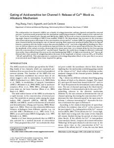
Haem binding to Pfhrp-2 was studied by recording the difference spectra of the protein with and without haem (Figure 2A). Haem alone has an absorption maximum at 385 nm, which is shifted to 415 nm when it binds to Pfhrp-2. When chloroquine was added
Mechanism of malarial haem detoxification inhibition by chloroquine
335
2
3 1
4
Figure 2
Figure 1
Binding of haem to Pfhrp-2
(A) Bound haem is plotted against free haem for analysis using ‘ Easybound ’ software [23]. The inset shows the same data plotted with the x-axis on a log scale. (B) Direct plot of bound haem against total haem for analysis by the method of Dixon [24]. Details of data analysis are described in the Experimental section. For the titration, 20 µl of 11.73 µM Pfhrp-2 was added to the sample cuvette and haem was added in 1 µl increments from stock solutions of 250 µM and 1 mM. The amount of haem bound to the protein was measured from the difference spectra by monitoring the change in absorbance at 414 nm, using a millimolar absorption coefficient of 71.2 mM−1:cm−1 for the haem–Pfhrp-2 complex.
to the mixture of haem and Pfhrp-2, a significant reduction in haem–Pfhrp-2 complex formation was observed as shown by a fall in the absorption peak at 415 nm. Pre-incubation of chloroquine with haem, before addition to Pfhrp-2, resulted in almost complete loss of haem–Pfhrp-2 complex formation and the spectrum was indistinguishable from that of the haem–chloroquine complex. Removal of haem bound to Pfhrp-2 by chloroquine would interrupt malarial haem detoxification, which would be detrimental to the parasite. Haem–Pfhrp-2 interaction and the effect of chloroquine were confirmed by native PAGE analysis of complex formation (Figure 2B). Lanes 1–3 are stained with Coomassie Brilliant Blue, which detects the Pfhrp-2 protein. Movement of Pfhrp-2 alone (lane 1) was different from a mixture of haem and Pfhrp-2 (lane 2). When chloroquine was added to the reaction mixture along with haem (lane 3), the movement of Pfhrp-2 reverted back to normal. Haem runs ahead along with dye front during electrophoresis. To confirm the presence of haem–Pfhrp-2 complexes, we utilized peroxidase staining using DAB as the substrate (lanes 4–6). DAB specific-
Inhibition of haem–Pfhrp-2 complex formation by chloroquine
(A) Spectra of haem and Pfhrp-2 in the presence and absence of chloroquine. Haem alone (trace 1) has an absorption maximum at 385 nm that is shifted to 415 nm in the presence of Pfhrp2 (trace 2). Addition of chloroquine to the haem–Pfhrp-2 complex (trace 3) results in the loss of the haem–Pfhrp-2 complex. When chloroquine is added before addition of haem (trace 4), a dramatic reduction in haem binding is observed and haem–chloroquine complex formation is indicated. When chloroquine is added to the haem–Pfhrp-2 complex a significant decrease in complex formation was observed, indicative of chloroquine extracting the haem bound to Pfhrp-2 to form a haem–chloroquine complex. Details of spectral recordings and reactions are described in the Experimental section. (B) Native PAGE analysis of the haem–Pfhrp-2 complex and its interaction with chloroquine. Coomassie Brilliant Blue staining (lanes 1–3) and DAB staining (specific for haem/haemoproteins ; lanes 4–6). Lanes 1 and 4, Pfhrp-2 ; lanes 2 and 5, Pfhrp-2jhaem ; lanes 3 and 6, Pfhrp-2jchloroquinejhaem. The diffuse band in lane 2 indicated haem–Pfhrp-2 complex formation, which was confirmed by specific haem peroxidase staining in lane 5. Addition of chloroquine resulted in the loss of haem–Pfhrp-2 complex formation as evidenced by normal movement on the Coomassie Brilliant Blue-stained gel (lane 3) and loss of haem peroxidase staining (lane 6). Details of the experiment are described in the Experimental section.
ally stains haem or haemoproteins. Pfhrp-2 alone did not stain with DAB (lane 4) ; however, in the sample containing haem and Pfhrp-2 a distinct staining for haem peroxidase activity was observed (lane 5), confirming the presence of haem–Pfhrp-2 complexes. Addition of chloroquine in this reaction resulted in almost total loss of peroxidase staining (lane 6), indicating removal of haem from the haem–Pfhrp-2 complex, and confirming the results from the Coomassie Brilliant Blue-stained gel (lane 3). The presence of Pfhrp-2 in all samples was confirmed by Western-blot analysis of these samples using anti-(Pfhrp-2) antibodies (results not shown). By using specific staining for haem we were able to visualize haem–Pfhrp-2 complexes on a gel and distinguish them from native Pfhrp-2 in our experiments, since Pfhrp-2 itself does not have any haem group and thus does not show peroxidase activity. Loss of peroxidase stain on treatment with chloroquine provided evidence for the removal of haem from haem–Pfhrp-2 complexes, indicating a possible mechanism of chloroquine action.
Haem–chloroquine binding Haem–chloroquine binding was studied by monitoring the quenching of chloroquine fluorescence by haem. Data were plotted as the percentage quenching against the concentration # 2001 Biochemical Society
336
A. V. Pandey and others
1
2
1
2
Figure 3
Haem–chloroquine binding
(A) Direct plot of fractional quenching values against the haem/chloroquine ratio in solution. A typical curve for strong ligand binding is observed with a clear indication of saturation as a haem/chloroquine ratio of 2 is approached. (B) A Stern–Volmer plot of chloroquine fluorescence quenching by haem. Ifo is the initial fluorescence intensity and If is the fluorescence intensity after addition of haem at a given point. Binding constants were calculated from linear regions of the curve. The inset of (A) shows the plot of Ifo/If at low concentrations of chloroquine and haem. A Kd value of 37 nM was obtained for the haem–chloroquine complex from the slope of this plot. Deviation of the Stern–Volmer plot from linearity indicates the presence of more than one binding site, which was confirmed by results obtained from the direct plot shown in (A). For the titrations, 30 µl of chloroquine from a stock of 100 nM was taken in a 3 ml cuvette and titrated with 1 µl aliquots of 1 µM haem solution (freshly prepared prior to use). Other details of the experiment are given in the Experimental section.
of haem in solution (Figure 3A). The results are typical of strong binding reactions with clear demarcation of the saturation point. A haem\chloroquine ratio of 2 : 1 was indicated. Data were analysed based upon two haem binding sites per molecule of chloroquine as described previously [29]. A Stern–Volmer plot of fluorescence intensities of the control and the haem-containing samples also showed deviation from a straight line indicating more than one haem binding site per chloroquine molecule (Figure 3B). The experiments were performed at very low concentrations of haem and chloroquine in order to calculate binding constants (Figure 3A inset). A Kd value of 37 nM was obtained for haem–chloroquine complex formation from the slope of this plot.
Interaction of chloroquine with Pfhrp-2 Equilibrium dialysis experiments failed to detect any significant interaction between chloroquine and Pfhrp-2. We dialysed 47 µM # 2001 Biochemical Society
Figure 4 Pfhrp-2
Spectra of the haem–chloroquine complex and the effect of
(A) Spectra of haem before (trace 1) and after (trace 2) the addition of chloroquine (1 nmol of haem was mixed with 0.5 nmol of chloroquine). (B) Spectra of haem (trace 1) and haem–chloroquine (trace 2) in the presence of Pfhrp-2. Chloroquine was pre-incubated with Pfhrp-2 for 10 min before addition of haem.
Pfhrp-2 inside the dialysis tubing against 1 nM chloroquine outside. Even 100 : 1 binding would have resulted in an approx. 470-fold increase in chloroquine concentration inside the dialysis tubing containing Pfhrp-2. However, no significant increase in chloroquine concentration was detected indicating a lack of chloroquine binding by Pfhrp-2. In another experiment, where Pfhrp-2 preparations incubated with a 5 molar excess of chloroquine were dialysed against PBS and used for haem binding, no change in the amount of haem–Pfhrp-2 complex formation was detected (results not shown). This indicates that the haem binding sites of Pfhrp-2 are unaffected by chloroquine. Moreover, when haem–chloroquine binding was studied in the presence and absence of a 50-fold molar excess of Pfhrp-2, no change was observed in the haem–chloroquine interaction (Figure 4). These results indicate that haem has a higher affinity for chloroquine compared with Pfhrp-2. The observation is supported by a Kd value of 3.7i10−) M for the haem–chloroquine complex as compared with a Kd value of 9.4i10−( M for the haem–Pfhrp2 complex.
Mechanism of malarial haem detoxification inhibition by chloroquine Table 1 Inhibition of malaria cysteine protease activity by haem, and its protection by Pfhrp-2 Malaria cysteine protease activity was measured by monitoring the release of fluorescent AMC from the specific peptide substrate Z-Phe-Arg-AMC. Concentrations of additives were as follows : haem, 50 µM ; Pfhrp-2, 30 µM ; chloroquine (CQ), 50 µM ; E-64, 50 µM. The assay was performed as described previously [25]. All additives were incubated with the enzyme for 30 min before addition of the substrate. E-64 is a highly potent and specific inhibitor of cysteine proteases. It was used as a positive control in the reaction.
Sample
∆ Fluorescence intensity (arbitrary units/min)
Control Controljhaem ControljPfhrp-2 ControljPfhrp-2jhaem ControljPfhrp-2jCQjhaem ControljCQ ControljCQjhaem ControljE-64
12.5p1.5 1.5p0.7 12.1p0.8 7.9p0.8 1.4p0.5 11.7p1.2 1.2p0.5 0.0
Inhibition of cysteine protease by haem, and its antagonism by Pfhrp-2 Haem is a potent inhibitor of malaria cysteine protease activity [25]. Free haem released following haemoglobin digestion is detoxified by polymerization\degradation. The presence of haem-binding substances in parasite vacuoles would serve the purpose of haem detoxification and reduce the damage caused by haem. A drug that could interfere with haem detoxification will perturb this co-existence leading to haem toxicity. To test this hypothesis we assayed malaria cysteine protease (a crucial parasite enzyme known to be inhibited by haem) in the presence and absence of haem and chloroquine. A specific peptide substrate for malaria cysteine protease (Z-Phe-Arg-AMC) was used for the assay, and cleavage of fluorescent AMC from the peptide was monitored fluorimetrically. Haem inhibited the cysteine protease activity as shown by the reduction in the increase in fluorescence per minute. Inhibition of the malaria cysteine protease by haem in the presence of Pfhrp-2 was remarkably less than the control samples without Pfhrp-2 (Table 1). The presence of chloroquine in the reaction mixture reversed this effect and the inhibition returned to its normal level, indicating disruption of the haem detoxification function of Pfhrp-2. The presence of both haem and chloroquine without Pfhrp-2 caused somewhat higher inhibition. Under normal circumstances, toxicity of haem would be countered by haembinding proteins, however, in the presence of a drug such as chloroquine that has a higher affinity for haem, this protection would be lost and lead to the disruption of parasite metabolism by haem\haem–drug complexes.
Conclusions In the present study we have examined the binding of haem to Pfhrp-2, the major haem-binding protein present in the malaria parasite. For proteins with very high affinities for haem, such as haemopexin and haemoglobin, only approximate estimates of Kd values are reported, which mostly fall in the pM range. The Kd value calculated for the haem–Pfrp-2 complex is 0.94 µM. The strong haem-binding ability of this protein could provide the malaria parasite with an important source with which to combat haem toxicity, by storage of toxic haem until it can be processed by other means [30,31].
337
Malaria pigment has long been associated with the action of chloroquine. The first morphological effect of chloroquine treatment on the malaria parasite is the clumping of haemozoin pigment [32,33]. Our results described in the present study suggest that chloroquine interacts with haem bound to Pfhrp-2 and during this process a haem–chloroquine complex is formed. Since very little haem is sufficient to disrupt the biochemical processes of the parasite, even a small shift in the haem detoxification process towards free haem would be enough to cause damage to the parasite [34]. Moreover, haem bound to Pfhrp-2 may lose its toxic effects on parasite enzymes as observed by us in cysteine protease experiments. A protein such as Pfhrp-2 that has a large number of haem binding sites would serve as an ideal sink for the free haem and protect the parasite from its toxicity. It is probable that the malarial parasite might be using such proteins as a first line of defence against haem toxicity. Our results showing antagonism of haem damage to the cysteine protease by Pfhrp-2 and the loss of this protection in the presence of chloroquine seem to support this hypothesis. The observations that chloroquine can extract haem bound to Pfhrp-2, the major protein involved in haem detoxification, have provided an insight into the molecular interactions involved in the mechanism of action of this drug. Inhibition of haem–Pfhrp2 complex formation could be used for the screening of potential inhibitors of the haem detoxification system of the malaria parasite. We thank Dr D. E. Goldberg (Department of Molecular Microbiology, Howard Hughes Medical Institute, Washington University School of Medicine, St Louis, MO, U.S.A.) for providing the Pfhrp-2 gene, Dr Diane Taylor (Department of Biology, Georgetown University, Washington DC, U.S.A.) for providing monoclonal antibodies against Pfhrp-2, and Dr Peter Hedlund (Department of Molecular Biology, The Scripps Research Institute, La Jolla, CA, U.S.A.) for providing the ‘ Easybound ’ software for binding analysis. We thank Drs Leann Tilley (La Trobe University, Australia), Hagai Ginsburg (Hebrew University, Israel), Chetan Chitnis, Sanjay Singh, Andrew Lynn and R. M. Joshi, and Pawan Malhotra (ICGEB, India) for helpful suggestions and discussions.
REFERENCES 1 2 3 4
5
6 7 8 9
10
11 12 13
Pandey, A. V. and Chauhan, V. S. (1998) Heme polymerization by malarial parasite : a potential target for antimalarial drug development. Curr. Sci. 75, 911–918 Wellems, T. E. (1992) How chloroquine works. Nature (London) 355, 108–109 Foley, M. and Tilley, L. (1998) Quinoline antimalarials : mechanisms of action and resistance and prospects for new agents. Pharmacol. Ther. 79, 55–87 Rosenthal, P. J. and Meshnick, S. R. (1998) Hemoglobin degradation by malaria parasite. In Malaria : Parasite Biology, Pathogenesis and Protection (Sherman, I. W., ed.), pp. 145–158, ASM Press, Washington DC Chou, A. C., Chelvi, R. and Fitch, C. D. (1980) Ferriprotoporphyrin-IX fulfills the criteria for identification as the chloroquine receptor of malaria parasites. Biochemistry 19, 1543–1549 Slater, A. F. G. and Cerami, A. (1992) Inhibition by chloroquine of a novel heme polymerase enzyme activity in malaria trophozoites. Nature (London) 355, 167–169 Chou, A. C. and Fitch, C. D. (1992) Heme polymerase : modulation by chloroquine treatment of a rodent malaria. Life Sci. 51, 2073–2078 Chou, A. C. and Fitch, C. D. (1993) Control of heme polymerase by chloroquine and other quinoline derivatives. Biochem. Biophys. Res. Commun. 195, 422–427 Raynes, K. J., Stocks, P. A., O‘Neill, P. M., Park, B. K. and Ward, S. A. (1999) New 4-aminoquinoline Mannich base antimalarials. 1. Effect of an alkyl substituent in the 5h-position of the 4h-hydroxyanilino side chain. J. Med. Chem. 42, 2747–2751 Egan, T. J., Hunter, R., Kaschula, C. H., Marques, H. M., Misplon, A. and Walden, J. (2000) Structure-function relationships in aminoquinolines : effect of amino and chloro groups on quinoline-hematin complex formation, inhibition of β-hematin formation and antiplasmodial activity. J. Med. Chem. 43, 283–291 Ignatushchenko, M. V., Winter, R. W. and Riscoe, M. (2000) Xanthones as antimalarial agents : stage specificity. Am. J. Trop. Med. Hyg. 62, 77–81 Egan, T. J., Ross, D. C. and Adams, P. A. (1994) Quinoline antimalarial drugs inhibit spontaneous formation of β-hematin (malaria pigment). FEBS Lett. 352, 54–57 Dorn, A., Stoffel, R., Matile, H., Bubendorf, A. and Ridley, R. G. (1995) Malarial hemozoin/β-hematin supports haem polymerisation in the absence of protein. Nature (London) 374, 269–271 # 2001 Biochemical Society
338
A. V. Pandey and others
14 Pandey, A. V. and Tekwani, B. L. (1996) Formation of haemozoin/β-hematin under physiological conditions is not spontaneous. FEBS Lett. 393, 189–192 15 Blauer, G. and Akkawi, M. (2000) On the preparation of β-haematin. Biochem. J. 346, 249–250 16 Sullivan, D. J., Gluzman, I. Y. and Goldberg, D. E. (1996) Plasmodium hemozoin formation mediated by histidine rich proteins. Science (Washington, D. C.) 271, 219–221 17 Wellems, T. E. and Howard, R. J. (1986) Homologous genes encode two distinct histidine rich proteins in a cloned isolate of Plasmodium falciparum. Proc. Natl. Acad. Sci. U.S.A. 83, 6065–6069 18 Pandey, A. V., Joshi, R. M., Tekwani, B. L., Singh, R. L. and Chauhan, V. S. (1997) Synthetic peptides corresponding to a repetitive sequence of malarial histidine rich protein bind heme and inhibit hemozoin formation in vitro. Mol. Biochem. Parasitol. 90, 281–287 19 Pandey, A. V., Singh, N., Tekwani, B. L., Puri, S. K. and Chauhan, V. S. (1999) Assay of β-hematin formation by malaria parasite. J. Pharm. Biomed. Anal. 20, 203–207 20 Padmanaban, G. and Rangarajan, P. N. (2000) Heme metabolism of Plasmodium is a major antimalarial target. Biochem. Biophys. Res. Commun. 268, 665–668 21 Studier, F. W., Rosenberg, A. H., Dunn, J. J. and Dubendorff, J. W. (1990) Use of T7 RNA polymerase to direct expression of proteins. Methods Enzymol. 185, 60–89 22 Thomas, P. E., Ryan, D. and Levin, W. (1976) An improved staining procedure for the detection of the peroxidase activity of cytochrome P-450 on sodium dodecyl sulfate polyacrylamide gels. Anal. Biochem. 75, 168–176 23 Hedlund, P. B. and von Euler, G. (1999) EasyBound – a user friendly approach to nonlinear regression analysis of binding data. Comput. Methods Programs Biomed. 58, 245–249 Received 22 August 2000/9 November 2000 ; accepted 10 January 2001
# 2001 Biochemical Society
24 Dixon, M. (1972) The graphical determination of Km and Ki. Biochem. J. 129, 197–202 25 Pandey, A. V., Tekwani, B. L., Singh, R. L. and Chauhan, V. S. (1999) Artemisinin, an endoperoxide antimalarial, disrupts the hemoglobin catabolism and heme detoxification systems in malarial parasite. J. Biol. Chem. 274, 19383–19388 26 Shenai, B. R., Sijwali, P. S., Singh, A. and Rosenthal, P. J. (2000) Characterization of native and recombinant falcipain-2, a principal trophozoite cysteine protease and essential hemoglobinase of Plasmodium falciparum. J. Biol. Chem. 275, 29000–29010 27 Stoscheck, C. M. (1990) Quantitation of protein. Methods. Enzymol. 182, 50–68 28 Choi, C. Y., Cerda, J. F., Chu, H. A., Babcock, G. T. and Marletta, M. A. (1999) Spectroscopic characterization of the heme binding sites in Plasmodium falciparum histidine-rich protein 2. Biochemistry 38, 16916–16924 29 Balasubramanian, D., Mohan Rao, C. and Panijpan, B. (1984) The malaria parasite monitored by photoacoustic spectroscopy. Science (Washington, D. C.) 223, 828–830 30 Loria, P., Miller, S., Foley, M. and Tilley, L. (1999) Inhibition of the peroxidative degradation of haem as the basis of action of chloroquine and other quinoline antimalarials. Biochem. J. 339, 363–370 31 Ginsburg, H., Famin, O., Jhang, J. and Krugliak, M. (1998) Kinetics of inhibition of glutathione mediated degradation of ferriprotoporphyrin IX by antimalarial drugs. Biochem. Pharmacol. 56, 1305–1313 32 Macomber, P. B. and Sprinz, H. (1967) Morphological effects of chloroquine on Plasmodium berghei in mice. Nature (London) 214, 937–938 33 Homewood, C. A., Warhurst, D. C., Peters, W. and Baggaley, V. C. (1972) Lysosomes, pH and the anti-malarial action of chloroquine. Nature (London) 235, 50–52 34 Orjih, A. U., Banyal, H. S., Chevli, R. and Fitch, C. D. (1981) Hemin lyses malaria parasites. Science (Washington, D.C.) 214, 667–669