DOCTOR OF MEDICAL SCIENCE
DANISH MEDICAL BULLETIN
Mechanisms of cellular synchronization in the vascular wall Mechanisms of vasomotion Vladimir V. Matchkov
This review has been accepted as a thesis together with 9 previously published papers by the Faculty of Health Sciences, University of Aarhus at 7. April 2010 and defended on 13. august 2010 Official opponents: Iain Greenwood & Steen Dissing & Søren K. Moestrup Correspondence: Department of Physiology and Biophysics, University of Aarhus, Aarhus, Denmark E-mail:
[email protected]
vasomotion can be used as a “readout” for intercellular communication. Using this approach I demonstrated that inhibition of + + the ouabain-sensitive Na /K -ATPase uncouples smooth muscle cells in the vascular wall and suggested the mechanism responsible for this electrical uncoupling. In my studies on the role of the + + ouabain-sensitive Na /K -ATPase for vascular function I suggested + + the presence of Na /K -ATPase-based signalosome which also + 2+ includes the Na /Ca -exchanger, gap junctions and the ATP+ dependent K channels. These studies provide a useful tool for manipulations intercellular communication in the small arteries. The thesis includes the following previous publications:
Dan Med Bull 2010;57: (10)B4191
1. PREFACE This thesis is based on the work carried in the Vascular Smooth Muscle group at the Institute of Physiology and Biophysics, Aarhus University. This thesis is focused on a mechanistic understanding of cellular synchronization in the small resistance arteries. I have started this work because of my general interest in vasomotion, a phenomenon of synchronized activity in the vascular wall which has been known for more than 150 years. In spite of the long history and suggestions that vasomotion is important for pathological states the studies of vasomotion have been mostly descriptive. Development of new experimental techniques 2+ such as small artery myography, intracellular Ca imaging and electrophysiological approaches brought new possibilities to the studies of cellular mechanisms of vascular synchronization. I have used these advanced methods to characterize vasomotion in detail and have suggested and tested a model for generation of vasomotion in the rat mesenteric artery. The suggested model is one of several models of vasomotion but it has strong experimental support and is supplemented by the mathematical modeling published by our group. Two key elements for the synchronized oscillation in the mesenteric small arteries a cGMP-dependent Ca2+-activated Cl- current and the electrical intercellular communication were further explored in my research. I have character2+ ized the cGMP-dependent Ca -activated Cl current suggested by our model for vasomotion and demonstrated this current in different vascular beds. Using a novel siRNA approach I have then shown the association between this current and bestrophin-3 protein expression in vivo and in vitro. Based on these results I suggested the molecular identity of this current and its significance for smooth muscle cell synchronization by a membrane potential-dependent mechanism. The studies of intercellular communication in the vascular wall are lacking specific and effective tools to manipulate these intercellular contacts. I have performed comprehensive studies to analyze the action of the most commonly used gap junction blockers and demonstrated that
I. Peng H, Matchkov V, Ivarsen A, Aalkjaer C, Nilsson H. Hypothesis for the initiation of vasomotion. Circ Res. 2001; 88(8): 810-815. II. Rahman A, Matchkov V, Nilsson H, Aalkjaer C. Effects of cGMP on coordination of vascular smooth muscle cells of rat mesenteric small arteries. J Vasc Res. 2005; 42(4):301-311. III. Matchkov VV, Aalkjaer C, Nilsson H. A cyclic GMP-dependent calcium-activated chloride current in smooth-muscle cells from rat mesenteric resistance arteries. J Gen Physiol. 2004; 123(2): 121-134. IV. Matchkov VV, Aalkjaer C, Nilsson H. Distribution of cGMP2+ dependent and cGMP-independent Ca -activated Cl conductances in smooth muscle cells from different vascular beds and colon. Pflugers Arch. 2005; 451(2): 371-379. V. Matchkov VV, Larsen P, Bouzinova EV, Rojek A, Boedtkjer DM, Golubinskaya V, Pedersen FS, Aalkjaer C, Nilsson H. Bestrophin-3 (vitelliform macular dystrophy 2-like 3 protein) is essential for the cGMP-dependent calcium-activated chloride conductance in vascular smooth muscle cells. Circ Res. 2008; 103(8): 864-872. VI. Matchkov VV, Rahman A, Peng H, Nilsson H, Aalkjaer C. Junctional and nonjunctional effects of heptanol and glycyrrhetinic acid derivates in rat mesenteric small arteries. Br J Pharmacol. 2004; 142(6): 961-972. VII. Matchkov VV, Rahman A, Bakker LM, Griffith TM, Nilsson H, Aalkjaer C. Analysis of effects of connexin-mimetic peptides in rat mesenteric small arteries. Am J Physiol. 2006; 291(1): H357-H367. VIII. Matchkov VV, Gustafsson H, Rahman A, Briggs Boedtkjer DM, Gorintin S, Hansen AK, Bouzinova EV, Praetorius HA, Aalkjaer C, + + + 2+ Nilsson H. Interaction between Na /K -pump and Na /Ca exchanger modulates intercellular communication. Circ Res. 2007; 100(7): 1026-1035. DANISH MEDICAL BULLETIN
1
IX. Glavind-Kristensen M, Matchkov V, Hansen VB, Forman A, Nilsson H, Aalkjaer C. KATP-channel-induced vasodilation is modulated by the Na,K-pump activity in rabbit coronary small arteries. Br J Pharmacol. 2004; 143(7): 872-880. 2. ACKNOWLEDGMENTS The present doctoral dissertation is based on the studies performed at the Institute of Physiology and Biophysics, University of Aarhus, where I began as assistant research professor in 1999 and then continued as associate professor in 2007. The work in this stimulating and supportive environment has had a great influence on my life and research carrier. There are many individuals who have been or are presently a part of the Institute of Physiology and Biophysics to whom I would like to express my gratitude. Firstly, the members of the Vascular Smooth Muscle and Epithelium groups. I would like to give my special thanks to Professor Christian Aalkjær and Professor Holger Nilsson for their support through all these years, for sharing with me their great experience and knowledge in science and life. I am indebted for having had the privilege to work with these great scientists. I would like to express my special thanks for the excellent technical assistance and for creating a warm lab environment to Jørgen Andresen, Susie Mogensen, Jane Rønn, and Kirsten Skaarup. I thank my previous collaborators, together with whom we began the studies included in this dissertation. My acknowledgements go to Dr. Hongli Peng, Dr. Andres Ivarsen, Dr. Awahan Rahman, Dr. Veronika Golubinskaya, Dr. Zahra Nourian, and Dr. Per Larsen for their enthusiasm in our projects and inspiring scientific discussions. I would also like to thank Dr. Donna Bødtkjer for sharing daily research activities, constructive thinking and for stimulating discussions. I am thankful to Professor Helle Praetorius for her inestimable criticism and for her consistent eagerness to help. I would like to thank my former and present colleagues Thomas Hansen, Anne Kirstine Hansen, Ebbe Bødtkjer, Torbjørn Brøgger, Nina Møller-Nielsen, Vibeke Secher Nielsen, and Kate Møller for their enthusiasm and curiosity, which made many difficult projects possible. I am thankful to Finn Marquard for the maintenance and repair of equipment; to Kristian Klærke and Per Holm for their IT infrastructural support, and the printing office of the Faculty of Health Sciences for their efficient and professional work. Special thanks to Carina Mikkelsen, Inga Edney, Dorte Abildskov, Inge Eggert, and Else Marie Sørensen for keeping the administrative and financial procedures in proper order. I also would like to give special thanks to my colleagues at the Department of Pharmacology where I began my scientific carrier in Denmark. I am especially thankful to Professor Michael Mulvany and Professor Ulf Simonsen for providing their invaluable support, for their constructive suggestions and for sharing their expertise. I would also like to thank my colleagues from the Water and Salt Research Center and the Department of Anatomy. Special thanks to Professor Jeppe Praetorius for his expertise and help in immunohistochemistry. I am grateful to Inger Merete Paulsen (Department of Anatomy) and Helle Zibrandtsen (Department of Pharmacology) for their wonderful technical assistance in the protein studies. I would like to give special thank to Professor Niels-Henrik Holstein-Rathlou and Associate Professor Jens Christian Brings
Jacobsen from University of Copenhagen for our long-lasting collaboration and for complimenting my experimental results with mathematical modeling. I would like to express my sincere gratitude to my first laboratory at the Lomonosov Moscow State University, where I was introduced to the field of cardiovascular physiology. I am especially thankful to Professor Ivan M. Rodionov, Professor Olga S. Tarasova, and Professor Vladimir B. Koshelev for introducing me to the scientific world and for having the courage to be my supervisors. Dear Olga, thank you for your continued support in my life and persuit of scientific interest after my move to Denmark. I would like to give special thanks to Professor Rudolf Schubert (University of Heidelberg), who introduced me to the patch clamp technique during my stay at the Rostock University; Professor Tudor Griffith (Cardiff University), and Professor Alun Hughes (Imperial College, London) for enjoyable and productive collaborations. My special thanks go to my friend and colleague Edgaras Stankevicius and his family for being supportive in scientific as well as in non-scientific issues. I would like to thank all my friends, who have been a great support and practical help to me and my family through many years. My thanks go to Tatiana K., Max, and Natasha Jørgensen, Natalia Sanotskaya, Oleg, Isabella, and Alexandra Balakirievy, Elena, Vladimir, and Sasha Stolba. I would also like to express my appreciation to Larissa, Dasha, and Katja (former Polekarevy), and their families as well as to Joana Matos. I would like to thank my parents and great grandparents for always being on my side, for teaching me to be focused on my work, to not be afraid of difficulties and to make important decisions. I am thankful to my family in-law for their belief in me, endless support, and for our warm relations through many years. I would like to thank my children Victor and Nikolai for their respect and understanding of my busyness and last, but not least, I am greatly thankful to my wife, my friend, and my colleague Elena V. Bouzinova for being with me, for creating and keeping the environment, which made my work possible, and for daily understanding and support. Finally, I thank the Faculty of Health Sciences at Aarhus University for their willing help and financial support. My acknowledgement also goes to the Center for Psychiatric Research at the Aarhus University Hospital for new opportunities and promising collaboration. My projects have been supported by the Water and Salt Research Center, which is established by the Danish National Research Foundation (Danmarks Grundforskningsfond); the Danish Research Council; the Danish Heart Foundation, the Novo Nordisk Foundation, and the Lundbeck Foundation. 3. LIST OF ABBREVIATIONS AA arachidonic acid ADP adenosine diphosphate ANO1 anoctamin-1; see also TMEM16A ATP adenosine triphosphate AVP arginin-vasopressine BAPTA 1,2-bis(o-aminophenoxy)ethane-N,N,N',N'tetraacetic acid 2+ + BK big-conductance Ca -activated K channels 2+ [Ca ]i intracellular calcium concentration 2+ CaCC Ca -activated Cl channels DANISH MEDICAL BULLETIN
2
CAMKII cAMP cGMP CICR Cx DIDS EDHF EGTA ER GAP GJ IAA-94 ICl(Ca) ICl(Ca,cGMP) IK IP3 IP3-R KATP KCa MAPK MLC NCX NO NPY PKG PKC PLC PLA2 RYA-R siRNA SK SMCs SR TMEM16A TRP channel VDCCs VMD
calmodulin kinase II cyclic adenosine monophosphate cyclic guanosine monophosphate 2+ 2+ Ca -induced Ca release connexin 4,4'-diisothiocyanostilbene-2,2'-disulfonic acid endothelium-derived hyperpolarizing “factor” ethylene glycol tetraacetic acid endoplasmic reticulum connexin-mimetic peptides gap junctions R(+)-[(6,7-Dichloro-2-cyclopentyl-2,3-dihydro-2methyl-1-oxo-1H-inden-5-yl)-oxy]acetic acid 2+ Ca -activated Cl current 2+ cGMP-dependent Ca -activated Cl current + intermediate-conductance K channels inositol 1,4,5-trisphosphate IP3-sensitive channels + ATP-dependent K channels 2+ + Ca -activated K channels mitogen-activated protein kinase myosin light chain + 2+ Na /Ca exchanger nitric oxide neuropeptide Y protein kinase G protein kinase C phospholipase C phospholipase A2 ryanodine-sensitive channel or receptor small interfering RNA + small-conductance K channels smooth muscle cells sarcoplasmic reticulum transmembrane protein 16A, see also ANO1 transient receptor potential channel 2+ voltage-dependent Ca channels vitelliform macular dystrophy
4. INTRODUCTION A blood circulation system in complex, multicellular organisms should satisfy the metabolic demands of all cells in the body. This demand varies widely with location of the tissues and with time, and is affected by changes in environmental and internal parameters over a considerable range. Therefore, it is important to have a very precise regulation of blood flow that is achieved by the combined effects of multiple interacting mechanisms, including sensitivity to pressure, flow rate, metabolite levels, and neural signals. Flow regulation requires the sensing of metabolic and hemodynamic conditions, and the main effectors of this regulation are the arterioles and small arteries, which are located proximally to the tissue that they supply. Arterial pressure falls mark1 edly while passing these vessels , which demonstrates that they are responsible for a significant part of total vascular resistance in 2-4 the circulation . These small arteries are therefore known as resistance arteries. Abnormal changes in peripheral vascular resistance were shown to be associated with a number of pathological conditions including hypertension and diabetes, which underlines the importance of understanding their function. Arterial resistance is under constant control of numerous regulatory systems, such as neurogenic and hormonal influences as well as a broad range of local and intrinsic factors. These regulatory
mechanisms are not functioning independently but rather are deeply integrated into each other, modulating the final vascular responses. Nevertheless, the final effect of all these regulations is the change in the vessel diameter, i.e. vascular resistance, which depends on the contractile status of smooth muscle cells in the vascular wall. Whether smooth muscle cells are relaxed or constricted depends on the level of myosin light chain (MLC) phos2+ phorylation by MLC kinase activated by the Ca -calmodulin com5 plex . Thus, the contractile status of smooth muscle depends on 2+ the intracellular calcium ([Ca ]i) level as well as on the sensitivity 2+ to [Ca ]i of proteins involved in the dynamic process of MLC phosphorylation-dephosphorylation. Many agonists and local stimuli, e.g. noradrenaline and transmural pressure, act in both 2+ directions: by increasing [Ca ]i via membrane influx and release 2+ from intracellular Ca stores, and by the sensitizing the contrac2+ 6-9 tile apparatus to prevailing Ca level . 2+ [Ca ]i and membrane potential in smooth muscle cells are in a 10 reciprocal relation , i.e. membrane depolarization opens the 2+ voltage-dependent L-type Ca channels which are the major 2+ 11 2+ pathway for Ca influx , while increase in [Ca ]i stimulates a 2+ Ca -dependent Cl conductance on the smooth muscle cell mem12-15 brane . In contrast to some other tissues, e.g. skeletal muscles 16 , Cl in smooth muscle cells is not distributed passively across the plasma membrane, but accumulates actively inside the cell 17;18 . This makes the equilibrium potential for Cl less negative than resting membrane potential in smooth muscle cells. There2+ fore, Ca -activated increase in Cl conductance will lead to Cl efflux across the plasma membrane and depolarize smooth mus19-24 . Although the degree to which the resulting cle cells (SMCs) depolarization contributes to contraction of smooth muscles is 25 2+ not known , the depolarizing Ca -activated Cl conductance 2+ + counterbalances to a certain extent a Ca -activated K current 12;26-31 which tend to hyperpolarize and relax smooth muscle cells. Although various external signals changing SMCs contractility are obviously important for both long-term and short-term regulation of arterial diameter, an internal ability of SMCs to alter the vascular wall tone in response to physical factors at least as important 2;32;33 . In reality, the combination of myogenic and non-myogenic factors creates the final vascular tone, which can be both stable and varying over time. Rhythmic changes in the vascular tone, known as vasomotion, were observed in different vessels but are 34 clearly more prevalent in small arteries and arterioles . Vasomotion is one of the most mysterious and fascinating vascular responses although only very limited information regarding the 35 generating mechanism was available until recently . The importance of such knowledge is obvious since the changes in the rhythmic activities in the vascular wall have been associated with 34 several pathologies . Thus, it has been shown that vasomotion is more prevalent or pronounced in hypertension. Studies on both animal models and humans indicate a tight coupling between the 36-39 high blood pressure and the ability of vessels to oscillate . 40 Vasomotion is reduced in different forms of diabetes . It is noteworthy that certain oral antidiabetics (e.g. metformin) markedly 41 stimulate vasomotion in diabetes . Altogether this suggests that vasomotion is of pathophysiological, and tentatively of prognostic, interest. Recent studies significantly improved our understanding of vaso34;35;42-44 motion . Several models for initiation of vasomotion were 45-55 suggested and received experimental support (I and ). We have suggested a model for vasomotion in the mesenteric small arteries which is based on synchronization of intracellular Ca2+ oscillations by a membrane potential related phenomenon (I). The key elements for this synchronization are the depolarizing DANISH MEDICAL BULLETIN
3
2+
-
2+
Ca -activated Cl current which projects changes in [Ca ]i into membrane potential oscillations and gap junctions which enables spreading of the depolarization between the smooth muscle cells. The following detailed studies of these key players provide a better understanding of their role in the generation of vasomotion and their molecular identities (V, VII and VIII). This brings us to a new molecular level in our understanding of the phenomenon of vasomotion. 5. THE MODEL FOR THE GENERATION OF VASOMOTION IN RAT MESENTERIC SMALL ARTERY (PAPERS I AND II) An outstanding motor phenomenon in the vasculature: 150 years of research Rhythmic contractions, which are known for many organs from the heart to the gastrointestinal and urinary tracts, are also described in blood vessels where they are termed ‘vasomotion’. Vasomotion is sometimes used as a broad term which describes 56 any vasomotor response, i.e. a change in the vascular diameter , but it is also used exclusively to describe spontaneous, rhythmical changes in the vascular diameter or tone. Although both applications of the term are still in use, the majority of vascular physiologists prefer to confine the term vasomotion to the rhythmic oscil34;44;57-60 latory behavior of the vascular wall . This “outstanding motor phenomenon observed in peripheral 61 vascular structures” was first described in 1852 in vivo in study 62 of bat wing circulation . This observation of rhythmic contraction and dilatation was ascribed to a natural state of veins while the ability of arteries to oscillate on its own was seriously doubted. The evidence accumulated during the following 100 years proved, however, that vasomotion is a phenomenon com63;64 mon for both arteries and veins , and this led to the classical 61 study on vasomotion by Nicoll and Webb in 1955 . This study postulated that SMCs function during vasomotion as independent effectors modulated by changes in their immediate environment 61 . Nicoll and Webb made a large effort to study these regulatory factors which they subdivided into the nerve impulses, the specific or general chemical substances, and physical phenomena, such as temperature and pressure. They concluded that all these factors have only modulatory function and regulate the frequency 61 and characteristics of vasomotion which has an intrinsic nature . This conclusion is still valid and there is no doubt that vasomotion 34;42;60;65 is an intrinsic function of the vascular wall . During the last years vasomotion has been observed by many researchers in many, if not all, vascular networks under certain 61;62 conditions. Being essentially characterized in vivo , vasomotion research remained to be quite descriptive due to technical limitations over a long period of time. A significant advancement was made by the development of modern techniques for both in vivo and in vitro studies, such as myography of small (few hun44;57;66-69 dred micrometer diameter) vessels , electrophysiological approaches for membrane potential measurement and patch 70-74 ), intracelclamping of single ionic currents (I, VI, VII, VIII and 43;53;54;75lular ion imaging and confocal microscopy (VII, VIII and 80 81 ), laser-Doppler flowmetry , immunohistochemistry and mo74;82-85 lecular biological methods (VII and ). In spite of great progress the cellular mechanism for vasomotion remained a matter for debate. The fact that this discussion has 150 years’ history indicates the many problems which researchers have had and still have in the experimental studies of vasomotion. Vasomotion is often unpredictable, making it difficult to standardize results and to draw generalized conclusions. This has led to intense scientific
debates between research groups whether some treatment really stops or induces vasomotion, or just brings the vessel to a state 86;87 where oscillations in tone are not possible . The appearance of vasomotion depends on the type of blood vessel, the nature of stimulation and is also very sensitive to the experimental procedure, i.e. form of anesthesia, solutions, preparations and physical 35;65 conditions . Is it possible at all to generalize the appearance of vasomotion? Being regulated by multiple factors which in variable combinations can give different results, vasomotion is difficult to evaluate by analogy to many other biological responses where an intensity of stimulus can be correlated to the strength of the response. The fact that the same artery under certain conditions can develop different types of oscillations, makes the situation even more complicated. As described previously, the inhibition of one oscillator in the vascular wall will not necessary lead to elimination of vasomotion. On the contrary, this can unmask another oscillator, which was suppressed by the ‘dominating’ oscillator and this will 87;88 initiate vasomotion with other characteristics than before . Thus, several oscillators in the vascular wall are interacting with each other in a complicated manner. The final outcome of these interactions might depend on experimental conditions. Caution should be therefore taken when different reports on vasomotion are compared and a number of different parameters should be taken into account. Interestingly, non-invasive in vivo measurements detect several different types of oscillations simultaneously in the same vascular 89 bed . Although it was previously suggested that these oscillations have different origin, e.g. cardiac, respiratory, myogenic, 90 neurogenic and endothelial types , they may also represent different types of intrinsic myogenic or myoendothelial oscillations which can be seen in vitro depending on experimental con87;88 ). In vivo oscillations termed myogenic have ditions (I, II and the same frequency as vasomotion normally observed in vitro on 90 the arterial segment (I and ) but other types of oscillation can also be induced. It is obvious that the studies of vasomotion in vivo have great physiological significance but are limited in the possibilities to provide mechanistic insight. In vitro experiments can give the mechanistic insight although the meaning of ‘physiological conditions’ is significantly reduced in vitro. Isolated arterial segments provide the possibility to study vasomotion without mechanical, hormonal and neurogenic influence from the rest of body. Oscillators in the vascular wall. The mechanism of vasomotion may vary between different species and within the same species between different vascular beds. Several models for the generation of vasomotion have been suggested and are receiving strong experimental support (I and 34;35;42;43;46-48;50-52;55;91 ). It is necessary to accept that the complexity of the vascular wall makes it impossible to exactly reproduce vasomotion by theoretical modeling. On the other hand, the modeling of the process helps to highlight the major components which are important for vasomotion and also it helps to suggest 46;47 and predict possible interventions . Virtually all existing models for the generation of vasomotion are 35;92 based on the presence of oscillators . It is generally accepted 2+ that the release of Ca from intracellular stores and the following synchronization through coupling of oscillations in SMCs are the basis of vasomotion. With respect to the mechanism, the putative oscillators can be subdivided into cytosolic and membrane oscillaDANISH MEDICAL BULLETIN
4
34;42
. As it can be appreciated from the name, the cytosolic tors oscillator originates from the cytoplasm. The current view is that 2+ low concentrations of an agonist can induce transients of [Ca ]i increases which are not necessarily associated with membrane 78;93 potential changes (I and ) but strictly depend on the SR func35;42;60;65 tion . Depending on the vessel studied and the type of 2+ stimulation, Ca is released either via inositol 1,4,5-trisphosphate 2+ (IP3)-induced Ca release and/or via ryanodine-sensitive channels. 2+ This localized initial rise in [Ca ]i appears in specific regions of the 48 cell and propagates along the cytoplasm in a wave-like manner . 2+ 2+ The Ca waves do not represent simple diffusion of Ca but 2+ 2+ 2+ require regeneration by Ca -induced Ca release (CICR). The Ca 48;48;79;94 waves appear spontaneously under resting conditions and when low concentrations of contractile agonists are applied (I 43;95-97 and ). Stimulation of adrenoreceptors causes initially tran2+ sient Ca waves, with a typical frequency of 0.01-0.2 Hz (I, II and 2+ VII). These Ca waves are uncoordinated between neighboring cells and show a considerable heterogeneity between different 48 2+ SMCs in the arterial wall (I, II and ). When [Ca ]i is integrated 2+ over an entire cell with time, these Ca waves appear as rhythmi2+ cal oscillation in [Ca ]i but due to their asynchrony have little 2+ effect on the global [Ca ]i changes across the entire arterial wall or on tension (I). 2+ The CICR allows [Ca ]i to propagate over substantial distance 98;99 without decrement in strength . Both IP3- and ryanodinesensitive channels are theoretically suitable for the CICR and 78;97;99 ). There is a these have received experimental proof (I and general suggestion that the IP3 channels stimulated by IP3 pro2+ duced by agonist stimulation are essential for the initial [Ca ]i rise which then can propagate by means of IP3- or ryanodine 34;42;60;65 channels, or by interaction of both types . Thus, in rabbit 97 75 inferior vena cava , in cultured aortic SMCs and in rat portal 100 2+ vein the blockade of IP3-channels stops Ca waves. Similarly, 2+ acute inhibition of ryanodine channels blocks the Ca waves in 75 rat mesenteric artery (I), in cultured aortic SMCs , in rat tail 99 94 artery and in rabbit inferior vena cava . Interestingly, chronic downregulation of the ryanodine channels in rat tail artery did 2+ not affect Ca waves while acute application of ryanodine 101 2+ stopped it suggesting that one source of Ca release can be 2+ sufficient for propagation of Ca waves and can compensate for the lack of another. 2+ The transience of the Ca waves is based on the following inhibi2+ tion of the Ca release. This is ascribed to a number of mecha2+ 102;103 nisms, such as inhibition of IP3 channels with the high [Ca ]i 2+ 104 and/or by low luminal SR Ca , an adaptive inactivation of 105 ryanodine channels and a time-dependent inactivation of both 106 IP3 and ryanodine channels . The temporal characteristics of the inhibition determine the frequency of oscillations. This is 2+ supported by the observation that the frequency of Ca oscillations has normally a limit and does not increase continuously with 48;94 . increasing agonist concentration An increase in agonist stimulation increases the number of SMCs 2+ responding with the Ca waves and leads to SMCs synchroniza48;78;94;99;107;108 tion . Synchronization of SMCs within the vascular 2+ wall gives rise to global oscillations in [Ca ]i and vasomotion (I 43;96;108 2+ and ). The global Ca oscillations represent a uniform rise 2+ in [Ca ]i throughout the cell. Significant changes in membrane 2+ potential are essential to induce such global synchronized Ca 2+ influx through the voltage-dependent Ca channels (VDCCs). Consistent with this, vasomotion was shown to be associated with oscillations in membrane potential in all vessels where it has been
7;71;72;86;93;109-111
measured (I, VI and ) with the exception of irideal 68;74 arterioles . To be synchronized SMCs need to be coupled to allow a coordinating signal to quickly spread between the cells. There is no doubt that intercellular gap junctions are the key elements for such synchronization. It has been documented experimentally 2+ that interruption of gap junctions desynchronizes Ca transients and membrane potential oscillations and stops vasomotion, but is 2+ 112;113 ). This without effect on the Ca waves (VI, VII, VIII and suggests an essential role of gap junctions in synchronization and 2+ 45;47;48 entrainment of the Ca oscillations . The nature of this signal which spreads through the gap junctions is, however, debatable. The synchronization can be mediated by transfer of small signaling molecules between SMCs. Depending on the model for 46;47 2+ 45 ) or [Ca ]i have been sugsynchronization, current (I and 2+ gested as major candidates. The movement of [Ca ]i between 2+ SMCs seems to be small since the Ca waves in one cell were not 2+ shown to initiate the Ca waves in other, neighboring cells (I and 48;94;96 ). This can be due to limited number of gap junctions be114;115 tween SMCs in the vascular wall or due to a low (a few hundred nanomolar) concentration gradient (i.e. driving force) of 2+ [Ca ]i between two SMCs in comparison to the gradients be2+ 2+ tween cytosol and extracellular Ca or Ca stored in the SR. A high buffering capacity of the cytosol will also prevent spreading 2+ 2+ of the Ca signal between the cells. Thus, Ca flux between two 2+ cells is unlikely to significantly affect the global [Ca ]i. The electrical current is therefore the more likely candidate to substantially 2+ affect the membrane potential and induce massive Ca influx 46;47 through the VDCCs (I and ). Based on the current knowledge one of three generalized mechanism for generation of vasomotion can be suggested by combining the parameters discussed above. In all suggested models the 2+ Ca release from the SR is essential for vasomotion. In one rare 68;74 case, seen only for irideal arterioles , no voltage-dependent membrane channels are involved. The critical dependence of such voltage-independent vasomotion on phospholipase C and A2 pathways suggested their function as oscillators (Fig. 1A). The feedback loop will result here in oscillations due to biphasic regu2+ 68 lation of the IP3 channels by [Ca ]i . These oscillations are independent of membrane voltage but can induce oscillations in membrane potential on a secondary basis. This suggests that SMCs are not synchronized by means of voltage but coupled by 45 movement of second messengers . Whether the kinetics of second messenger movements is consistent with the speed sufficient for information transfer between the SMCs necessary for vasomotion is unclear. Alternatively, in most of other blood vessels vasomotion is volt2+ age-dependent because the influx of Ca through the VDCCs is essential for the synchronization of individual oscillators. This synchronization can arise from an interplay between membrane conductances (membrane oscillators) or between cytosolic and membrane oscillator. The first might be due to temporary shifted 2+ 2+ activation by [Ca ]i of the Ca activated Cl channels and the 2+ + Ca -dependent K channels. This is possible because of different 2+ + voltage-, Ca - and time-dependence of Cl and K channels (Fig. 26-31;116 1B) . This suggestion is based on the observation that in + hamster cheek pouch arteries inhibition of K membrane conduc117 tance abolishes vasomotion . It is important to note that the + involvement of K and Cl is not mandatory and several other membrane transporters have been suggested to act as membrane + + 66 118 oscillators, e.g. the Na /K -ATPase and TRP channels . Finally, oscillations can appear due to activation of a depolarizing 2+ current which is stimulated by oscillating [Ca ]i. This depolarizing DANISH MEDICAL BULLETIN
5
2+
current will lead to the VDCCs opening, Ca influx and synchroni2+ zation of the global Ca oscillations by membrane potential (Fig. 1C). Although significant discrepancies between different groups were reported, this model provides suitable explanation for a large part of reports on vasomotion in rat mesenteric arteries (II 43;54;66;69;76;78;86;87;96;110;112;119;120 and ). I dedicated my research to improve the understanding of this oscillation type in the rat mesenteric small arteries.
B
A Contractile agonist
IP3-R
PKC Ca2+
PLA2
PLC PLC
IP3
IP3-R
Ca2+
AA metabolites
KCa
synchronization
SR based cytosolic oscillator
IP3
SR based cytosolic oscillator
PLC
Contractile agonist
ClCa
depolarization
Membrane oscillator
hyperpolarization
synchronization
C Contractile agonist Ca2+ waves - cytosolic oscillator PLC PLC
IP3
IP3-R
localized Ca2+ release
Ca2+
IP3-R / RYA-R
endothelium NO / cGMP
?
SERCA
ClCa
VDCCs
Membrane oscillator
depolarization synchronization
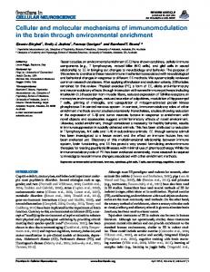
Figure 1 Sequences of events suggested for three different general models for the initiation of vasomotion. Panel A illustrates the voltage-independent model where the interaction between phospholipase C (PLC), phospholipase A2 (PLA2) and protein kinase C (PKC) amplifies the IP3 signal. The elevated IP3 level can induce oscillation in [Ca2+]i due to biphasic regulation of the IP3 channels by [Ca2+]i. SMCs can be synchronized by the movement of second messengers between the cells. AA is arachidonic acid. Panel B shows the pathway suggested for voltage-dependent oscillations. CICR can 2+ + affect two membrane conductances (for example, Ca -activated K channels (KCa) and Ca2+-activated Cl- channels (CaCC)) which have opposite effect on the membrane voltage. Neighboring SMCs will be then synchronized by membrane potential changes. Panel C shows an interaction between cytosolic and membrane oscillators. IP3 induces the local Ca2+ release which gives rise to Ca2+ waves through CICR. Transiently elevated [Ca2+]i stimulates a depolarizing membrane current, possibly the Ca2+-activated Cl- current. The following depolarization opens VDCCs, induces a global Ca2+ influx which in turn affects the membrane potential and enhances the possibility of oscillations. This model has experimental support where vasomotion was shown to be endothelium-dependent (I and II). This can be due to steep cGMP2+ 121 dependence of the Ca -activated Cl current (III, IV and ). This endotheliumdependence is however still matter of debate 44.
Hypothesis for the initiation of vasomotion in rat mesenteric small arteries (Paper I) Agonist-induced responses of rat mesenteric small artery in vitro Rat mesenteric small arteries are popular for studies the structure and function of resistance arteries, due to their easy accessibility and a large number of long branches of different diameters 2;115;122;123 . However, these arteries have unique properties in comparison to other small arteries. They have virtually no intrinsic myogenic tone which is often observed in other resistance 9;124-129 arteries . Mesenteric small arteries contribute, neverthe130-132 less, significantly to the total peripheral resistance where
the sympathetic nervous control of smooth muscle contraction is 80;122;133 . Mesenteric small arteries are heavof major importance ily innervated and sensitive to sympathetic neurotransmitters 134-137 [ATP, noradrenaline (NA) and neuropeptide Y (NPY) ] as well as to a number of other contractile agonists, such as vasopressin 138;139 140 141 , endothelin , thromboxane and some vasoactive pep142 tides . This agonist-induced receptor-coupled stimulation of 2+ vascular contractility involves elevating [Ca ]i as well as a sensiti2+ 143 2+ zation of myofilaments to [Ca ]i . [Ca ]i, elevated either by 2+ transmembrane Ca influx, or by release from the SR, can then either directly activate the contractile filaments or indirectly alter cell excitability by affecting ion channel activity in the plasma 65;144;145 membrane . In the mesenteric small arteries noradrenaline is the most often used contractile agonist (I, II, VI and 7;43;54;57;66;70;86;87;96;119;120;140;146-148 ). Development of myograph technique revolutionized the experi149 mental use of small arteries in vitro . Before this technique was developed, in vitro research was limited to strips and rings of 122 large, conduit arteries . Development of small vessel myographs allowed arteries with diameter of few hundreds microme2 ters and below to be studied . In myographs changes in the wall tension or diameter are measured under isometric or isobaric conditions, respectively, to evaluate the vascular response to the stimulation. Although many researchers suggest that the isobaric conditions more closely resemble situation in vivo, in practice the difference between these two methods is not so dramatic: the arteries show similar passive pressure-diameter characteristics, although under isobaric conditions they are more sensitive to 150 agonist stimulation . Nevertheless, the isobaric conditions (i.e. pressure myograph) are preferable for studies of vascular wall autoregulation. Moreover, under these conditions researchers receive the possibility to monitor changes in different parts of arterial segment independently. Thus, pressure myograph was a suitable technique for our study of partial synchronization in the vascular wall (II). Wire myography has an advantage over isobaric myography in experiments where the accurate and reproducible determination 2 of basal tension is essential . This method allows normalization of the arteries in each experiment by determination of the passive length-tension relationship and then setting the internal diameter to a value that gives maximal force development. Thus, the normalization sets all vessels in the same standard conditions which are utilized for almost all studies employing isometric myography 2 of resistance arteries . In our studies (I, II, VI-IX) the vessel diameter was set to 90 % of the value vessel would have had in vivo 149 under transmural pressure of 100 mmHg . These standardized isometric conditions are ideal for interventional studies of vasomotion. The observation that submaximal stimulation by different contractile agonists can induce rhythmic oscillations in tone suggests a primary role of SMC activation for initiation of vasomotion rather than a specific effect from a certain receptor. Vasomotion in rat mesenteric small arteries has been seen with electrical field stimulation of sympathetic nerves, where they are suggested to 80 be due to noradrenaline release , in response to administration of exogenous noradrenaline (I, II, VI and 7;43;54;57;66;70;86;87;96;119;120;140;146-148 151 137 ), vasopressin and NPY (Fig. 2). Although stimulation with the thromboxane analog U46619 or endothelin-1 is reported to fail to induce vasomotion in rat mesenteric small arteries 140, the presence of endothelin-induced 152 vasomotion in other vascular beds, e.g. cat arterioles , and our unpublished observation (Fig. 2C) in the rat mesenteric small arteries could suggest an importance of different experimental DANISH MEDICAL BULLETIN
6
conditions. The inconsistence could also be due to the steepness of the concentration-response curves for U46619 and endothelin1 which makes it difficult to achieve a reasonable submaximal level of tone.
3 µM NA
1.5 µM NA
1 µM NA
1 min
1.3 µM NA
10 mN
A
8 nM AVP
2.7 nM AVP
0.8 nM AVP
0.27 nM AVP
1 min
0.08 nM AVP
10 mN
B
300 pM endothelin-1
100 pM endothelin-1
30 pM endothelin-1
1 min
10 pM endothelin-1
10 mN
C
Figure 2 In spite of the different kinetics of contraction, vasomotion induced by different contractile agonists has a similar pattern. Panel A shows a cumulative stimulation with increasing concentrations of noradrenaline (NA). Panel B shows vasomotion in response to arginin vasopressin (AVP). Panel C shows response to endothelin-1. Arteries were studied in vitro under isometric conditions. Vasomotion is normally seen over nearly the entire spectrum of vascular tone, though their characteristics may change with the tone. This is especially true for the amplitude of oscillations while the frequency does not change much at different levels of tone. Since maximal amplitude is achieved at about 50% of maximal tone, this is a standard level of contraction where vasomotion is 87;110 normally being studied (I, II and ). 2+
[Ca ]i imaging in the vascular wall in vitro Development of new techniques, first of all live fluorescence microscopy, improved our understanding of the sequence of events leading to vasomotion (I) (Fig. 1C). The possibility of loading the arterial wall with fluorescent dyes was greatly improved with development of acetoxymethyl ester (AM) dye forms. Prior introduction of the fluorescent dyes into the cells was a harmful procedure including temporary membrane disruption with deter153 gents or voltage pulses . The membrane permeable AM-form
becomes an impermeable, hydrophilic form inside the cell after the AM group is cleaved away by endogenous esterases. Available fluorescent dyes have various properties making them useful for 2+ different applications. Thus, we have used the Ca ratiometric (dual excitation) dye Fura-2 (I, VI and VII) which is a practical tool 2+ for the continuous real-time monitoring of global [Ca ]i events 154 . The ratiometric properties allow a conversion of the fluores2+ cence ratio signal into [Ca ]i (VI) although this calibration does not necessary contribute further important information and often calibration is not done (VII). The fluorescence ratio depends on several parameters, e.g. temperature, pH and ionic strength, 2+ which modify the dissociation constant for Ca binding to Fura-2. This uncertainty is a disadvantage to the calibration method and it is necessary to assume that the dissociation constant is unchanged during the study. 2+ To record [Ca ]i changes with Fura-2 we used a conventional epifluorescent microscopic technique which does not allow moni2+ toring of [Ca ]i dynamic at the cellular and subcellular levels. This techniqual limitation can be overcome with the laser confocal microscopy approach. Combining a high numerical aperture objective and ability to move the focal point this approach makes it possible to record live images of the individual SMCs in the vascular wall mounted in the specially designed wire myograph over 48;146 ). The narrow focal plane complicates time (II, VII, VIII and 2+ recording of [Ca ]i during even slight movement, i.e. the region of interest can move out of focus when the artery constricts. The movements can be inhibited chemically, e.g. wortmannin inhibits the myosin light kinase and therefore contraction, or by sustained + hyperpolarization, e.g. pinacidil opens the ATP-dependent K channels. These methods of inducing stabilization however limited the ability to study vasomotion, i.e. the oscillation in tension. Therefore, most of the confocal data in our studies were obtained without these drugs because under the isometric conditions the movements are negligible (I, II, VI-VIII). Due to techniqual limitations (lack of suitable excitation wavelengths) we were not able to use Fura-2 dye in our confocal stud2+ ies. The non-ratiometric Ca dye Calcium Green-1 was used instead (I, II, VI-VIII). Calcium Green-1 increases in intensity upon 2+ binding to Ca without a shift in the wavelength where emission is seen. This increases the probability of interference from movement artifact. In the experiments where this risk was espe2+ cially high, e.g. measurement of subcellular Ca dynamic in very 2+ small region of interest, we combined two Ca dye indicators to 2+ perform semi-ratiometric [Ca ]i measurements (VIII). Thus, ele2+ vated [Ca ]i results in increased fluorescence intensity of Calcium Green-1 and decreased fluorescence intensity of Fura Red. The combination of these two calcium indicators allows ratiometric 2+ analysis of [Ca ]i changes relatively independent from movements. 2+
[Ca ]i transients in smooth muscle cells induced by an intracellular oscillator 2+ Our paper by Peng et al. (I) clearly illustrates that Ca waves within the individual SMCs precede synchronized oscillations in 2+ 2+ [Ca ]i and vasomotion (Fig. 3). Similar asynchronous [Ca ]i waves preceding the rise in tension were previously seen in rat tail ar99 94 tery and rabbit vena cava SMCs. We have also detected 2+ 48 [Ca ]i waves in some SMCs of un-stimulated arteries . Increasing noradrenaline concentration recruits SMCs into an oscillatory 2+ mode. The frequency of these asynchronous [Ca ]i waves varied 48 between SMCs (I and II) but was constant over time . This means that even during repeated stimulation the characteristic freDANISH MEDICAL BULLETIN
7
100
80 60 40
100
5 sec
[Ca2+]i wave
80 60 40 5 sec
75 Global [Ca2+]i oscillations
50 25 0 10-7
0
10-6
10-5
[noradrenaline], log M
Fluorescence emission
Wave velocity, µm/s
C
Quiescent cell
100
0.98 sec
Fluorescence emission
50 µm
Fluorescence emission
B
A
100 80 60 40 5 sec
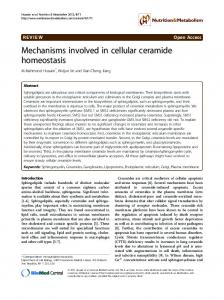
Figure 3 2+ 2+ [Ca ]i events in SMCs in rat mesenteric small artery studied using confocal microscopy. Panel A shows [Ca ]i image of arterial wall 2+ 2+ loaded with Calcium Green 1/AM. Panel B illustrates different stages of [Ca ]i in one SMC from panel A. [Ca ]i was measured in regions of interest (ROI) placed in two distant (about 40 µm) points within the cell as indicated by corresponding colors. Upper panel shows the 2+ quiescent state of cell before noradrenaline administration. Middle panel illustrate [Ca ]i waves stimulated with noradrenaline before synchronization occurred. The 0.98 sec delay in the peak fluorescence gives wave velocity of 40.8 µm/s. Lower panel shows the global 2+ [Ca ]i oscillations observed when the synchronization occurred. Panel C shows increase in the wave velocity with increasing noradrenaline concentration. An average of 4 independent experiments, at least 5 cell in each. quency of individual cells remained fairly constant. This observation indicated the phenotypic heterogeneity of SMCs in the vascu2+ 48;79 . The source for this lar wall with respect to the Ca dynamics 46;48 2+ . Ca waves also differ in direction heterogeneity is unclear and dynamics (I and 48;79). The waves in individual SMCs move in different directions, they can be initiated in the cell end or somewhere near the center and spread to non-excited parts of the cell, 2+ but they do not spread between the cells. The Ca waves spread with different velocities between 12 and 175 µm/s with a median of 36 µm/s (I) (Fig. 3C) and their frequency increases with the noradrenaline concentration. This frequency is usually slower although overlap with the frequencies of synchronized oscillations consistent with the suggested model (I). 2+
It is generally accepted that Ca wave generation needs a func35 tional SR . We have shown that this is also the case for mesenteric small arteries. Interruption of the SR function stopped the 2+ 88 Ca waves (I and ). In line with this observation, the increase in noradrenaline concentration and, thus, IP3 production increases 2+ the velocity of the Ca waves (Fig. 3C). In contrast, we found that 2+ 2+ another source for [Ca ]i rise, an extracellular Ca influx is not
2+
2+
necessary for the appearance of Ca waves (I). Inhibition of Ca 2+ influx with VDCCs inhibitors or Ca -free bath solution preserves 2+ 2+ Ca waves for some period of time. Ca waves disappeared 2+ eventually after 10 to 60 minutes of Ca influx inhibition (I). This 2+ was probably due to loss of some Ca from the cell by pumping across the membrane. Similar conclusions were made previously 99 94 by other groups in studies on rat tail artery and rabbit venous 2+ SMCs. Interestingly, the Ca waves were also preserved in SMCs + hyperpolarized by opening the ATP-dependent K channels with 2+ pinacidil (I) suggesting its independence not only from the Ca influx but also from the membrane potential. Thus, based on the 78;94;99 experimental facts, consistent with other reports , we can 2+ conclude that the Ca waves are initiated by an intracellular oscillator and propagated by an intracellular mechanism (I). The 2+ Ca waves are seen in the absence of synchronization between SMCs, i.e. when vascular tone is static. These asynchronous waves 2+ shift to global [Ca ]i oscillations when SMCs synchronize and vasomotion appears (I).
DANISH MEDICAL BULLETIN
8
2+
Transition from waves to global [Ca ]i oscillations In agonist-stimulated rat mesenteric small arteries spontaneous synchronization occurs after a variable period of time (normally within 30 seconds – 5 minutes), if the endothelium layer is intact (I) or if sufficient concentration of a membrane-permeable analog of cGMP (II) is present in the bath. During synchronization the 2+ 2+ Ca waves change to global Ca oscillations, which are seen in all 48 SMCs in the arterial wall (I and ). These synchronized oscillations 2+ 2+ in [Ca ]i lead to the rhythmic contractions. Analysis of [Ca ]i 2+ dynamics showed that during these global [Ca ]i oscillations (I 48 2+ and ) [Ca ]i rose simultaneously (within the limits of the temporal resolution) throughout the whole cell (Fig. 3). In spite of significant difference in the dynamic characteristics, we 2+ concluded that the Ca waves function as pacemakers for the 2+ 66;76 global Ca oscillations and, thus, vasomotion (I and ). This is similar to a number of other reports which demonstrate that 2+ when either release or uptake of Ca into the SR is inhibited 2+ 2+ vasomotion, the synchronized Ca oscillation and the Ca waves 68;74;88;93;155;156 2+ . In contrast to the Ca waves, the global disappear 2+ 2+ Ca oscillations are dependent on Ca influx; the blockade of 2+ VDCCs, immersion into the Ca -free bath and hyperpolarization + with the ATP-dependent K channel opener all stopped vasomotion in the rat mesenteric arteries and, even more interesting, 2+ transformed the global Ca oscillation back to the asynchronous 2+ Ca waves (I). Thus, the synchronized activity in the mesenteric artery wall is voltage-dependent (I and II). Vasomotion and syn2+ chronized Ca oscillations are accompanied with rhythmic changes in membrane potential with the same frequency that 7;54;70;86 ) were observed in rat mesenteric arteries (II, VI, VII and 71;72;74;93;109 and many other blood vessels . Moreover, the oscillations in membrane potential are shown to precede the oscilla2+ 35 tions in [Ca ]i and the wall tension (I and ). Although each cycle of contraction in vasomotion starts from 35 2+ depolarization following by a rise in [Ca ]i which leads to smooth muscle contraction, the means by which transition from 2+ 2+ Ca waves to global oscillations in Ca and membrane voltage occurs remains to be explained. Based on the previously reported 7;54;70-72;74;86;93;109 ) there is a little measurements (II, VI, VII and doubt that SMCs synchronize by membrane potential dependent mechanism. An electrical signal is probably the only signal fast enough to synchronize SMCs over a long arterial segment. It is important to mention that the mesenteric small artery wall is 115;157-160 equipped with low-resistance channels of gap junctions . The gap junctions between of SMCs undoubtedly mediate the electrical coupling between cells which has been documented experimentally by different techniques. In the studies using putative blockers of gap junctions vasomotion was shown to be inhib96;112;113 2+ ited after uncoupling SMCs (VII and ) and the global Ca 2+ oscillations were replaced by unsynchronized Ca waves (VII). The observation of an irregular arterial vasomotion in cremaster muscle arterioles in connexin 40 knockout mice is also consistent with the importance of intercellular communication in synchroni59 zation . It is obvious in this context that the regulation of gap junctional conductance could be an important regulator of vasomotion. Little is known, however, about such regulation, although some potential regulatory elements have been suggested, e.g. cGMP, interaction with other membrane transporters such as + 2+ + + + Na /Ca -exchanger, K channels and Na /K -ATPase (II, VIII and 66;70 ). The key element for our model for vasomotion in the rat mesenteric small arteries is the suggestion that the intracellular and membrane oscillators interact with each other to produce phase locking of the individual cells (I). This is possible because the
2+
intracellular oscillations in Ca induce rhythmic changes in mem2+ brane potential. We suggested (I) that Ca activates a depolarizing current on the cell membrane. The consequent depolarization 2+ increases Ca influx through the VDCCs and the likelihood for CICR, which amplifies the depolarization that spreads through the gap junctions to the neighboring SMCs. This depolarizing current 2+ enhances Ca influx in the neighboring cell which increases the 2+ 2+ probability of Ca release and, thus, entrains the Ca release and forms the basis for vasomotion. The relaxing part of the vasomotion cycle might be caused by hyperpolarizing current generated 2+ + by the Ca -sensitive K channels which have slow activation 2+ kinetics, strong voltage-dependence and low sensitivity to [Ca ]i 30;145;161-167 and therefore activate substantially only when the 2+ membrane depolarizes and [Ca ]i is significantly elevated. It is + important in this respect that the K channel blockers have only 66 modulating effect on vasomotion (I and ). This could be because several different types of channels are involved in the repolarization. Another reason for the repolarization could be a slow-down 2+ of Ca release due to emptying of the SR in the combination with 2+ 97 an active removal of Ca from the cytoplasm . A decrease of 2+ Ca release from the SR could be due to a refractoriness of the 2+ 98 Ca -release channels . 2+ The transition of Ca waves to the synchronized oscillations in the rat mesenteric small arteries is only possible if the endothelial layer is intact or if a sufficient concentration of cGMP is present (I, 87 II and ). The fact that a fixed concentration of intracellular cGMP could, at least partially, compensate for the absence of endothelium suggests that oscillations originate in SMCs and that the endothelium is not directly involved in the generation of oscilla70;88;108;168;169 ). A similar tions under normal conditions (II and importance of endothelium was previously observed in other 89;170-175 vessels , while in some vessels, e.g. rabbit mesenteric and 155;175-178 , vasomoear arteries, aorta and hamster cheek pouch tion was shown to be potentiated by endothelium removal or NO production blockade. The reason for such inconsistencies is unknown and can be ascribed to significant differences in experimental protocols or to the variability in the mechanism of vasomotion in different vasculatures. It is quite natural to suggest that several endothelium-derived factors have influences on vasomotion and the role of each of the factors depends on the vasculature and the experimental conditions. Our studies together with other groups reports show a promoting role of the NO/cGMP pathway for vasomotion in the rat mesenteric small arteries (II 70;88;96;108;168;169 and ). The importance of cGMP for the entrain2+ ment of the intracellular Ca oscillations via a membrane potential changes led us to suggest the presence of cGMP-dependent 2+ Ca -activated depolarizing membrane conductance (I). This suggestion was supported by our membrane potential measure2+ ments demonstrating that caffeine-induced Ca release from the SR could depolarize SMCs only if the endothelium was present or if the loss of endothelium was compensated by a membrane permeable-analog of cGMP (I). We later identified this conduc2+ tance as the cGMP-dependent Ca -activated Cl conductance (III 146;179 and ). The model for generation of vasomotion Our comprehensive study of vasomotion in the rat mesenteric small arteries together with other studies in this field lead us to suggest a detailed model for the initiation of vasomotion in this vessel (I). Our model (Fig. 4) suggests that agonist stimulation of 2+ SMCs induces intermittent release of Ca from the SR which 2+ activates a Ca -dependent depolarizing Cl channel in the memDANISH MEDICAL BULLETIN
9
IP3
G
G
agonist
(arbitrary units)
2
IP3
1 0
Ca2+
Ca2+
IP3-R / RYA-R
IP3
G
G
agonist
IP3
2 1 0
agonist
cGMP
Ca2+
IP3-R / RYA-R
IP3
G agonist
G
IP3
agonist
(arbitrary units)
IP3-R / RYA-R
0.2 2
(N/m)
+
+ Ca2+
20 s
ClcGMP-Ca
Ca2+ Ca2+
[Ca2+]i
Ca2+ Ca2+
Tension
ClcGMP-Ca
20 s
0.2
(N/m)
Ca2+
[Ca2+]i
Ca2+
Tension
Ca2+
IP3-R / RYA-R
(arbitrary units)
agonist
Ca2+
cGMP
0.2
(N/m)
IP3-R
Tension
Ca2+
IP3-R
[Ca2+]i
Ca2+ Ca2+
1
20 s 0
Figure 4 Model for the generation of vasomotion we suggest based on our experimental data (I, II). Left panel represents the schematic sequence of events during the initiation of vasomotion, the right panel shows changes in [Ca2+]i in two individual SMCs (shown in two colors), the global [Ca2+]i in the vascular wall (black line) and changes in the wall tension. Initial stimulation with contractile agonist increases IP3 production and stimulates localized Ca2+ released from the SR (upper panel). This is not accompanied with any changes in the global [Ca2+]i and in force. This local Ca2+ release can initiate the Ca2+ waves amplified by the CICR (middle panel) causing unsynchronized Ca2+ transients but not contractions. In the presence of a substantial amount of cGMP a rise in [Ca2+]i can stimulate the cGMP-dependent Ca2+-activated Cl- channel (bottom panel) and depolarize the 2+ membrane. Membrane depolarization opens the VDCCs, induces global Ca oscillation and synchronizes SMCs via gap junctions. This leads to vasomotion.
brane only when cGMP is present. While this may occur randomly in different SMCs, the individual cells become synchronized by 2+ entrainment of Ca release from SR in individual cells. When this occurs in a sufficient nu mber of cells, a depolarization is evoked which spreads through 46;47 the gap junctions (I and ). A complex action of cGMP in the generation of vasomotion (Paper II) In accordance with our model for the generation of vasomotion the presence of a sufficient amount of intracellular cGMP is nec2+ essary for transition of the Ca waves to synchronized oscillation in the vascular wall (I). This role of the endothelium/cGMP pathway in the synchronization of SMCs is somewhat controversial: cGMP/endothelium has been shown to have both potentiating (I
70;170;171
67;180
and ) and suppressing action, depending on the vasculature type and the experimental protocols. In an attempt to understand this controversy we tested whether cGMP has a more complex role in the initiation of vasomotion (II) than those suggested by our initial model (I) where some of the parameters 46-48 were simplified . Our detailed study of vasomotion “beating” and the concentration-dependent effect of 8Br-cGMP on the oscillation parameters, e.g. the frequency and amplitude of oscillations, led us to two major conclusions (II). The first finding is that a state of partial coupling between SMCs is possible and that intermediate concentrations of cGMP can provide it. The second 2+ is that the stimulation of Ca -activated Cl channel is only one of several different targets of cGMP in its complex modulation of vasomotion (II). The partial coupling of SMCs in the vascular wall was seen as a ‘beating’ oscillation in endothelium-denuded arterDANISH MEDICAL BULLETIN
10
ies under isometric conditions after addition of intermediate concentrations of 8Br-cGMP (30-100 µM) (II). The phenomenon can be explained by the presence of two or more regions oscillating with different frequencies. This was confirmed in isobaric experiment with pressurized arteries where different segments of a denuded artery oscillated with different frequencies at intermediate cGMP concentrations (II). Importantly, this phenomenon is rarely seen in the endothelium-intact rat mesenteric small 181 arteries . Moreover, the frequencies between different pieces of the endothelium-intact vessels are the same even when these 110 to the pieces are not coupled to each other . The partial synchronization was accompanied by a dissociation of oscillations in membrane potential and tension (II). When a glass electrode was implanted in one SMC the membrane potential oscillations often differed from the ‘summarized’ force oscilla2+ tions. Similar observation was also made for the [Ca ]i dynamics (II). At the state of intermediate cGMP concentration islands of 2+ cells having synchronous oscillations of [Ca ]i were observed (II 48 and ). Even in the presence of a high (300 µM) concentration of 8Br-cGMP some SMCs were quiescent during synchronous oscilla2+ 48 tions in [Ca ]i in their neighbors . Other SMCs had an extra transient which were not synchronized and was in the form of a 2+ 2+ Ca wave (II). The observed extra [Ca ]i waves were not seen in arteries with intact endothelium suggesting that endothelium provides more than NO/cGMP. The endothelium can also provide additional means of synchronization in the vascular wall, which is also suggested by the observation that vasomotion is not always 57 stopped by blockade of NO production (II and ). This might be explained by an essential role of an endothelium-derived hyperpolarizing factor because its inhibition by charybdotoxin and 57 apamin blocked vasomotion . This was, however, not the case in 66 another study and in our own experiments (unpublished observation). The cGMP molecule has multiple effects on vascular function. Thus, cGMP-dependent phosphorylation of connexins 37 and 43 182 reduces intercellular coupling but cGMP enhance the intercel183-185 . It is therefore possilular communication via connexin 40 ble that cGMP affects vasomotion via an effect on intracellular 115 coupling . It has been shown previously that the coupling resistance between SMCs is much higher than that between endothe186 lial cells . Thus, the removal of endothelium also abolishes a low-resistance pathway for current synchronization and therefore potentially makes it more difficult to entrain synchronized islands of SMCs located distantly. In our cGMP concentration-effect study we also observed that 2+ cGMP had a direct effect on [Ca ]i dynamic by reducing the num2+ ber of oscillating cells and the frequency of Ca release (II). The exact reason for this suppressing action of cGMP was not studied, but it has been reported previously that cGMP inhibited SR2+ 2+ dependent Ca transients via stimulation of Ca extrusion 2+ 187 + 2+ mechanisms, i.e. the Ca -ATPase and the Na /Ca -exchanger 188 189;190 , as well as through direct inhibition of the IP3 pathway . 2+ Increased Ca extrusion could explain the reduction in the frequency of oscillation. Thus, similar to our findings, stimulation of NO/cGMP production by acetylcholine and sodium nitroprusside 2+ in tail artery reduces [Ca ]i dynamics even in hyperpolarized 191 2+ arteries . We suggested that Ca waves, being the pacemaker for vasomotion, have a strong influence on the frequency of the synchronized oscillations. It is therefore possible to expect that cGMP, by affecting the frequency of the Ca2+ waves, will also reduce the frequency of vasomotion. We confirmed this experimentally by showing that the frequency of vasomotion decreased gradually with an increasing 8Br-cGMP concentration (II).
Electrophysiological approaches in the studies of vasomotion Registration of electrical events over the cell membrane is one of the essentials for understanding vasomotion. The measurements of membrane potential in isolated arterial segment supported the 2+ suggested theory about transition of [Ca ]i waves into the syn2+ chronized global Ca oscillations by means of a membrane potential dependent mechanism (I and II). Moreover, simultaneous 2+ measurements of membrane potential, [Ca ]i and isometric force showed the initial role of membrane potential changes in the 35 sequence of events leading to the rhythmic contractions . The great advantage of this method is an ability to record membrane 7 potential without isolating the cells . Thus, a sharp glass electrode can be impaled in smooth muscle cell located in its natural environment in the vascular wall. It is therefore possible to combine membrane potential recording with isometric force and 2+ [Ca ]i measurements, although the problems with movement artifacts are also relevant for this method. Although the myosin light chain kinase inhibitor wortmannin can be used for stabilization, normally recordings under isometric conditions are made without wortmannin. We controlled the location of the electrode tip by continuous recording of electrode resistance (input resistance) by current pulse injection (VI and VII). The recorded input resistance is an indicator of both the membrane resistance of the impaled cell and the resistance of intercellular contacts within the vascular wall. We have, therefore, used input resistance for qualitative evaluation of intercellular coupling within the intact vascular wall (VI and VII). 2+ The suggested presence of a cGMP-dependent Ca -activated depolarizing current based on the membrane potential measurements (I) received support in our voltage clamp studies where we characterized the current (I, III-V). Under voltage clamp conditions the current injected to clamp the potential reflects the ionic 192;193 . A major disadvantage of current across the cell membrane voltage clamp is the difficulty of applying it to vascular smooth muscle cells in situ. In comparison with the conventional sharp electrode used for membrane potential measurements, the voltage clamp electrode has low tip resistance and “patch” the cell membrane forming a very high-resistance seal with the mem194 brane surface . The formation of a gigaohm seal is essential for successful patch clamp experiments and this means that only 195 “clear” membrane surfaces can normally be used . Vascular wall cells are not “clean” and additional procedures, e.g. enzyme 195;196 “shaving” of vessels surface are necessary . Even if it will be possible to patch the vessel without enzymatic digestion (as it is the case for some arterioles) this will not give much advantage over the conventional membrane potential measurement due to the “space phenomenon”, i.e. inability to control the voltage over 192;195;197 a piece of tissue containing more than a few cells . This problem can however be solved by chemical uncoupling the cells 196;198 in the vascular wall . In summary, some modification of the conventional technique provides the possibility to patch SMCs in the vascular wall but the necessary manipulations will significantly modify the cells. The cells for conventional patch clamp can be isolated by enzymatic digestion of the vessel. The method of digestion depends on the type of artery and the type of conductance which is studied. There is no one ideal isolation technique which is suitable for all studies. Different combinations of collagenase, elastase and a broad-spectrum protease, papain, are normally used to digest different vessels 195. Papain is know to damage some types of Ca2+ + 199 conductances but is good for studies of K currents . Although 2+ Ca conductances are resistant to the collagenase/elastase digesDANISH MEDICAL BULLETIN
11
tion, the isolated cells show hyperreactivity and are constrict to 200 any stimulus which makes difficult to patch them . After the gigaseal is obtained several patch clamp configurations 195 can be formed . Rupture of the membrane under the tip forms a low resistant pathway between the intracellular environment and the pipette solution. The configuration I have mostly used in my studies is the “whole-cell” technique because it enables me to record the current from the entire cell membrane. Under the whole-cell configuration the contents of the cell and the pipette exchanges within a few minutes which allows control of the intra201 cellular environment . This property is helpful for effective isolation of single ionic conductances in macroscopic mode. I have dialyzed the cells with a cesium rich solution to establish more + complete block of K conductances which otherwise could contaminate the recordings (III and IV). Similarly the intracellular messengers and other big signaling molecules can be added via the pipette in defined concentrations, e.g. I applied intracellularly membrane-impermeable cGMP (III-V), ATP (IX), protein kinase G 2+ 2+ (III) and controlled intracellular Ca by Ca -chelators (III, V, VIII). Importantly, diffusion also occurs from the cell to the pipette, and significant loss of important cellular component can be observed 201-203 . This is the explanation for rundown of several membrane 2+ 204 currents, e.g. Ca -activated Cl current . Washing out of diffusible second messengers explains also the disruption of receptor12 channel coupling seen in the whole-cell experiments . This problem with the loss of intracellular constituents is partially overcome using the perforated patch clamp where the membrane patches only permeabilized for monovalent ions by anti205 fungal drugs, nystatin and amphotericin (I and ). Permeabilized patch does not allow addition of bigger molecules via the pipette 2+ solution resulting in pure control of [Ca ]i, ATP/ADP ratio, etc. Noteworthy, the conventional patch clamp configuration does not seem to provide a complete control over larger organic molecules (e.g. cAMP and ATP) which have low washout rates partially 206;207 because of their size and partially because of the microdo208;209 main structures . Moreover, ion concentration was also suggested to be poorly controlled in some subcellular compart2+ ments. Thus, the possibility to change local [Ca ]i is strongly 2+ dependent on the type of Ca chelator and the distance to the 210 signaling target . Other modifications of patch clamp allow direct measurements of the conductance of a single channel and provide detailed information on channel biophysical properties which can not be obtained from the whole-cell macroscopic recordings. This can be done in “cell-attached” mode when other cellular constitutes can 121 directly modulate channel behavior . Only poor control of voltage over the patch membrane and no control of the intracellular environment are limiting the use of this method. A singlechannel can also be studied in a cell-independent manner when the membrane patch is completely isolated from the cell in “in195;211 side-out” and in “outside-out” modes . These methods permit identification of distinct channel subtypes when the mac195;211 roscopic currents are not easy to distinguish . Under these conditions there is a tight control of the environment on both sides of the membrane which is a clear advantage but a drawback at the same time since single channels in excised patches behave differently because of lack of many intracellular constituents which play regulatory and modulatory roles. Therefore, caution should be taken when relating the patch clamp data to functions of the intact vessels. Patch clamp is used not only for studies of membrane currents but also for evaluation of intercellular communication. Evaluation of electrical coupling can be seen under conditions when visual
72;212
dye coupling fails to detect intercellular communication . Electrical coupling between two cells can be determined by dual 213;214 . This technique is more challenging than conpatch clamp ventional single electrode recording. Alternatively, monitoring the membrane capacitance of coupled cells by single electrode patch 215;216 clamp can give insight to the cell coupling (VII, VIII and ). Cell membranes store a charge as a capacitor. Therefore, when cells are electrically coupled the total electrically coupled membrane surface is increased and membrane capacitance increases likewise (VIII). Uncoupling of coupled cells decreases capacitance. The great advantage of this method is that it enables intercellular coupling to be estimated under conditions where only one of the 215;216 . Importantly, this way to estimate coupled cells is patched electrical coupling can not be used easily for quantitative measurements because of non-linear relation between the measured 217 total capacitance and intercellular resistance . Nevertheless, intercellular resistance changes can be estimated by a compli217-219 cated recalculation of the measured parameters . -
6. A CL CHANNEL IN VASOMOTION? (PAPERS III, IV, V) 2+
The characterization of a unique cGMP-dependent Ca activated Cl current in SMCs from rat mesenteric small arteries (Paper III) 2+ A cGMP-dependent Ca - activated depolarizing conductance was predicted by the model for the initiation of vasomotion in rat mesenteric small arteries (I).We recorded this cGMP-depended current initially in permeabilized whole-cell mode (I). This type of recording makes it difficult to manipulate the intracellular environment and to characterize the conductance in detail. We chose, therefore, the conventional whole-cell patch-clamp method which allowed us to manipulate both intracellular and extracellular environments. Using this method we characterized the cGMPdependent current in detail (III). Piper and Large published a description of the same conductance at the single channel level 121;220 . 2+
[Ca ]i-dependence Similar to the permeabilized patch-clamp experiments, caffeine2+ induced Ca release induced under the conventional ruptured patch-clamp conditions a few hundred pA inward current only when micromolar cGMP was added into the pipette solution (I 2+ and III). This cGMP-dependent, Ca -activated inward current can 2+ be also stimulated by increasing [Ca ]i by other means, e.g. 2+ stimulation of extracellular Ca influx (III) or dialysis of the cell 2+ with high-Ca solution (IV and V). Importantly, this current can also be stimulated by noradrenaline via the G-protein-coupled 121 2+ adrenoreceptor in the cell-attached mode . Chelating Ca with a low concentration of BAPTA or EGTA abolished the cGMP2+ dependent current induced by caffeine (III) suggesting that Ca 210 can be released at a distance (>100 nm) from the channel and that there is no need for a very close interaction between the SR and the channel. 2+ The cGMP-dependent inward current was steeply Ca depend121 ent, as was shown with the single channel recordings . The 2+ current was detectable already at 50 nM [Ca ]i and reached its maximum at 100 nM with half-maximal activation at 74 nM. This 2+ [Ca ]i-dependence was shown to be mediated via calmodulin which increased the open probability of the channel in a calmodu220 lin kinase II (CaMKII) independent pathway . The surprisingly high sensitivity for [Ca2+]i suggests that either under the physio2+ logical conditions Ca sensitivity is under control of second mesDANISH MEDICAL BULLETIN
12
2+
sengers, as it is known for other Ca -activated ion channels, or 2+ the channel is regulated by local spatially restricted Ca changes 2+ rather than global [Ca ]i. We observed sometimes during caffeine stimulations a superimposed outward current due to an activation of big-conductance 2+ + Ca -activated K channels (BK) (III), since it was abolished by 221 specific blockers, e.g. charybdotoxin or iberiotoxin . It also has 2+ + been shown previously that Ca -activated outward (K ) and 27-29 inward (Cl ) conductances can superimposed on each other . The appearance and prevalence of the BK current depended on the membrane voltage and became significant at voltages posi+ tive to -60 mV, when it was distant from the K equilibrium potential (EK=–78.5 mV). To avoid complications in the analyses of the + Cl conductance, we had K channel blockers, charybdotoxin and barium chloride, in the bath during almost all our experiments. + Another reason to have these K blockers in the bath was to compensate for the non-specific effects of niflumic acid on the 222 membrane conductance (IV and ) (see below). An absolute requirement for cGMP There is no doubt that caffeine induces the inward current by 2+ elevation of [Ca ]i only in the presence of intracellular cGMP (III). This requirement for cGMP was absolute under whole-cell configuration but a small inward current was seen under permeabilized conditions in the absence of 8Br-cGMP (I). This can be due to remaining endogenous cGMP. It is, therefore, important that the single channel recordings clearly indicate an absolute require121 ment for cGMP . The sensitivity to cGMP (in micromolar range) was not significantly different under whole-cell (III) and single 121 channel configurations . Although the detected sensitivity for cGMP is similar to that described for cyclic-nucleotide-gated 223;224 channels this channel is not one of them, because we have shown that the channel is activated via phosphorylation by a cGMP-dependent protein kinase (PKG) which is not the case for 121 the cyclic-nucleotide-gated channels (III and ). Key evidence for this was that a cGMP-independent constitutively active catalytic 225 2+ fragment of PKG Iα activated the Ca -activated inward current in the absence of cGMP (III). The catalytic subunit of PKG I was 225 produced by trypsinization but it is not possible to specify to which extent it depends on the two PKG I variants, i.e. PKG Iα and 226 PKG Iβ . The role of the variants is, however, important since 227 the dominating smooth muscle variant, PKG Iα , is highly sensi226 tive to cGMP with half-maximum activation at 0.1 µM . This is much higher than the sensitivity detected in our studies and that 121 of Piper & Large (III and ). Thus, since PKG Iβ is also expressed in SMCs and is at least 10-times less sensitive to cGMP, PKG Iβ is the most likely candidate for the activation of the cGMP2+ dependent Ca -activated inward current. The two PKG variants 228;229 are involved in distinct signaling pathways . Thus, it is possible that cGMP pathways divide into the well-known NO-mediated relaxation via cGMP with involvement of PKG Iα, and another pathway via PKG Iβ which is associated with rhythmic membrane 2+ depolarization via the cGMP-dependent Ca -activated depolarizing current (I). This is consistent with previous suggestions that 2+ both Ca and cGMP signaling are spatially restricted in SMCs 65;228-232 . A novel cGMP-dependent Ca2+-activated Cl- current + Since the equilibrium potential for K was negative to the holding + potential in our experiments, K cannot be a charge carrier for the + 2+ inward current, but Na , Ca and Cl ions are candidates (ENa=+68 2+ mV; ECa=+88 mV; ECl=0 mV) (III). However, neither Ca -free ex-
2+
tracellular solution nor Ca channel blockers affected the cGMP+ dependent current. Na was also excluded from the candidates based on substitution experiments (III). In contrast, the close correlation between the equilibrium potential for Cl (ECl) and the current reversal potential (EREV) indicates that the cGMP2+ dependent Ca -activated inward current is carried by Cl (III and 121 ). Er ev 20
10
-20
-10
10
20 ECl
-10
-20
Figure 5 EREV is plotted against calculated ECl. The dashed line is the identity line, expected for ideal Cl- channel. Bath solution contained the following cations (in mM) 6 K+, 145 Na+, 0.1 Ca2+. Pipette solution contained (in mM) 132 K+, 10 Na+, 0.01 Ca2+. Cl- concentration was varied as described previously (III). Vertical bars indicate SEM. Note the difference of the experimental results from the identity line. This difference disappeared when symmetrical CsCl solution was used instead (III).
To estimate the anion selectivity in the whole-cell configuration 2+ + we used CsCl solutions to suppress the Ca -activated K channels 2+ and buffering [Ca ]i at 900 nM. Under these conditions, EREV 24;233;234 corresponds fairly well to ECl indicating a pronounced, although not perfect, selectivity for anions (III). It is not apparent, however, whether some deviations from the identity line reflect cations passing through the channel, or if they result from an insufficient control of intracellular ion concentrations. In general, a Cl channel displays a 5-50 time selectivity for anions over cations. This is the case for, e.g. cystic fibrosis transmembrane 235 236 conductance regulator (CFTR) , GABAA and glycine receptors 237-239 and the voltage-gated Cl channels . However, a more ap2+ propriate name might be a Ca -activated anion channel. + + 2+ When we used complex solutions, containing K , Na , Ca and Cl ions, the correlation between ECl and EREV was no longer ideal (Fig. 5), indicating less than perfect anion selectivity. The downward shift from the identity line seen under these conditions indicates + a significant influence of K conductance which has a negative EK relative to ECl. A relatively poor selectivity for anions over for 2+ cations was previously described for the Ca -activated Cl chan240 nels (CaCCs) . More information about the ion conducting properties comes from the studies of relative halide permeability. This was normally evaluated under bi-ionic substitutions, i.e. when Cl on one side is substituted for another anion, and this 240;241 results in the EREV change . The relative halide permeability of most CaCCs increases with the - ionic radius and normally follows the sequence I >Br >Cl >F . The Cl channels are also permeable to polyatomic non-halide anions, such as thiocyanate (SCN ), acetate and aspartate. SCN is usually more permeable than Cl , while acetate and aspartate are known 240;242 to be less permeable due to their size . To enter the channel DANISH MEDICAL BULLETIN
13
pore, anions have to be partly dehydrated and the energy necessary for dehydration must be supplied by a binding site within the 243 pore. Wright and Diamond suggested that a binding site with a low electrical field strength (where the interaction of ions with the channel is weaker than interaction with water) provides this energy and, thus, defines the typical permeability sequence for the anion channel. In the simple terms this would indicate that I , which is the large ion, binds better within the pore than F , which is small, and, therefore, is more permeable. Importantly, the relative permeability can not be used as a measure of an ion flux and describes only an ability of an ion to enter the channel pore. The flux of ions is determined mainly by the strength of interaction within the pore. High-affinity binding slows down ion flux (the ion “sticks” in the pore) and reduces channel conductance. Conductance is evaluated from the current-voltage relationship 240 but is rarely studied in details. When it has been studied, I was - 235;244 either shown to be more conductive than Cl or no differ245;246 ence was detected . This indicates that I binds in the channel pore with either similar or weaker strength than Cl . Keeping this in mind, we identified both the relative conduc2+ tances and permeabilities for the cGMP-dependent Ca -activated Cl current (ICl(Ca,cGMP)) in SMCs (III). The sequences were similar to 2+ those of the ‘classical’ Ca -activated Cl current (ICl(Ca)) but not - identical, e.g. we found Br >I . This is possible when permeabilities 243 for I , Br and Cl are close which was the case in our study (III). This might also be the reason for the inconsistency with results achieved in the single channel recordings, where the relative - permeability sequence was Cl >I , but again the I /Cl ratio was 121 close to 1 . When the channel permeability was placed against the hydration energy of the ions (Fig. 6) the slope was shallow 15;240;241;247 compared to that reported for other CaCCs . This indi240;241;247 cates that in contrast to other CaCCs , the hydration energies is not a major determinant of the ion permeability. Which other parameters are of significance for the cGMPdependent CaCC permeability and conductance remains unclear, 248 but we failed to find a correlation with ion dimensions . Also the binding affinity could not explain the observations since the recalculated relative affinities for the binding site in the channel were equal for all halides (III). Obviously, ionic radius is playing a role when ion dimensions become very big, as is the case with aspartate. Acetate, in contrast, is still permeable in accordance with its hydration energy. These results suggest that the channel pore is bigger than 4.7 Å (acetate size) but smaller than 5.8 Å 248 (aspartate size) .
10
P x/PCl
1
SCN F-
Cl-
Br-
Acetate0.1
0.01 -450
I-
Aspartate -
-400 -350 -300 Hydration energy, kJ/m ol
-250
Figure 6 The permeability of anions (PX) measured relative to Cl- permeability (PCl) (III) corre-
lated reasonably, but with a shallow slope, to the hydration energy of the anions . -
Another interesting observation we made was that SCN has high permeability but low conductance suggesting a high affinity to the 240 channel binding site (III and ). Similar properties were previ240 ously shown for the native ICl(Ca) in the Xenopus oocytes and, surprisingly, for GABA receptors where SCN induced pore block236 ade . The latter observation might suggest a possible structural similarity between the cGMP-dependent CaCC and the GABAA receptors (V). The voltage and time independence 2+ In addition to [Ca ]i, the open probability of many CaCCs is determined by membrane voltage. This was not, however, the case in our voltage-clamp experiments where the ICl(Ca,cGMP) changed linearly with the membrane voltages (III and IV). Similar results 121 were obtained from single channel recordings . It is possible 2+ that the high [Ca ]i we used in the studies (III and IV) can be the 2+ reason, since voltage-dependence is known to be [Ca ]i2+ dependent; current rectification is prominent at lower [Ca ]i but 2+ 15;241;247 is almost absent at micromolar [Ca ]i . We tested the 2+ possibility of Ca dependence of the voltage-dependency and found that this was not the case: the ICl(Ca,cGMP) was voltage2+ independent through the entire [Ca ]i range (V). The observation 2+ that the cGMP-independent ‘classical’ ICl(Ca) had the [Ca ]i sensitive outward rectification under these experimental conditions suggests that our finding is not an artifact of the experimental protocol but a unique property of the channel. Some previously described ICl(Ca) also have a weak or even absent the voltage2+ 249-251 dependence at submaximal [Ca ]i . The reason for these discrepancies is unknown. The ICl(Ca,cGMP) has no obvious time dependence (III and IV): as 2+ soon as [Ca ]i and cGMP are sufficient for activation, the channel is activated and the ICl(Ca,cGMP) depends only on the driving force for Cl . Therefore, voltage steps in SMC dialyzed with cGMP and 2+ high [Ca ]i produce an immediate Cl current which is not different between the early and the instantaneous phase (III and IV). The absence of a tail current after repolarization also demonstrates that the gating mechanism is time- and voltageindependent. Sensitivity to blockers None of several putative blockers, known to block other Cl conductances, were found to be specifically effective for the ICl(Ca,cGMP). Thus, DIDS and IAA-94 were without effect on the ICl(Ca,cGMP) (III) when applied in concentrations known to inhibit 15;241;247 other Cl conductances . The most common blocker of the 20;252-254 ICl(Ca) , niflumic acid, also had no effect on the ICl(Ca,cGMP) (III, 121 IV, V and ). This is consistent with previous reports on insensi2+ 255 tivity of some of Ca -activated Cl currents and even a poten116;256-258 tiating action of niflumic acid on the Cl conductance . This dual effect of niflumic acid on the ICl(Ca) is of interest because 2+ recent data suggest an overlapping pharmacology of the Ca 2+ + 116;222 activated Cl and the Ca -activated K (BK) currents . It has been shown recently that niflumic acid can potentiate both the 2+ BK current and, under certain conditions, the sustained Ca 116;256-258 activated Cl current (III, IV and ), while the BK channel 2+ 258 modulators can also affect the Ca -activated Cl current . Therefore, some similarities in the molecular structure have been 116 suggested , although the effect of a physical interaction between the two proteins is also possible. Importantly, these nonspecific effects appeared only when the blockers were applied DANISH MEDICAL BULLETIN
14
separately. When charybdotoxin is applied first, niflumic acid produces effective inhibition of the ICl(Ca). Thus, the combination of blockers is needed to achieve inhibition of the ICl(Ca). In our experiments (III-V), the bath solutions contained charybdotoxin to 2+ avoid the potentiating effects of niflumic acid on the BK and Ca activated Cl currents. The inhibition of the classic ICl(Ca) supports action of niflumic acid on this conduction (III-V). Since several Cl conductances were previously reported to be sensitive to multivalent cations, we tested the effect of divalent 2+ cations on the ICl(Ca,cGMP) and found that micromolar Zn effec2+ tively inhibited the current while Co was without effect (III and 121 ). Zinc was previously used to inhibit voltage-gated Cl channels 259;260 but at higher concentrations . Interestingly, the sensitivity to 2+ micromolar Zn is one of the fingerprints for GABAA sensitive Cl 261;262 conductance . Zinc inhibits GABAA channels by binding to the 263;264 selectivity rings in the channel pore again suggesting a possible similarity in the pore structure between putative cGMPdependent CaCC and the GABAA channel. 2+
(III). The protocol was then carefully tested for unwanted effects 2+ of the blockers, e.g. niflumic acid, cGMP and Zn (IV and V). 2+ 121 The [Ca ]i sensitivity of the ICl(Ca,cGMP) is surprisingly high in 15;241;247 comparison to previously published values for other ICl(Ca) . Most of the ICl(Ca) reported in the SMCs have half-maximal activa2+ tion at 0.2-0.6 µM of [Ca ]i and achieve maximal activation at 2+ 23;246;265;275 0.6-1 µM of [Ca ]i . This broad range can probably be explained by a variability of mechanisms (a direct activation or by 2+ phosphorylation via Ca /CaM-dependent protein kinase II) for 2+ Ca -activation within the heterogeneous group of the CaCCs 247;276;277 , although this has not been studied systematically. In our experiments we dialyzed SMCs with a solution containing 2+ ~0.9 µM free [Ca ]i, which was enough for activation of both 2+ Ca -activated Cl conductances (III and IV). When sensitivities of the two types of the Cl current were directly compared, both Cl 2+ currents were maximally activated at 0.3-0.9 µM [Ca ]i, but the 2+ ICl(Ca,cGMP) showed a higher [Ca ]i sensitivity (V). No current reduc2+ tion was seen with supra-maximal [Ca ]i, as it was shown previ220 ously for single channel recording .
-
Two distinct Ca -activated Cl currents co-exist in SMCs (Papers III and IV) The current conductance The characterization of the ICl(Ca,cGMP) clearly indicated that the current we described (III) was unique in comparisons to other 15;241;247 . The unique position of known, so called “classical”, ICl(Ca) the ICl(Ca,cGMP) in the group of the ICl(Ca) was strongly supported by the finding that both types of current are present in the same SMCs (III and V). Also single channel recording revealed the pres121;265 ence of both currents . The classical Cl conductance was much smaller (~3 pS) than the ICl(Ca,cGMP) conductance which was 121 ~20 pS . This interesting feature is in accordance with previous observations indicating that at least two distinct types of anion conductances can be detected in the cell membrane. The lowconductive CaCCs is the most reported type. This conductance is 265-268 . The reports detected in the various tissues including SMCs 2+ on the large Ca -activated Cl conductance are sparser. In the beginning of the 1990’s Benos and co-authors identified in bovine trachea a membrane protein constructed of 38- and 64kDa polypeptides which incorporated in a lipid bilayers gave 25269 30 pS ICl(Ca) without any voltage-dependence . Interestingly, this reconstituted channel did not need any other intracellular mes2+ sengers but Ca for its activation and was completely inhibited by 100 µM DIDS. In contrast, other authors reported a large ICl(Ca), which depended on protein kinase G activation, and explained the Cl- current rundown by washout of intracellular messengers 270-272 . No voltage-dependence was shown for these ICl(Ca). It should be noted, that CaCCs with conductance >100 pS was previously reported too, although most of these conductances be273;274 long to intracellular Cl channels . The difference in pharmacology 2+ The cGMP-dependence was used to distinguish between the Ca activated Cl currents when they were activated in the same SMCs 121;220 (III-V and ). This differentiation was possible also based on distinct pharmacological properties. Thus, the ICl(Ca,cGMP) had low sensitivity to niflumic acid in concentrations which inhibited the classical ICl(Ca) (III). In contrast, the classical ICl(Ca) was insensitive to 2+ 121;220 micromolar Zn , the blocker of the ICl(Ca,cGMP) (III-V and ). This allowed us to design a protocol where the two currents were measured and evaluated during the same voltage clamp of SMC
Distribution of the currents in SMCs of different origin 2+ The two different Ca -activated Cl conductances might be caused by the same protein where phosphorylation by PKG changes the channel properties. PKG can thus act as a switch between different channel modes. This is, however, not the case, since the 2+ cGMP-dependent and the classical Ca -activated Cl currents have different expression profiles in SMCs. This is also supported 2+ by differences between these two Ca -activated Cl conductances in pharmacology, single-channel conductance and ion selectivity. The ICl(Ca,cGMP) was first characterized in SMCs freshly isolated from the rat mesenteric small arteries (III). It was then detected in SMCs isolated from other vascular beds and in visceral SMCs (IV). In most vessels, e.g. aorta, tail artery, femoral artery and vein, middle cerebral artery, renal artery, portal vein, superior mesenteric artery and colon, the ICl(Ca,cGMP) was accompanied with the classical ICl(Ca) in variable proportions (IV). Importantly, due to different voltage-dependences the relative size of two currents was strongly dependent on the membrane voltage at which the currents were measured (IV). The classical ICl(Ca) showed a strong outward rectification. Thus, for the comparison of the two Cl currents, it is important to define at which membrane voltage the currents are compared. It seems more relevant to compare the currents under conditions close to physiological membrane potentials. We, therefore, compared the 2+ Ca -activated Cl currents at membrane voltages close to the resting membrane potential of SMCs (IV and V). Under these conditions SMCs from cerebral arteries and femoral vein had dominating ICl(Ca,cGMP), while in the myocytes from tail and mesenteric arteries and portal vein these two currents are not different. Importantly, the pulmonary arteries had virtually no ICl(Ca,cGMP) (III). It remains unclear whether this has physiological significance, but we have used this observation to search for the gene responsible for ICl(Ca,cGMP) (V). -
The Cl conductance in the SMCs If Cl is distributed passively across the plasma membrane intracellular Cl concentration should be ~10-15 mM. This is consistent 16 with the data in skeletal muscles , but not the smooth muscles. 14;278-281 36 Measured with intracellular electrode , chloride efflux 282;283 284 and a fluorescent dye in different SMCs, the [Cl ]i is surprisingly high within the range between 30 and 50 mM. Thus, the estimated ECl will be between -40 and -25 mV which is above DANISH MEDICAL BULLETIN
15
resting membrane potential in the majority of smooth muscles . This suggests active transport of Cl into the SMCs. There are at least two transport mechanisms which contribute to intra14;287 + + cellular Cl accumulation: Cl /HCO3 exchange and the Na -K 288;289 Cl -co-transport . In addition to these well-known types of Cl transports, a third form of transport, the so called ‘pump III’, is 290-292 suggested from the work of Chipperfield and co-authors . Neither the molecular identity nor the driving force for the ‘pump III’ is known. Opening of Cl channels will depolarize SMCs and, therefore, induce smooth muscle contraction. Indeed, a role of Cl efflux for agonist-induced contraction has been shown in several types of 19-24 . If Cl is smooth muscle and the role of CaCCs was emphasized important for contraction, the contraction should depend on the Cl gradient across the plasma membrane. Several studies have shown that replacement of extracellular Cl with non-permeate anions can amplify agonist-induced depolarization and contrac25;125;293 tion , but this is not always the case. Substitution of extracellular Cl was shown to have only mild effects on both resting 294 and stimulated membrane potentials and no significant effect 87 on contraction in rat mesenteric arteries . A growing number of reports suggest a variable significance of Cl for SMC contraction 293 depending on the type of vasculature and stimulation . 285;286
2+
-
Ca -activated Cl conductance: an important function of unknown origin 295-297 The CaCCs, first described more than 25 years ago , are found at the functional level in most SMCs where they have been characterized electrophysiologically and pharmacologically 15;247;276 . Despite their obvious importance and the great interest in this group of channels their molecular identity is still under 247;277;298 debate . Why does the question of molecular identity remain open? Several problems have made it difficult to pinpoint the molecular counterparts of endogenous ICl(Ca). • Heterologous expression has often been used to show that a protein is a bona fide CaCC. This technique has several drawbacks and the major of them is the absence of an expression system that has no endogenous Cl conductance. This endogenous Cl conductance often seems to be upregulated by over-expression of a candidate protein, resulting in false positives. Thus, the most widely used expression system, the Xenopus oocytes, expresses a huge amount of an endoge2+ nous Ca -activated Cl conductance and is therefore difficult to use for characterizing a candidate. • Compounds blocking or activating the conductance of interest are needed for differentiation of the ICl(Ca) from other membrane currents and, especially, other Cl currents. How116;299 ever, available drugs are lacking specificity . There are currently attempts to overcome this problem by using molecular biological tools, such as antisense DNA, knockout models, siRNA knock-downs, and specific antibodies. • Most of the currently known membrane channels are multimers composed of several different subunits. Some of the subunits form the channel pore, while others are involved in the regulation, but all of them may be essential to produce the current. This means that the heterologous expression may be unable to reproduce the endogenously observed current. This is a well-known problem even for the Cl channels that have already been cloned, such as CFTR, GABAA receptors and ClC channels.
Finally, a broad spectrum of CaCC properties are found 15;241;247 . This variability suggests that the CaCCs are a heterogeneous group of different protein families. Three to four dif277 ferent classes of CaCCs have been suggested . Some CaCCs are directly activated by physiological concentrations of 2+ 239 [Ca ]i, while others are modulated by CaMKII . The Cl channels with large, over 100 pS channel pore conductance 270;273;274;300 form possibly a special class of the CaCCs .The cGMP-dependent CaCC can be considered another independent class, although it shares many similarities with the group 2+ 121;277 of channels directly regulated by [Ca ]i (III, V and ). The problems described above complicate the search for the molecular identity of the CaCCs and indicate that only a complex strategy can be successful. It suggests also that caution should be taken with respect to conclusions made based upon only one research strategy, since it can be misleading as it has been re301 cently shown in an example with CLCA candidate gene . •
2+
-
The potential candidates for the role of the Ca -activated Cl channel CLCA In spite of these problems, some molecular candidates for CaCCs have been suggested over the past years. The CLCA protein was 255;302 the first notable candidate to mediate an ICl(Ca) . The first member of the CLCA family was cloned from a bovine tracheal cDNA library by immunoscreening with an antibody against a Ca2+-activated anion channel 255. The following evidence for its 255;302-305 : function as a CaCC was collected • Heterologous expression of various isoforms of CLCA gener2+ ated membrane currents which activated by high [Ca ]i. • The heterologously expressed current was sensitive to the classic Cl channels blockers such as niflumic acid and DIDS. • The heterologously expressed CLCA protein carried Cl ions, + + but this was shown under Na and K free conditions. This evidence is, however, not enough to accept CLCA as a valid candidate for the CaCC structure, especially because several inconsistencies were also identified. • The channel structure remains unclear. Although mutagenesis analyses identified potential protein domains which might be responsible for forming a pore, the deletion of these domains 306 did not affect the current . • There was a number of concerns regarding membrane localization of CLCA, and CLCA also has been suggested to function 307-309 as an adhesion molecule or a secreted protein . • There are significant phenotypical differences between characteristics of the classical ICl(Ca) and the current induced by 310 heterologous expression of CLCA . • The CLCA-associated current has been shown to be activated 2+ by [Ca ]i in much higher concentrations than are known to 310 activate the endogenous ICl(Ca) . Greenwood and colleagues have solved this problem by co-expressing the CLCA protein 2+ + 311 with the β-subunit of the Ca -activated K channel . This 2+ co-expression ‘normalized’ the Ca sensitivity and voltagedependence suggesting the lack of a regulatory subunit in the CLCA expression system. • Importantly, the correlation of ICl(Ca) and the CLCA expression in different tissue is not convincing. For example, mouse Ehrlich tumor ascites cells have the ICl(Ca) but do not express CLCA 310 . However, in spite of these concerns, it is too early to exclude the CLCA protein family from the list of potential candidates for CaCCs. DANISH MEDICAL BULLETIN
16
ClC channels 239;312 . ClC channels are the family of voltage-gated Cl channels One member of this family, ClC-3, was shown previously to be 2+ 313;314 regulated by [Ca ]i via CaMKII activation . The expression of ClC-3 produces an outward-rectifying, slightly time-dependent ICl(Ca). It has been shown, however, that although the CaMKIIactivated Cl conductance is gone in ClC-3 knockout mice, the ICl(Ca) 315 is still present . This suggests that ClC-3 could be responsible for the CaMKII-activated Cl current, but another pore-forming 2+ protein conducts the directly-Ca -activated Cl current. TWEENTY The heterologous expression of mammalian homologues for Drosophila flightless gene TWEENTY has been shown previously 316 to induce the ICl(Ca) . The TWEENTY-associated Cl channels have an unusually high conductance of >100 pS and, therefore, belong to the distinct group of maxi-Cl channels. The maxi-Cl currents, 270 274 which were found endogenously in neurons , kidneys and 273;300 skeletal muscles have biophysical properties similar to the TWEENTY-associated Cl conductance. There is, however, no correlation between the TWEENTY-homologous gene expression 2+ and the appearance of smaller Ca -activated Cl conductances. TMEM16A TMEM16A gene belongs to a family assembled from bioinformat317;318 and has a number of different names, e.g. ics analyses TAOS2, DOG-1, OVRAOV2 and NGEP, which are given because of 319 its association with different forms of cancer . After identifica320-322 tion as a putative CaCC , the protein produced by TMEM16A gene was termed ‘anoctamin-1’ (ANO1) because of the anion 320-322 . selectivity of this eight transmembrane domain structure The characterization of ANO1 was received as a breakthrough in 298;319 the search for the CaCCs identity because this protein most closely reproduces the native properties of endogenous ICl(Ca). Several strong arguments support it. • ANO1 was predicted to have eight transmembrane domains and was shown to be localized in the plasma membrane 320;322 . • ANO1 has been shown directly to produce Cl current in response to G-protein-coupled receptor stimulation (via endothelin, angiotensin II, muscarinic, histamine and purinergic re320-322 . ceptors) • The biophysical properties and halide conductance (I > Br > 319-322 Cl > F ) of the ANO1-overexpressed current are similar 15;241;247;321 to the endogenous ICl(Ca) . The ANO1-associated Cl current shows time-dependent activation and outward recti2+ fication which reduces with increase in [Ca ]i. The detected 320 single-channel conductance of ANO1 Cl current (~8 pS) corresponds to the well-known small conductances of the 241;247 . classical ICl(Ca) • The overexpressed ANO1-associated Cl current has been shown to be sensitive to low concentrations (10 µM) of clas320 sical Cl channel blockers, such as DIDS and niflumic acid . These concentrations are, however, somewhat different from the sensitivities of native ICl(Ca). Thus, in many tissues the sensitivity to DIDS and niflumic acid was shown to be significantly 15;116;241;247;276 higher or lower . Interestingly, measuring Cl conductance as I influx, Caputo and co-authors reported sensitivity of ANO1 to niflumic acid much closer to endogenous 321 ICl(Ca) sensitivity . Finally, Schroeder and co-authors reported the concentrations inhibiting half of the ANO1associated Cl current to be ~30 µM for niflumic acid and ~24 322 µM for DIDS , which again is consistent with previous re15;116;241 ports .
-
2+
The Cl current associated with ANO1 is sensitive to [Ca ]i, 2+ although [Ca ]i inducing half-maximal activation (2.6 µM) is 320 15;241;247 higher than that described for many native ICl(Ca) . Schroeder and colleagues also evoked the ANO1-associated 2+ ICl(Ca) in oocytes permeabilized with a Ca -ionophore by in2+ creasing extracellular Ca to 5 mM but detailed characteriza2+ 322 tion of the Ca sensitivity was not done . In contrast, other groups have showed a significant activation of the ANO12+ associated ICl(Ca) already at 0.2-0.6 µM [Ca ]i, although the complete concentration-response study has not been per319;321;322 formed . Nevertheless, native ICl(Ca) with half-maximal 2+ activation by [Ca ]i >1 µM were reported previously as well 2+ as current activated half maximally at ~0.3-0.6 µM [Ca ]i 15;241;247 2+ . In addition, similar to the native ICl(Ca), the Ca sensitivity of the ANO1 current was voltage-dependent and 2+ 320 the current was inhibited at extreme (>10 µM) [Ca ]i . • Mutagenesis analyses identified the potential protein region which forms the channel pore. Mutations in the putative ion selectivity filter or agents modifying sulfhydryl groups af320;321 . fected ion conductance • ICl(Ca) was significantly reduced after downregulation of ANO1 320;321 expression with siRNA . • ANO1 expression was shown in the tissues, such as salivary and submandibulary glands, pancreas, lungs, airway epithelia, 320kidney, retina and neurons, where the ICl(Ca) is important 322 . Expression of ANO1 was shown to be associated with changes in the organ function. However, the downregulation of ANO1 with siRNA suppressed only part of secretory function ( I->Cl- (III)
permeability
-
- 121
Bestrophin-2
Bestrophin-3
Bestrophin-4
(VMD2)
(VMD2-like1)
(VMD2-like3)
(VMD2-like2)
Yes 325;326;336;339;347;351-353;363;364
Yes 324;325;337;339;344;351;364
Yes 326;354;358;362;364
Yes 326;347;364
I-> Br->Cl- 325;341;342;349
I-> Br->Cl- 324;337;338;348
activation;
Ca -sensitivity
(III, V and 121)
EC50≈75 nM
121
EC30≈140nM
341;345
directly, without
Voltagedependence
nM
351
324;337;339;348;349;351
inhibition at >1µM
binding to the CaM-like motif
;
351
,
, probably, direct action
Ca2+/CaMKII 220
might be phosphorylation 355
347;348
Yes (III)
No 341;342;355
Yes 337;338;344
No (mBest-1 351);
No (xBest-2 324, mBest-2
Small (hBest-1
337;338;344;348;351;358;364
Conductance block with SCN-
activation EC50≈200-400
; inhibition
345
no effect in the mouse heart 362
not tested
current run-down in whole cell325
>1µM Action of [Ca2+]i
I > Br->Cl- 354
activation352;361; possibly explain
EC50≈3-6 µM
activation 325;339;341;349;352;355;363 2+
I->Cl- 362 or -
or Cl >I
cGMPdependence
Bestrophin-1
No (III-V and
325;326;336;339;341;342;350;353;355;364 121
)
and
hBest-2 325;339); small
EC50≈200 nM 362
probably, direct action 347
No (mBest-3 343;354;358;362 and hBest-3 364); Inward rectification
dmBest-1 325;363); Inward rectifica- (hBest-2 364 and dmBest-2 (hBest-3 326 and mBest-3 354) tion (ceBest-1 325;326)
355
probably, no effect 347
activation 349;354;358
No (for mBest-3) 354;362
,
not tested
EC50≈200 nM
347
probably, direct action 347;348 not tested
Small (hBest-4 326;364
); Inward
rectification (hBest4 354)
)
No effect of 100 µM; Sensitivity to niflumic acid
100% inhibition by 1 ~50% inhibition by 100 µM 350;353
EC50≈10 µM 348
mM (III and 121)
~65% inhibition by 100 µM (mouse) 348
not tested
No effect of 200 µM; 60-80% inhibition by 100 µM
Sensitivity to 80% inhibition by 1 DIDS
325;350-353;361
EC50≈3 - 100 µM, voltagedependent block
337;338;351
~70% inhibition by 100 µM (mouse)
358;362
not tested
mM (III) Sensitivity to Zn2+ Cell localization
EC50≈2-6 µM (III-V and 121) membrane current (III-V
121;220
)
No 361
not tested
not tested
not tested
Cell membrane325;341;342, many
cell membrane (partially)
cell membrane (partially)
cell membrane
324;328;337;348;349
326;343;362
(partially) 326;349
intracellular
326;328;339;349
RPE; lung; submucosal glands;
(≥5 splice variants) heart; testis; RPE; retina; lung; gut;
endothelium; SMCs; neurons; Tissue expression
vascular and colonic SMCs (IV)
exocrine glands; kidney; lung; liver; spleen; airways and
airways, colonic and kidney epithelia; heart; testis
331;348;350-
352;362;365
Bestrophin-3 is important for vasomotion. Although we have shown that Best-3 is expressed in rat mesenteric small arteries and is associated with the ICl(Ca,cGMP) (V), the important question remains to be answered: what is the function of this Best-3-associated Cl current? Since our essential interest for the ICl(Ca,cGMP) arises from the model of vasomotion (I), we hypothesized that this channel might be important for synchroni46;47 zation of SMCs by membrane potential changes . This suggests a Cl dependence of vasomotion. Indeed, this was the case 87 . Substitution of Cl with impermeable anions abolished vasomotion in the mesenteric small arteries, consequent with a critical
liver; pancreas; spleen; lymph
colon 329;331;363
olfactory epithelia 324;331;348;350;351;362
node; brain; intestine; skeletal muscles 331;348;350;358;362
role of membrane potential for the global synchronized oscillations in the vascular wall. Thiocyanate was previously shown to inhibit the ICl(Ca,cGMP) (III) and we demonstrated later that SCN abolished vasomotion by stopping oscillations in membrane 2+ 87 potential and desynchronizing [Ca ]i oscillation . Surprisingly, we have also seen inhibition of the classical ICl(Ca) in the inward direction by SCN , which is in contrast to previous reports showing - 366-368 stimulation of the ICl(Ca) with full substitution of Cl by SCN . This inconsistence prevents us from distinguishing the importance 2+ of the two Ca -activated Cl conductances for vasomotion. The 2+ use of the Cl conductance blockers, DIDS and Zn , in the myog87 raph experiments did not make the picture more clear . We expected that these blockers would inhibit one or both types of DANISH MEDICAL BULLETIN
20
2+
-
the Ca -activated Cl currents, but instead vasomotion changed properties and became endothelium-independent. This endothelium-independent vasomotion was still inhibited by SCN suggest87 ing an importance of a Cl conductance . Other Cl conductance blockers can not be used because of their unspecific inhibitory effect on the vessel contractility. Thus, pharmacological tests and 2+ anion substitutions demonstrated the importance of Ca 87 activated Cl conductance for the generation of vasomotion . The lack of specific tools makes it, however, impossible to differentiate between the role of the classic ICl(Ca) and the ICl(Ca,cGMP). 2+ One of the ways to differentiate between the two Ca -activated Cl conductances will be a specific removal of the channel from 369 cell membrane in situ . We reported previously the successful downregulation of Best-3 in the mesenteric small arteries in vivo which was accompanied with significant inhibition of the ICl(Ca,cGMP) (V). When arteries, downregulated for Best-3, were tested in an isometric myograph (unpublished), no change in contractility was seen. However, the arteries with downregulated Best-3 had significantly suppressed amplitude of vasomotion without changes in oscillation frequency. Vasomotion suppressed by Best-3 downregulation was endothelium-dependent and addition of 8BrcGMP to the endothelium-denuded arteries reproduced vasomotion, but still with a significantly lower amplitude. These unpublished results are consistent with our initial model for the genera46;47 tion of vasomotion . 7. INTERCELLULAR COMMUNICATION SYNCHRONIZES SMOOTH MUSCLE CELLS IN THE VASCULAR WALL (PAPERS VI AND VII) Gap junctions Gap junctional communication, in contrast to the chemical synapse, involves direct exchange of information between adjacent 370 cells . Cell-to-cell communication through gap junctions (GJ) exists in most animal cells and is essential for many important biological processes where synchronization of cells is important including rapid transmission of electric signals to coordinate contraction of cardiac and smooth muscle and the intercellular 2+ propagation of Ca waves within a tissue. GJs consist of two hemichannels, aggregates of six transmembrane connexin proteins, that dock to each other forming a chan157;158;371;372 nel between the adjacent cells . Recombinant gap junction channels revealed that there is a tight seal between the 373;374 two hemichannels . The wall of each hemichannel creates an approximately 2 nm-diameter aqueous pore that allows diffusion of molecules of about 1 kDa between the cytoplasm of adjacent 375;376 cells . The commonly used nomenclature distinguishes connexins (Cx’s) by their molecular mass deduced from their respective cDNAs (www.genenames.org/genefamily/gj.php). Hemichannels are homomeric, when composed from a single Cx type, or heteromeric, when they contain different Cx’s. Furthermore, homotypic and heterotypic GJ channels were shown to be formed depending on the types of hemichannels in two interact157;158;377 ing cells . Given that at least 24 mammalian Cx’s have been characterized to date, a number of structurally and, thus, potentially physiologically distinct GJ channels may be formed to provide a diversity of intercellular communication. The diversity of Cx combinations confers distinct regulatory and biophysical 377 properties to GJ channels . Heteromeric hemichannels and heterotypic GJ channels have been shown in expression systems 378-381 and in some cell cultures but whether they are present in vivo remains unknown. Some electron microscope studies suggested the presence of heteromeric hemichannels and hetero-
382-384
typic GJ between endothelial cells but, unfortunately, these studies do not differentiate between real mixtures of Cx types in one truly heteromeric GJ channel and different homotypic GJ channels mixed in one GJ plaque. The potential heterogeneity in the GJ channels could have functional importance, for example to provide the base for asymmetrical transfer of dyes between 385 endothelial and smooth muscle cells . For technical reasons, most biophysical properties of intercellular communication are studied on homotypic GJ channels demonstrating symmetric properties. These properties are, however, 377 quite different depending on the type of Cx’s . In general, all homotypic GJ channels display voltage sensitivity. But Cx37 is 379;386 . more sensitive to the transjunctional voltage than Cx43 387 Cx40 is shown to have intermediate voltage sensitivity . For these “vascular” Cx’s the transjunctional voltage providing halfmaximal conductance is between 30 and 80 mV suggesting that the voltage sensitivity of GJ channels is not a major determinant of the dynamic changes in intercellular coupling under physiological conditions. The exception for the vascular Cx’s is Cx45 which has half maximal conductance already at 20 mV transjunctional 387 potential . When a transjunctional voltage is applied, the initial current decays with time to a steady-state level, indicating a 388 significant time-dependence of the conductance . Importantly, although the voltages modifying the GJ conductance are outside the usual physiological range, this can be modified by intracellular 389 second messengers and ligands . Interestingly, Cx’s have a broad spectrum of unitary conductances, which suggests different functions. Thus, for the ‘vascular’ Cx’s the sequence 377 Cx37>Cx40>Cx43>Cx45 (from ~400 pS to ~25 pS) is suggested . This makes Cx37 and Cx40 the more attractive candidates to 390 mediate the conductive vascular responses . Whether this suggests an involvement of the charge selectivity filter remains to be studied. Nevertheless, it has been shown that Cx43 forms rather non-selective ion pores while Cx40 and Cx45 have some 391;392 anion selectivity and, furthermore, the selectivity of Cx37 393 depends nonlinearly on ion concentrations . Although the biophysical properties of GJ formed by different Cx’s are quite variable and could suggest different functional properties they, probably, represent the real endogenous properties only to a certain extent. Of special physiological interest is the permeability of GJ channels + 2+ to such important ions as H and Ca . Both of these ions were shown in high concentration to close GJ channels but could per394;395 meate at low, physiological concentrations . There is evi2+ + dence that Ca and H can affect the GJ gating synergistically but 396 other studies suggest independent action of these ions . The following order of pH sensitivity for Cx’s was suggested (with 397 pKa~7) Cx45>Cx37>Cx43>Cx40 which is an intrinsic property of 398 the GJ hemichannels . Some data suggest that low intracellular 2+ pH affects gating via an increase in [Ca ]i which in turn modulates 399;400 the GJ gating by activation of calmodulin . Although the 2+ + question regarding Ca and H sensitivity is still open, it is clear that the sensitivity is dependent on the Cx composition of the GJ 2+ channel. Thus, Ca blocks Cx43-formed GJ channels in a concen401 tration-dependent manner in the range from 150 to 600 nM . The inhibition was later shown to be dependent on calmodulin 2+ but not on CaMK II, with a half-maximal inhibition at 310 nM Ca 2+ which makes Ca a potent regulator at physiological conditions 402 . In addition to mediating electrical continuity, GJ channels are also 389 known to be permeable to a variety of cytoplasmic molecules . Being aqueous intercellular channels, GJ provide partial cytoplasmic continuity between cells. GJ channels were shown to be DANISH MEDICAL BULLETIN
21
freely permeable to second messenger molecules. Thus, hemichannels formed of Cx43 and Cx43 homotypic GJ channels have been shown to be permeable to ATP and ADP, cAMP, 2+ 403-405 . Interestingly, even siRNA molecules adenosine, IP3 and Ca 406 were shown to be transferred through the GJ . The general view that large channels, such as Cx’s, act as molecular sieves and discriminate between molecules by their size, contradicts with the simple comparison of Cx’s pore diameters and permeabilities 389 of uncharged molecules . Thus, the permeability cannot be accounted by the permeating molecule’s size and some affinity of 407 the relevant molecules to the GJ channel pore is likely . It is important that the same second messengers, which permeate through GJ channels, have been shown to modulate intercellular 408 communication between coupled cells . Thus, it remains unclear whether this permeability has significance for organ function. Several factors, such as a short lifetime of second messengers and their small concentration gradient, suggest that they 389 would not diffuse rapidly between the cells . Some publications suggest that oscillatory changes in signal molecule concentration 2+ (primarily Ca ) observed in coupled cells occur due to spread of 85;409;410 the oscillatory signal through GJ channels (I and ) and although diffusion of signaling molecules can be involved in the synchronization of some slow processes, spread through GJ’s of an electrical signal seems to be the best candidate (see chapter about vasomotion), at least in vascular tissue (I). Intercellular communication in the vascular wall Communication between cells is important for coordination of cellular behavior in the vessel wall. Cx’s have a major influence on vascular function and are key factors in integration and regulation of intercellular communication. Signaling via GJ channels spreads along the same type of cells in the vascular wall (homocellular coupling of either SMCs or endothelial cells) or from one cell layer to another (heterocellular coupling between SMCs and endothe159;182;411 . lial cells). There is evidence for both types of signaling While longitudinal, homocellular signaling is important for vascular conductive responses playing a role in blood flow redistribu182 tion , heterocellular myoendothelial gap junctions are suggested to be responsible for an endothelium-derived 412;413 hyperpolarizing “factor” (EDHF) . Also regular oscillations of 85 vascular tone or diameter – vasomotion (I, VI and ) – require intercellular communication, and GJ channels are of key importance for these. At least three type of Cx’s (Cx37, 40 and 43) have been identified 159;182;414;415 in the vasculature (VII and ). Although Cx45 has been 414;416-419 suggested to be expressed in the vasculature this is quite 420 controversial , and it should be noted here that some Cx45 421 antibodies have been shown to cross-react with Cx43 protein . Although only a limited number of Cx subtypes are expressed in the vasculature, their distribution varies through the circulation and between different cell types, as well as between species 158;159;182;414;422 . The mapping of Cx in the vascular wall is difficult and is mostly studied using specific antibodies. It should also be noted that immunohistochemistry is not sufficient and immunoelectron microscopy is the only definitive method available to 383;384 demonstrate Cx’s at morphologically defined gap junctions . Identification of expressed Cx can be affected by plaque size 382;384 ; the larger the number of colocalized GJ channels the more visible they will be on staining with an antibody. Indeed, big and numerous GJ plaques between vascular endothelial cells are often 83;414 described but the GJ between SMCs appear as single chan422-424 nels or in smaller plaques . Therefore, GJ channels between
SMCs are rarely identified on a morphological basis and information about Cx’s in SMCs is relatively limited compared to the extensive and varied Cx expression in endothelial cells of different 182;414;420;425 . vessels A pattern of Cx distribution can be suggested, at least for larger 384;414;426 vessels . Thus, Cx40 is mostly found in the endothelium, while its appearance in SMCs is rarely reported. Cx37 is found to 426-428 ). The distribution be more uniformly distributed (VII and and prevalence of Cx43 has been most extensively studied. Cx43 is expressed in both endothelial and SMCs and is the most abun418 dant vascular Cx . The expression of Cx43 falls in endothelium and disappears in the media with reduction of arterial size along 429 422 including rat mesenteric (VII and ) different vascular beds 430;431 and renal circulations . Similar observations made with 432 mouse Cx40 suggests that this is a general phenomenon for Cx 422;426 distribution with the vascular size . The suggestion that endothelial cells are more extensively linked 433;434 via GJ channels than the accompanying SMCs is supported by functional data showing that the vascular cells do not communicate electrically to a similar extent. It has been suggested that the endothelial layer may provide a more efficient pathway for a longitudinal electrical signal because of the anatomical shape and 435 the orientation of a single cell . The intensity of functional intercellular coupling is difficult to evaluate using dye transfer technique. Nevertheless, dye movement was seen between both 424;436 SMCs and endothelial cells in most studies , whereas in some reports it was limited to endothelial or myoendothelial GJ 71;385;385;437 . Coupling between SMCs has been directly channels confirmed by measurements of electrical conducted responses 427;438 along endothelium denuded arteries , although the importance of SMC homocellular GJ channels for the conducted response is questioned and probably depends on the type of stimu439-441 . lus Searching for specific inhibitors of intracellular communications (Papers VI and VII) Drugs specifically affecting GJ conductance are essential for demonstration of a functional role of intercellular communication. Although a number of different compounds have been suggested to have a specific action on GJ channels, the effects of most of them are questioned. GJ channels are regulated at many levels including conductance, trafficking, synthesis and downregulation. On this basis many drugs and compounds have been suggested to 372;408;442-445 modulate the GJ activity . Thus, drugs affecting the + 2+ + 2+ intracellular ion concentration, e.g. Na , Ca , H and Mg , have 445;446 been shown to modulate GJ conductance . Inhibition of the + 2+ + + Na /Ca exchanger or Na , K -pump has been suggested to act 447;448 via ionic changes on GJ conductance (VIII and ). Since endogenous amines, e.g. histamine, adrenaline and noradrenaline were also shown to modulate GJ conductivity via regulatory pathways, drugs acting on the cAMP- and cGMP-activated protein kinases, protein kinases C, mitogen-activated protein kinase, tyrosine kinase and others can potentially modulate gating of GJ 449-451 channels . Although these types of drugs are broadly used in the research of modulation of intercellular communication, it is obvious that they have no specificity and, therefore, are not always useful tools in studies of GJ’s. Several other groups of chemicals are suggested to have direct action of the GJ channels, possibly without involvement of complicated regulatory pathways. A group of lypophylic drugs includes the inhalation narcotics halothane and isoflurane, as well as long-chain alcohols such as heptanol and ochtanol, and a numDANISH MEDICAL BULLETIN
22
ber of organic acids and their derivates: glycyrrhetinic, myris408;446 . The action of these toleic, decaenoic and palmitoleic acids lypophylic drugs is explained by their incorporation into lipid bilayers leading to impairment of GJ conductance. Thus, heptanol has been reported to reduce coupling by reducing the open probability of GJ channels through conformational changes induced by 452;453 the interaction between connexin and plasma membrane . The inhibitory action of heptanol (KD ~160 µM) was shown to be 454 reversible . Heptanol is commonly used to block intercellular 455-457 communication in the vasculature , in spite of a considerable number of reports questioning the specificity of its action at 113;186;458-460 physiologically relevant concentrations . For this reason, we studied in detail the action of heptanol in the mesenteric small arteries (VI). We found that a concentration of heptanol (150 μM), which has insignificant effect on intercellular communi+ cation, increases K membrane conductance, induces hyperpolari2+ 2+ zation, directly inhibits the Ca current, reduces [Ca ]i and lowers tension development. Only at concentrations above 200 μM heptanol significantly affected intercellular communication between SMCs but also depolarized SMCs (VI). Consistent with the previous reports we found both heptanol-induced depolarization 461 127 and hyperpolarization , and suggested that nonjunctional effects of heptanol depended on both heptanol concentration and the vasculature type. Also derivatives of glycyrrhetinic acid, 18α- and 18β-glycyrrhetinic acid, which are commonly used to block GJ’s have been suggested 462;463;463-466 to have nonjunctional effects . 18α-glycyrrhetinic acid 467;468 in concentrations has been used as a gap junction inhibitor 469 of ~50 µM and 18β-glycyrrhetinic acid has been suggested to 470 be 10 times more potent . We have also found that 18βglycyrrhetinic acid at micromolar concentrations inhibited inter113;372;471 cellular communication (VI), similar to previous reports . 18β-glycyrrhetinic acid has, however, several nonjunctional ef2+ fects; it depolarizes the membrane, reduces [Ca ]i and relaxes the arteries (VI). This is consistent with observations made by 463;466;472 other researchers . Effects of heptanol and 18βglycyrrhetinic acid on membrane conductance could be explained by a lypophylic action of these drugs independent of their effect on intercellular communication since it was also seen under single 127;465;473 cell conditions (VI). In contrast to previous reports , we did not find any significant nonjunctional effects of 18αglycyrrhetinic acid on SMCs (VI). We suggested, however, the 18α-glycyrrhetinic acid acts specifically on the myoendothelial GJ, 83;467;474 similar to previous studies . Another approach to block GJ channels is the use of peptides that 375 mimic the extracellular loops of Cx’s . The synthetic connexinmimetic peptides (GAP) are peptide fragments corresponding to 475 short sequences of one of the extracellular loops of Cx’s . It was found that sequences of 10 to 14 amino acids are the most efficient to specifically inhibit intercellular communication while 476 longer peptides show less efficiency and specificity . Despite GAP peptides having been intensively used in the functional studies of GJ communication, the mechanism underlying their action remains unresolved. It has been shown that GAP peptides do not affect expression, synthesis and de novo formation of GJ 477-479 channels . This suggests that GAP peptides should act directly on the GJ channel. Although prolonged exposure with GAP peptides was shown to disturb docking of GJ channel formed 480;481 from Cx40 and Cx43 in lymphocytes , this is a slow process which is unlikely responsible for a quick (within minutes) func479 tional effects of GAP peptides . One of the fast responses, which have been suggested to be involved in the GAP peptide action, is an assembly/disassembly of connexons into GJ channels.
GAP peptides could disturb the docking of two hemichannels either sterically or electrostatically. This could either prevent new 482 docking of the GJ channels (Cx’s have a rapid turnover ) or dissociate already coupled Cx’s, leading to GJ channel breakdown. Alternatively, GAP peptides may directly interact with GJ channels and alter channel gating. It is important to notice here that different types of Cx’s were suggested to dock each other to form 158;378 heteromeric channels . How GAP peptides preserve specificity in this situation remains unknown. A number of functional reports support either one or another possible mechanism of GAP peptide action. Thus, GAP peptides 469;479 inhibit transfer of fluorescent dye between cells , electrical 483 communication (VII and ) and propagation of Ca2+ waves 484 across groups of cells (VII and ), which is consistent with the specific blockade of intercellular communication. If GAP peptides uncouple already formed GJ channels, their effect will be seen immediately. This seemed dubious, however, since time was 485 always needed to achieve the uncoupling effect . Nevertheless, we reported previously that GAP peptides uncouple electrically coupled cultured SMCs within minutes and explained the longer time of action in the vascular wall (15 to 40 minutes) by the time necessary for diffusion of the big peptide molecules into the arterial media (VII). Another interesting observation in this study is that uncoupling by GAP peptides is accompanied with an in2+ crease in [Ca ]i and depolarization of SMCs. This cannot be just an unspecific effect, as was the case for 18β-glycyrrhetinic acid (VI), because GAP peptides do not affect membrane conductance of single SMCs (VII). It has been shown that under physiological 486 conditions uncoupled GJ connexons are closed . The docking of the extracellular loops of the Cx protein to extracellular loops of another Cx protein opens connexons and leads to GJ channel forming. It is, therefore, possible that GAP peptides mimic its action and by uncoupling of GJ channels create transiently open connexons on the cell membrane. These ‘leaky’ connexons close with time, possibly by some environmental stimuli, for example 2+ 486-488 elevation of Ca . This hypothesis is not fully consistent with the observation that GAP peptides block ionic conductance 485;489 . Although through the single, “non-paired” connexons these experiments were made under special experimental conditions, which provoke opening of connexons, they suggest that GAP peptides could have some other actions on the connexons, possibly through a steric block of the channel, which is not spe485 cific to a certain type of connexins . Unfortunately, the question regarding the specificity of GAP peptides is still a very complicated. Negative results are unfortu85 nately not often published, although there are exceptions . Several others refer to potential nonjunctional effects of GAP 84;85;85;425 peptides . It is, however, difficult to define functionally such nonjunctional effects, since some of them, e.g. depolariza2+ tion or changes in [Ca ]i, might be consequences of junctional actions of GAP peptides (VII). Finally, caution should be taken in the analyses and explanations of data received with GAP peptides, since their action depends not only on Cx expression, type of tissue etc., but also on experimental conditions, origin of the peptides, the way the peptides were cleaned and the buffers in which they were stored. Functional consequences of SMCs uncoupling are consistent with the model for generation of vasomotion In spite of the debate regarding the specificity of GAP peptides, they still remain the currently most specific compounds available for studies of GJ communication in the vascular wall. The GAP DANISH MEDICAL BULLETIN
23
peptides have been used to inhibit an EDHF response , a conducted response along the artery, and syn85;113 chronization of SMCs in the vascular wall (VII and ). We have used a triple combination of GAP peptides because three different types of Cx’s were found expressed in the mesenteric small arteries (VII). Such ‘combined’ treatment was recently 490 shown to be effective in hepatic arteries from rat . Consistent 113 with the previous findings we have shown that GAP peptides 2+ stop vasomotion. A study of [Ca ]i dynamics indicated that this 2+ was not due to an effect on intracellular Ca handling (VII). Consistent with the model for vasomotion (I), inhibition of intercellular communication abolished synchronization of SMCs, although 2+ asynchronous Ca waves were still present in the SMCs. Also endothelium-dependent hyperpolarization was diminished in the presence of the GAP peptides suggesting inhibition of myoendothelial gap junctions (VII). 84;85;412;425;490
8. REGULATION OF INTERCELLULAR COMMUNICATION BY THE + NA -PUMP (PAPERS VIII AND IX) Modulatory protein interactions with connexins Recent studies have shown that Cx’s can have either direct or indirect interactions with other plasma membrane ion channels or membrane transport proteins with important functional consequences. Although the precise molecular nature of these interactions has yet to be defined, their consequences may be critical for normal tissue homeostasis. Thus, Cx43 and Cx45 have been suggested to be parts of a multiprotein complex containing CFTR 491 where Cx’s are regulated by TNF-α via the tyrosine kinase c-Src 492 . Cx’s have also been shown to interact with aquaporins: Cx45 and Cx56 are consistently collocalized with aquaporin-0 in lens 493 epithelial cells , and Cx43 interacts functionally with aquaporin494 4 in the mouse brain . In the vasculature Cx’s comprising GJ channels are recognized in close spatial association with small and intermediate conductance 2+ + 84;495;496 . This interaction Ca -activated K channels (SK and IK) was suggested to be of specific functional importance for endo425 thelium-dependent hyperpolarization . Endothelial cells in the mesenteric small arteries are coupled by Cx37, Cx40 and Cx43. All three Cx type homocellular endothelial GJ channels were found spatially close to SK, while for myoendothelial GJ Cx37 was found 84;495;496 associated with IK . Interestingly, the SR and its IP3 channels were also found closely associated with vascular GJ channels 425 + + . Finally, the Na , K -ATPase was recently also shown to be 495 colocalized with GJ channels . Importantly, intercellular com+ munication was reported to be reduced by blockers of the Na , + 54;66;497-500 K -ATPase (VIII and ). +
+
Na , K -ATPase - membrane transporter and regulator of cell activity + + + The Na , K -ATPase, also called the Na -pump, can be dubbed “an enzyme of life” because of its essential role for cell life and death. + The Na -pump is a ubiquitous membrane transport protein re+ + sponsible for establishing and maintaining high K and low Na in the cytoplasm which is required for normal resting membrane + + potential. The ionic homeostasis maintained by the Na , K ATPase is critical for numerous cellular functions and processes, such as cell growth, differentiation, movement, secretion and volume regulation. The list of the cellular tasks which are possible because of the Na+-pump is constantly growing. Thus, recently a + central role of the Na -pump for the different forms of cell death
501
was recognized . There is also a growing list of evidence for + functions of the Na -pump as a signal transducer and activator of 502;503 gene transcription . Whether the pumping and signal trans+ + duction activities of the Na , K -ATPase are independent functions of the same protein molecules or they are integrative is a matter of debate. +
Regulatory microdomains containing Na -pump in vascular SMCs. + 504;505 . Ouabain is a well-known specific inhibitor for Na -pump + The dominant Na -pump in rodent SMCs is the α1 isoform, which 506 is relatively ouabain-resistant ; micromolar concentrations of 507 ouabain block only the ouabain-sensitive α2- and α3-isoforms , 508-510 which are expressed at low levels in rat vasculature . It is generally accepted that all living cells express at least α1 and one + 511-513 more isoform of the Na -pump . Thus, skeletal, cardiac and smooth muscle cells co-express α1 and α2 isoforms, while neu+ ronal tissues expresses α1 and α3 isoforms of the Na -pump. Similarly, although it has been found that renal epithelia expresses mostly the α1 isoform, the expression of the α2 isoform 514 has recently been shown . Specific functions of the different α isoforms are suggested by the findings that α2/α3 isoforms are localized in plasma membrane microdomains, i.e. spatially re502;511;515 stricted areas in the plasma membrane . These α2/α3 isoform/containing membrane microdomains were previously shown to be associated to the “junctional” sarcoplas+ 2+ mic/endoplasmic reticulum (SR/ER) and include the Na /Ca exchanger (NCX) as the one of key players in the microdomain 504 + function . The α1 isoform of the Na pump, in contrast, is more widely and homogeneously distributed in the plasma membrane, 516 but is apparently excluded from the microdomains . The SRassociated microdomain was previously shown to constitute a 65;78;517-519 under the plasma spatially-restricted environment 520 + membrane that acts as a functional unit . The restriction of Na 2+ and Ca diffusion enables the appearance of concentration gra65;521 dients between these restricted spaces and the bulk cytosol . + Interestingly, the α2/α3 isoforms of the Na -pump have much + 522 lower affinities for Na than the α1 isoform . This suggests that + different α isoforms will rise [Na ]i more in restricted spaces + + controlled by α2/α3 isoforms of Na -pump than the global [Na ]i + which is under α1 isoform control. Thus, these α2/α3 Na -pump associated microdomains are well organized to control the local + 2+ Na electrochemical gradient which can influence Ca homeosta504;511 2+ sis via the co-localized NCX isoform 1 . This links cellular Ca + + to Na concentration; a spatially restricted rise in Na will lead to 2+ a localized elevation of Ca . Such interactions in ion metabolism 2+ 2+ do not only control local Ca but also affect global [Ca ]i via 2+ modulation of the SR/ER load. This microdomain-regulated Ca has many downstream signals, e.g. gene expression, cytoskeleton mobilization, membrane conductances, contraction and secretion. The interaction described above explains some well-known cellular effects of ouabain, such as arrhythmias and decrease in con447;448;523;524 duction velocity in the heart and SMC tone elevation 78;521;525;526 . Another type of ouabain-induced signaling independent of changes in intracellular ion concentrations was characterized recently in various cells, including SMCs and kidney epithelia 502;527-532 + + . In this pathway the Na , K -ATPase acts as a receptor for ouabain or endogenous ouabain-like compounds. Binding of ouabain induces activation of multiple signal transduction pathways, including the activation of Src kinase and tyrosine phosphorylation of the epidermal growth factor receptors and other DANISH MEDICAL BULLETIN
24
proteins, followed by the activation of Ras, the Ras/Raf/MEK/MAPK cascade, and increased production of reac502;529-533 . tive oxygen species + + The Na , K -ATPase is shown to be organized together with inter534-537 acting proteins in a microdomain called signalosome . These 538 signalosomes have been shown to be restricted to caveolae . + Exposure to nanomolar ouabain increased binding of both Na , + K -ATPase and Src kinase to the caveolae coating protein, caveo530 + + lin-1 . Moreover, the ouabain-bound Na , K -ATPase is able to recruit and assemble both Src and caveolin-1 into the signalo+ + some. Thus, the Na , K -ATPase is also an important signal transducer that not only interacts and regulates protein kinases, but also functions as a scaffold, capable of bringing the receptor and 533 effectors together to form functional signalosomes . Significantly, the activation of these regulatory responses by ouabain + + occurs at concentrations which exerts no inhibition of Na , K 534;539-541 + ATPase pumping activity . It has been shown that the Na , + K -ATPase dependent Src kinase activity is maintained in cells 542 expressing a pumping-null rat α1 mutant isoform . The data + + indicate that there is a pool of α1 non-pumping Na , K -ATPases interacting with the Src signaling cascade. + + It is important to point out that involvement of the Na , K ATPase in the signaling cascade does not exclude a role for its ion pumping function in ouabain-induced effects. Moreover, since + + + intracellular Na regulates the conformation of the Na , K -ATPase + (e.g., the E1 state), it is possible that changes in intracellular Na + + concentration could also regulate the formation of the Na , K 533 ATPase/Src complex, and thus cellular Src activity . This integrative hypothesis remains to be proved experimentally.
+
regarding the role of the ouabain-sensitive Na -pump in the regu2+ 516;521;544 lation of local [Ca ]i because of a close structural asso+ 2+ 508;525 ciation with the Na /Ca -exchanger . In accordance with the model we suggested for the interaction + + 2+ between the Na -pump, the Na /Ca -exchanger and gap junc+ tions, an inhibition of the Na -pump should lead to SMC uncoupling (VIII). This was not, however, the case when we attempted + to stop the pumping activity by omission of extracellular K . We therefore explored the possibility that a leak of K+ from the cyto+ plasm through KATP channels can provide K for the ouabain+ sensitive Na -pump, functionally linking these two transporters (VIII). Such an association requires that the proteins are physically located near to each other. This is strongly supported by our study on coronary arteries where we demonstrated the opposite interaction, i.e. that KATP channels are regulated by the ouabain+ + + sensitive Na ,K -pump either via modulation of the local K or the local intracellular ATP concentration (IX). Such a modulation can only be possible in spatially restricted subsarcolemmal areas. A similar interaction has been suggested previously to be present in 546 547 the renal proximal tubule , in pancreatic β-cells , in skeletal 548 549 muscle and in the heart .
IP
X : NC
a IP:
2N
3 p ump + pum in 4 n -1 a+ p nex eoli Na a3 Con Cav : IP: IP: IP
WB: NCX ~120 kDa
WB: a2 Na+pump ~100 kDa
WB: a3 Na+pump ~110 kDa
+
Ouabain-sensitive isoform of the Na -pump is involved in regulation of gap junction channels in the mesenteric small arteries 2+ Vasomotion and synchronized [Ca ]i oscillations can be used as a noninvasive way for evaluation of intercellular communication. It has been shown previously that blockade of GJ leads to desyn2+ chronization of [Ca ]i transients and inhibition of vasomotion (VI, 113;479 VII and ). Ouabain in micromolar concentration is also 110 known to abolish vasomotion even in rodent tissues. This means that the observed effect of ouabain must be mediated by + one of the two ouabain-sensitive isoforms of the Na -pump, α2 506 and/or α3 . We have detected expression of the α2 isoform of the Na+-pump in mesenteric small arteries (VIII), which is consis508;520;525;526;543;544 tent with previous findings . In our detailed study we found that the effect of ouabain on vasomotion is due to electrical uncoupling of SMCs from each other; single SMCs continued to oscillate in unsynchronized fashion (VIII). Low concentrations of ouabain have previously been shown to interrupt intercellular communication between cells in a number of tissues 500;545 including smooth muscles and cardiac myocytes where they 524 produce arrhythmias , but the mechanism of this action is unknown. Our study demonstrated that the uncoupling effect of ouabain is mediated through an interaction between the ouabain-sensitive + + 2+ Na -pump and the Na /Ca -exchanger (VIII). This conclusion is based on a patch-clamp study of electrically-coupled cultured aortic smooth muscle cells (A7r5). We further demonstrated that + 2+ the Na -pump modulates cell coupling via changes in the [Ca ]i 2+ (VIII). This is consistent with the high sensitivity to Ca of Cx43 abundantly expressed in A7r5 cells 479. Since micromolar concentrations of ouabain were not shown to affect global ion concen507 2+ trations we suggested the involvement of local Ca signaling in microdomains. This is consistent with previous suggestions
WB: Connexin 43 ~43 kDa
WB: Caveolin 1 ~22 kDa
Figure 8 Preliminary co-immunoprecipitation experiments suggested a physical interaction between the Na+/Ca2+ exchanger, α2 isoform of the Na+-pump and connexin 43 but not with the α3 isoform of Na +-pump and caveolin-1. The immunoprecipitates with each of these five proteins were tested on Western blot.
Lipid rafts and caveolae are present at high densities in endothe550-553 lium and smooth muscles . Caveolae/rafts harbor a subset of membrane proteins and signal transduction molecules, which 550 allows locally restricted, high-fidelity signaling . Interestingly, + 530 554 the ouabain-sensitive isoforms of the Na -pump , the NCX-1 , 555 556 KATP channels and gap junctions have all been shown to be localized in the caveolae/rafts, although their interactions within these microdomains have never been studied. We have recently studied this interaction directly by using immunoprecipitation assay (Fig. 8). Interestingly, our preliminary results suggest that + + 2+ the α2 isoform of Na -pump, Na /Ca exchanger, Cx43 interact physically with each other but not with caveolin-1. This suggests that this putative microdomain is located outside the caveolae, although it is possible that it may move to caveolae in response to 2+ 530 stimulation, e.g. increase in [Ca ]i . The dynamics of this interaction remain to be studied. Caveolae have also been implicated as sites of assembly and + + regulation of signalosomes organized by α1 Na , K -ATPase. However, recently both α1 and α2 isoforms have been localized in caveolae in SMCs from pulmonary arteries, although only the α1 + + isoform of the Na , K -ATPase was seen in the caveolin-free DANISH MEDICAL BULLETIN
25
557
membrane fraction . This functional study showed that the α2 + + isoform of the Na , K -ATPase plays a critical role in modulating 2+ + 2+ the local [Ca ]i via an interaction with the Na /Ca exchanger as + 2+ well as by some other ways, e.g. Na channels, L-type Ca chan+ + 557 nels and Na /H exchanger . 9. PERSPECTIVES The studies included in this thesis lead in general to a deeper understanding of mechanisms involved in smooth muscle cell synchronization in the arterial wall (II-IX). They illustrate the complexity of coordinated activities in the vascular wall and a dramatic need for specific tools which allow manipulation of single membrane transporters involved in the synchronization. Comprehensive studies are, therefore, often required when one or another membrane transporter function should be highlighted. 2+ For example, no truly specific blocker for Ca -activated Cl conductance is available currently. This means that pharmacological approaches attempting to demonstrate chloride channel function need to be combined with additional tests, e.g. patch-clamp or 87 molecular biological studies (V and ). A similar problem is seen during manipulations of intercellular communication where caution should be taken in interpreting the results of pharmacological experiments (VI-IX). RNA interference is a technique that has 360 great potential for solving these problems . Our previous study demonstrates the efficiency of siRNA to downregulate the mRNA, protein and function of the target of interest (V). The current use of siRNA in vascular physiology is, however, greatly limited by 558 difficulties in transfection of smooth muscle cells in situ . We were the first to suggest the technique for in vivo transfection of arteries with siRNA (V). Our recent experiments show, however, that caution should be taken in using this method because of different untargeted responses and serious optimization is necessary for each new gene target. Nevertheless, this technique seems to have great potential both for basic research and clinical applications. There is no doubt that it should be further optimized to be more effective and less traumatic for the tissues. This can then be used for studying specific membrane transporters in the + + vasculature, e.g. the role of different isoforms of the Na , K ATPase, the type of connexin forming gap junctions involved in smooth muscle cell synchronization and the proteins associated 2+ with Ca -activated Cl conductances. The great advantage of this method in comparison to other approaches, such as pharmacological tools and genetically modified animals, is that it is quite specific, and that it can be used for acute and reversible downregulation which limits compensatory changes in other proteins. In the future, it might also be used for correction of disease559 causing vascular abnormalities . + + The discovery that a specific form of the Na , K -pump is involved in the regulation of intercellular communication is of interest because of the potential role of endogenous ouabain-like com560;561 pounds for blood pressure regulation although this is still 562;563 + . Although our study indicated that the Na , under debate + 2+ K -pump is regulating intercellular communication via local [Ca ]i homeostasis, the detailed mechanism of this action remains 2+ unclear (VIII). It remains to be unraveled whether this local [Ca ]i acts directly on gap junctions or via other second messengers 502;511;516;521;532;564 pathways . It will also be important to clarify the time scale for ouabain action since the functional effects are quick and transient while ouabain binding has a much slower, 565 long-lasting profile (VIII and ). Several processes initiated by ouabain could be involved, e.g. an acute inhibition of gap junction conductance following changes in the membrane transporters’
500
expression and localization (VIII and ). Direct visualization of the membrane proteins will be necessary to solve this question. This could be helpful for understanding how ouabain-like sub566-568 stances are involved in elevating blood pressure . One has to accept that the identification of the protein responsi2+ ble for the Ca -activated Cl conductance is still lacking whilst the 277;298 number of potential candidates continues to grow . Future studies involving mutagenesis in the putative pore-structures, detection of membrane localizations and endogenous characteri276;330;332 zation remain to be done . It also remains to be shown how the potential protein candidates interact with each other and with other membrane proteins – for example, whether bestrophin and ANO1 proteins are both involved in the same type of 2+ Ca -activated Cl current or represent two distinct channel families. The accumulated knowledge suggests that transporters do not function independently but rather interact in a spatially and temporally restricted manner, organizing complicated cascades and functional pathways. These interactions may be either direct, via protein-protein interactions, or indirect, via interactions through signaling cascades or via modification of local ion concentrations. Most information presently available is based on functional data, which suggest that activity of one transporter locally can modulate the activity of another, but do not provide detailed insight into the mechanism of this interaction. It can therefore be suggested that future research will be focused on understanding these interactions. Such interaction of membrane transporters in functional and structural microdomains is a concept with substantial potential for enhancing our understanding of cell biology and is relevant probably for all cells in the body and hence for a variety of disciplines from the molecular biologic level to the clinic. 10. SUMMARY Although the function of rhythmic contractions in the vascular wall – vasomotion – is still under debate, it has been suggested to play a significant role for tissue oxygen homeostasis and under 35 pathological conditions where tissue perfusion is affected . Vasomotion has further been suggested to be important for blood pressure control and has been shown to be reduced in diabetes. Vasomotion is initiated by the coordinated activation of smooth muscle cells (SMCs) in the vascular wall leading to rhythmic contractions. We have suggested the model for generation of this rhythmic activity (I) and have shown that vasomotion initiates via interaction between intracellular calcium released from the sarcoplasmic reticulum and changes in membrane potential. Rhythmic changes in intracellular calcium induce, under certain conditions (in the presence of sufficient concentration of cGMP (II)), changes in membrane potential that lock the electricallyconnected SMCs into phase. Synchronized depolarization induces synchronous calcium influx and thus produces rhythmic contrac46-48;87 ). tion of blood vessels (I, II, VI-VIII and I have demonstrated and characterized a new chloride channel in vascular SMCs (I), which has properties necessary to coordinate SMCs in the vascular wall (III). Chloride channels have been investigated for many years but remained somewhat in the shadow of cation channels. We know now the molecular structures of some chloride channels, i.e. GABA receptors, ”cystic fibrosis transmembrane conductance regulator” (CFTR) and the ClC chloride channel family. There is one particular group of chloride channels, the calcium activated chloride channels (CaCCs), whose molecular structure is debated still. There are currently no pharmacological tools that activate or inhibit CaCCs with any significant selectivity. DANISH MEDICAL BULLETIN
26
The existence of CaCCs in almost all cells in the body has been known for many years based on electrophysiological and other functional studies. CaCCs have been suggested to be important for regulation of membrane potential and cellular volume, as well as for body homeostasis. CaCCs are well characterized in vascular 247 tissues but only at the functional level . The lack of their molecular structure makes it difficult to study the clinical significance of these channels. Based on patch clamp measurements of ion currents, I have previously characterized in SMCs a chloride current with unique properties (III). This chloride current activates by cGMP, has very high sensitivity to calcium and can be inhibited by low concentrations of zinc ions, while the traditional inhibitors of CaCCs affect this current only at very high concentrations. This cGMPdependent, calcium-activated chloride current has a linear voltage-dependence, which differs from previously characterized CaCCs, and it has characteristic anion permeability (III). This current has been detected in SMCs isolated from a number of different vascular beds but, importantly, it has not been detected in pulmonary arteries (IV). Moreover, this current has been shown in SMCs isolated intestine indicating its broad distribution. Based on unique characteristics I have suggested that the cGMPdependent calcium-activated chloride current can synchronize SMCs in the vascular wall and that bestrophin protein could be the molecular substrate for this current. Bestrophin has been characterized first as a gene in which mutations cause vitelliform macular dystrophy (VMD) or Best diseases 247 . Based on heterologous expression it has been suggested that 330 bestrophin is a chloride channel . This question is nevertheless controversial since caution should be taken in heterologous ex301 pression of calcium-activated chloride channel candidates . The presence of chloride channels in virtually all living cells is an essential problem as well as the dependence of ion channel properties on the complex interaction of many cellular proteins. I was the first who coupled the endogenous chloride current to one of four known bestrophin isoforms. PCR and Western blot studies on different blood vessels demonstrated the presence of bestrophin-3 protein with the exception of pulmonary arteries (V) (where the cGMP-dependent current is also absent (III and IV)). There was a strong indication that bestrophin-3 expression could be essential for the cGMP-dependent calcium-activated chloride current. To couple bestrophin-3 expression and this current I have used small interfering RNA (siRNA) technique to downregulate the expression of the candidate (bestrophin-3) and have studied the effect of this specific downregulation on chloride currents. I showed that bestrophin-3 expression is associated with the cGMP-dependent calcium-activated chloride current (V). This study does not tell us whether bestrophin-3 forms the channel or it is an essential subunit but the previous mutagenic experiments 247 suggested the first possibility. Electrical communication between SMCs is essential for successful synchronization (I) and depends on channels between the cells called gap junctions. The majority of cardiovascular diseases (e.g. hypertension and atherosclerosis) are associated with defects in intercellular communications or in gap junction regulation. The molecular mechanisms responsible for these defects are unknown because of lack of specific experimental tools. Our comprehensive study on the often used gap junction inhibitors heptanol and 18β-glycyrrhetinic acid demonstrated unspecific effects of these drugs at the concentrations where they have no or little gap junctions effects (VI). Other drugs, e.g. 18α-glycyrrhetinic acid and connexin-mimetic peptides are better to inhibit gap junctions but also have demonstrated unspecific effects (VI and VII).
Previous studies suggested that channels and transporters in the cell membrane do not function independently but interact as functional units in the spatially restricted areas of the cell. I have demonstrated a close functional interaction between gap junc+ + + 2+ tions and Na ,K -ATPase, Na /Ca -exchanger and ATP-dependent + K channels in the spatially restricted manner (VIII). I have shown + + that inhibition of the ouabain-sensitive Na , K -ATPase inhibits + 2+ calcium efflux by the Na /Ca -exchanger and this lead to the local elevation of intracellular calcium and inhibition of intercellular communications. This explains the inhibitory action of ouabain on + vasomotion (VIII). I have also found that the ATP-dependent K channel is an important player in this functional unit and this + + + interaction is reciprocal, since K channel supplies Na , K -ATPase + with K+ ions (VIII) while the ATP-dependent K channel current + + also regulates the Na , K -ATPase (IX). This dissertation is based on nine scientific publications where I have suggested the model for generation of vasomotion and characterized the essential elements of this model. References (1) Davis MJ, Ferrer PN, Gore RW. Vascular anatomy and hydrostatic pressure profile in the hamster cheek pouch. Am J Physiol 1986; 250(2 Pt 2):H291-H303. (2) Mulvany MJ, Aalkjaer C. Structure and function of small arteries. Physiol Rev 1990; 70(4):921-961. (3) Folkow B. Structural factors: the vascular wall. Consequences of treatment. Hypertension 1983; 5(5 Pt 2):III58-III62. (4) Mulvany MJ. Resistance vessel structure and the pathogenesis of hypertension. J Hypertens Suppl 1993; 11(5):S7-12. 2+
(5) Somlyo AP, Somlyo AV. Ca sensitivity of smooth muscle and nonmuscle myosin II: modulated by G proteins, kinases, and myosin phosphatase. Physiol Rev 2003; 83(4):1325-1358. (6) Buus CL, Aalkjaer C, Nilsson H, Juul B, Moller JV, 2+ Mulvany MJ. Mechanisms of Ca sensitization of force production by noradrenaline in rat mesenteric small arteries. J Physiol 1998; 510 ( Pt 2):577-590. (7) Mulvany MJ, Nilsson H, Flatman JA. Role of membrane potential in the response of rat small mesenteric arteries to exogenous noradrenaline stimulation. J Physiol 1982; 332:363-73.:363-373. (8) Jensen PE, Mulvany MJ, Aalkjaer C. Endogenous and exogenous agonist-induced changes in the coupling 2+ between [Ca ]i and force in rat resistance arteries. Pflügers Arch 1992; 420(5-6):536-543. (9) Matchkov VV, Tarasova OS, Mulvany MJ, Nilsson H. Myogenic response of rat femoral small arteries in 2+ relation to wall structure and [Ca ]i. Am J Physiol 2002; 283(1):H118-H125. (10) Nilsson H. Interactions between membrane potential and intracellular calcium concentration in vascular DANISH MEDICAL BULLETIN
27
smooth muscle. Acta Physiol Scand 1998; 164(4):559566.
activated chloride current in smooth muscle cells of rat portal vein. Pflügers Arch 1992; 421(2-3):125-130.
(11) Nelson MT, Standen NB, Brayden JE, Worley JF. Noradrenaline contracts arteries by activating voltage-dependent calcium channels. Nature 1988; 336(6197):382-385.
(24) Janssen LJ, Sims SM. Acetylcholine activates nonselective cation and chloride conductances in canine and guinea-pig tracheal myocytes. J Physiol 1992; 453:197-218.
(12) Greenwood IA, Large WA. Comparison of the effects 2+ of fenamates on Ca -activated chloride and potassium currents in rabbit portal vein smooth muscle cells. Br J Pharmacol 1995; 116(7):2939-2948.
(25) Lamb FS, Barna TJ. Chloride ion currents contribute functionally to norepinephrine-induced vascular contraction. Am J Physiol 1998; 275(1 Pt 2):H151H160.
(13) Suzuki H, Twarog BM. Membrane properties of smooth muscle cells in pulmonary arteries of the rat. Am J Physiol 1982; 242(5):H900-H906.
(26) Salter KJ, Turner JL, Albarwani S, Clapp LH, Kozlowski 2+ + RZ. Ca -activated Cl and K channels and their modulation by endothelin-1in rat pulmonary arterial smooth muscle cells. Exp Physiol 1995; 80(5):815824.
(14) Aickin CC, Vermue NA. Microelectrode measurement of intracellular chloride activity in smooth muscle cells of guinea-pig ureter. Pflugers Arch 1983; 397(1):25-28. (15) Large WA, Wang Q. Characteristics and physiological 2+ role of the Ca -activated Cl conductance in smooth muscle. Am J Physiol 1996; 271(2):C435-C454. (16) Hodgkin A, Horowicz P. The influence of potassium and chloride ions on the membrane potential of single muscle fibres. J Physiol 1959; 148:127-160. (17) Kitamura K, Yamazaki J. Chloride channels and their functional roles in smooth muscle tone in the vasculature. Jpn J Pharmacol 2001; 85(4):351-357. (18) Chipperfield AR, Harper AA. Chloride in smooth muscle. Prog Biophys Mol Biol 2000; 74(3-5):175-221. (19) Criddle DN, de Moura RS, Greenwood IA, Large WA. Effect of niflumic acid on noradrenaline-induced contractions of the rat aorta. Br J Pharmacol 1996; 118(4):1065-1071. (20) Kirkup AJ, Edwards G, Green ME, Miller M, Walker SD, Weston AH. Modulation of membrane currents and mechanical activity by niflumic acid in rat vascular smooth muscle. Eur J Pharmacol 1996; 317(1):165-174. (21) Parai K, Tabrizchi R. A comparative study of the effects of Cl channel blockers on mesenteric vascular conductance in anaesthetized rat. Eur J Pharmacol 2002; 448(1):59-66. (22) Pacaud P, Loirand G, Baron A, Mironneau C, 2+ Mironneau J. Ca channel activation and membrane depolarization mediated by Cl channels in response to noradrenaline in vascular myocytes. Br J Pharmacol 1991; 104(4):1000-1006. (23) Pacaud P, Loirand G, Gregoire G, Mironneau C, Mironneau J. Calcium-dependence of the calcium-
(27) Bakhramov A, Hartley SA, Salter KJ, Kozlowski RZ. + Contractile agonists preferentially activate Cl over K currents in arterial myocytes. Biochem Biophys Res Commun 1996; 227(1):168-175. (28) ZhuGe R, Sims SM, Tuft RA, Fogarty KE, Walsh JV, Jr. 2+ + Ca sparks activate K and Cl channels, resulting in spontaneous transient currents in guinea-pig tracheal myocytes. J Physiol 1998; 513:711-718. (29) Salter KJ, Kozlowski RZ. Differential electrophysiological actions of endothelin-1 on Cl and K+ currents in myocytes isolated from aorta, basilar and pulmonary artery. J Pharmacol Exp Ther 1998; 284(3):1122-1131. (30) Smani T, Iwabuchi S, Lopez-Barneo J, Urena J. 2+ Differential segmental activation of Ca -dependent + Cl and K channels in pulmonary arterial myocytes. Cell Calcium 2001; 29(6):369-377. (31) Cheong A, Quinn K, Dedman AM, Beech DJ. Activation thresholds of K(V), BK andCl(Ca) channels in smooth muscle cells in pial precapillary arterioles. J Vasc Res 2002; 39(2):122-130. (32) Folkow B. Myogenic mechanisms in the control of systemic resistance. Introduction and historical background. J Hypertens Suppl 1989; 7(4):S1-S4. (33) Bayliss WM. On the local reactions of the arterial wall to changes of internal pressure. J Physiol 1902; 28(3):220-231. (34) Nilsson H, Aalkjaer C. Vasomotion: Mechanisms and Physiological Importance. Mol Intervent 2003; 3(2):79-89. (35) Aalkjaer C, Nilsson H. Vasomotion: cellular background for the oscillator and for the synchronization of smooth muscle cells. Br J Pharmacol 2005; 144(5):605-616. DANISH MEDICAL BULLETIN
28
(36) Hollenberg NK, Sandor T. Vasomotion of renal blood flow in essential hypertension. Oscillations in xenon transit. Hypertension 1984; 6(4):579-585. (37) Bruner CA, Myers JH, Sing CF, Jokelainen PT, Webb RC. Genetic association of hypertension and vascular changes in stroke-prone spontaneously hypertensive rats. Hypertension 1986; 8(10):904-910. (38) Mulvany MJ. Resistance vessel structure and function in the etiology of hypertension studied in F2generation hypertensive-normotensive rats. J Hypertens 1988; 6(8):655-663. (39) Hollenberg NK, Sandor T, Holtzman E, Meyerovitz MF, Harrington DP. Renal vasomotion in essential hypertension: influence of vasodilators. Hypertension 1989; 14(1):9-13.
Transact A Math Phys Eng Sci 2008; 366(1880):34833502. (49) Tamura M, Harris TM, Phillips D, Blair IA, Wang YF, Hellerqvist CG et al. Identification of two cardiac glycosides as Na(+)-pump inhibitors in rat urine and diet. J Biol Chem 1994; 269(16):11972-11979. (50) Parthimos D, Haddock RE, Hill CE, Griffith TM. Dynamics of a three-variable nonlinear model of vasomotion: comparison of theory and experiment. Biophys J 2007; 93(5):1534-1556. (51) Griffith TM, Edwards DH. Mechanisms underlying chaotic vasomotion in isolated resistance arteries: roles of calcium and EDRF. Biorheology 1993; 30(56):333-347.
(40) Stansberry KB, Shapiro SA, Hill MA, McNitt PM, Meyer MD, Vinik AI. Impaired peripheral vasomotion in diabetes. Diabetes Care 1996; 19(7):715-721.
(52) Griffith TM, Edwards DH. Complexity of chaotic vasomotion is insensitive to flow and pressure but can be regulated by external control. Am J Physiol 1995; 269(2 Pt 2):H656-H668.
(41) Wiernsperger NF, Bouskela E. Microcirculation in insulin resistance and diabetes: more than just a complication. Diabetes Metab 2003; 29(4 Pt 2):6S776S87.
(53) Parthimos D, Edwards DH, Griffith TM. Minimal model of arterial chaos generated by coupled 2+ intracellular and membrane Ca oscillators. Am J Physiol 1999; 277(3 Pt 2):H1119-H1144.
(42) Haddock RE, Hill CE. Rhythmicity in arterial smooth muscle. J Physiol 2005; 566(Pt 3):645-656.
(54) Schuster A, Lamboley M, Grange C, Oishi H, Beny JL, Stergiopulos N et al. Calcium dynamics and vasomotion in rat mesenteric arteries. J Cardiovasc Pharmacol 2004; 43(4):539-548.
(43) Lamboley M, Schuster A, Beny JL, Meister JJ. Recruitment of smooth muscle cells and arterial vasomotion. Am J Physiol 2003; 285(2):H562-H569. (44) Koenigsberger M, Sauser R, Beny JL, Meister JJ. Role of the endothelium on arterial vasomotion. Biophys J 2005; 88(6):3845-3854. (45) Koenigsberger M, Sauser R, Lamboley M, Beny JL, 2+ Meister JJ. Ca dynamics in a population of smooth muscle cells: modeling the recruitment and synchronization. Biophys J 2004; 87(1):92-104. (46) Jacobsen JC, Aalkjaer C, Nilsson H, Matchkov V, Freiberg J, Holstein-Rathlou NH. Activation of a cGMP-sensitive calcium-dependent chloride channel may cause transition from calcium waves to wholecell oscillations in smooth muscle cells. Am J Physiol 2007; 293(1):H215-H228. (47) Jacobsen JC, Aalkjaer C, Nilsson H, Matchkov V, Freiberg J, Holstein-Rathlou NH. A model of smooth muscle cell synchronization in the arterial wall. Am J Physiol 2007; 293(1):H229-H237. (48) Jacobsen JC, Aalkjaer C, Matchkov VV, Nilsson H, Freiberg JJ, Holstein-Rathlou NH. Heterogeneity and weak coupling may explain the synchronization characteristics of cells in the arterial wall. Philos
(55) Koenigsberger M, Sauser R, Seppey D, Beny JL, Meister JJ. Calcium dynamics and vasomotion in arteries subject to isometric, isobaric, and isotonic conditions. Biophys J 2008; 95(6):2728-2738. (56) Tousoulis D, Davies GJ, Toutouzas PC. Vasomotion of coronary arteries: from nitrates to nitric oxide. Cardiovasc Drugs Ther 1999; 13(4):295-300. (57) Mauban JR, Wier WG. Essential role of EDHF in the initiation and maintenance of adrenergic vasomotion in rat mesenteric arteries. Am J Physiol 2004; 287(2):H608-H616. (58) Edwards DH, Griffith TM. Entrained ion transport systems generate the membrane component of chaotic agonist-induced vasomotion. Am J Physiol 1997; 273(2 Pt 2):H909-H920. (59) de Wit C, Roos F, Bolz SS, Pohl U. Lack of vascular connexin 40 is associated with hypertension and irregular arteriolar vasomotion. Physiol Genomics 2003; 13(2):169-177. (60) van Helden DF, Hosaka K, Imtiaz MS. Rhythmicity in the microcirculation. Clin Hemorheol Microcirc 2006; 34(1-2):59-66.
DANISH MEDICAL BULLETIN
29
(61) Nicoll PA, Webb R. Vascular patterns and active vasomotion as determiners of flow through minute vessels. Angiology 1955; 6(4):291-308.
(74) Hill CE, Eade J, Sandow SL. Mechanisms underlying spontaneous rhythmical contractions in irideal arterioles of the rat. J Physiol 1999; 521 Pt 2:507-516.
(62) Jones TW. Discovery that the veins of the bat's wing are endowed with rhythmical contractility and that onward flow of blood is accelerated by each contraction. Philos Trans R Soc Lond 1852; 142:131136.
(75) Blatter LA, Wier WG. Agonist-induced [Ca ]i waves 2+ 2+ and Ca -induced Ca release in mammalian vascular smooth muscle cells. Am J Physiol 1992; 263(2 Pt 2):H576-H586.
(63) Chambers R, Zweifach BW. The topography and function of the mesenteric capillary circulation. Am J Anat 1944;(75):173-205. (64) Clark ER, Clark EL. Observations of living preformed blood vessels as seen in a transparent chamber inserted in the rabbit's ear. Am J Anat 1932; 49:441474.
2+
(76) Gustafsson H, Nilsson H. Rhythmic contractions of isolated small arteries from rat: role of calcium. Acta Physiol Scand 1993; 149(3):283-291. (77) Peng HL, Jensen PE, Nilsson H, Aalkjaer C. Effect of 2+ acidosis on tension and [Ca ]i in rat cerebral arteries: is there a role for membrane potential? Am J Physiol 1998; 274(2 Pt 2):H655-H662.
(65) Lee CH, Poburko D, Kuo KH, Seow CY, van BC. Ca oscillations, gradients, and homeostasis in vascular smooth muscle. Am J Physiol 2002; 282(5):H1571H1583.
(78) Miriel VA, Mauban JR, Blaustein MP, Wier WG. Local and cellular Ca2+ transients in smooth muscle of pressurized rat resistance arteries during myogenic and agonist stimulation. J Physiol 1999; 518 ( Pt 3):815-824.
(66) Gustafsson H, Nilsson H. Rhythmic contractions in + isolated small arteries of rat: role of K channels and + + the Na ,K -pump. Acta Physiol Scand 1994; 150(2):161-170.
(79) Zang WJ, Balke CW, Wier WG. Graded alpha1adrenoceptor activation of arteries involves recruitment of smooth muscle cells to produce 'all or 2+ none' Ca signals. Cell Calcium 2001; 29(5):327-334.
(67) Lacza Z, Herman P, Gorlach C, Hortobagyi T, Sandor P, Wahl M et al. NO synthase blockade induces chaotic cerebral vasomotion via activation of thromboxane receptors. Stroke 2001; 32(11):2609-2614.
(80) Zang WJ, Zacharia J, Lamont C, Wier WG. Sympathetically evoked Ca2+ signaling in arterial smooth muscle. Acta Pharmacol Sin 2006; 27(12):1515-1525.
(68) Haddock RE, Hirst GD, Hill CE. Voltage independence of vasomotion in isolated irideal arterioles of the rat. J Physiol 2002; 540(Pt 1):219-229.
(81) Braverman IM, Keh A, Goldminz D. Correlation of laser Doppler wave patterns with underlying microvascular anatomy. J Invest Dermatol 1990; 95(3):283-286.
2+
(69) Gustafsson H, Bulow A, Nilsson H. Rhythmic contractions of isolated, pressurized small arteries from rat. Acta Physiol Scand 1994; 152(2):145-152. (70) Gustafsson H, Mulvany MJ, Nilsson H. Rhythmic contractions of isolated small arteries from rat: influence of the endothelium. Acta Physiol Scand 1993; 148(2):153-163. (71) Segal SS, Beny JL. Intracellular recording and dye transfer in arterioles during blood flow control. Am J Physiol 1992; 263(1 Pt 2):H1-H7.
(82) Sandow SL, Hill CE. Incidence of myoendothelial gap junctions in the proximal and distal mesenteric arteries of the rat is suggestive of a role in endothelium-derived hyperpolarizing factormediated responses. Circ Res 2000; 86(3):341-346. (83) Hill CE, Hickey H, Sandow SL. Role of gap junctions in acetylcholine-induced vasodilation of proximal and distal arteries of the rat mesentery. J Auton Nerv Syst 2000; 81(1-3):122-127.
(72) von der Weid PY, Beny JL. Simultaneous oscillations in the membrane potential of pig coronary artery endothelial and smooth muscle cells. J Physiol 1993; 471:13-24.
(84) Mather S, Dora KA, Sandow SL, Winter P, Garland CJ. Rapid endothelial cell-selective loading of connexin 40 antibody blocks endothelium-derived hyperpolarizing factor dilation in rat small mesenteric arteries. Circ Res 2005; 97(4):399-407.
(73) Dora KA, Sandow SL, Gallagher NT, Takano H, Rummery NM, Hill CE et al. Myoendothelial gap junctions may provide the pathway for EDHF in mouse mesenteric artery. J Vasc Res 2003; 40(5):480490.
(85) Haddock RE, Grayson TH, Brackenbury TD, Meaney KR, Neylon CB, Sandow SL et al. Endothelial coordination of cerebral vasomotion via myoendothelial gap junctions containing connexins37 and 40. Am J Physiol 2006; . DANISH MEDICAL BULLETIN
30
(86) Oishi H, Schuster A, Lamboley M, Stergiopulos N, Meister JJ, Bény JL. Role of membrane potential in vasomotion of isolated pressurized rat arteries. Life Sci 2002; 71(19):2239-2248. (87) Boedtkjer DM, Matchkov VV, Boedtkjer E, Nilsson H, Aalkjaer C. Vasomotion has chloride-dependency in rat mesenteric small arteries. Pflugers Arch 2008; 457(2):389-404. (88) Rahman A, Hughes A, Matchkov V, Nilsson H, Aalkjaer C. Antiphase oscillations of endothelium and smooth 2+ muscle [Ca ]i in vasomotion of rat mesenteric small arteries. Cell Calcium 2007; 42(6):536-547. (89) Kvandal P, Stefanovska A, Veber M, Kvernmo HD, Kirkeboen KA. Regulation of human cutaneous circulation evaluated by laser Doppler flowmetry, iontophoresis, and spectral analysis: importance of nitric oxide and prostaglandines. Microvasc Res 2003; 65(3):160-171. (90) Stefanovska A. Coupled oscillators. Complex but not complicated cardiovascular and brain interactions. IEEE Eng Med Biol Mag 2007; 26(6):25-29. (91) Griffith TM. Temporal chaos in the microcirculation. Cardiovasc Res 1996; 31(3):342-358. (92) Berridge MJ, Rapp PE. A comparative survey of the function, mechanism and control of cellular oscillators. J Exp Biol 1979; 81:217-279. (93) Haddock RE, Hill CE. Differential activation of ion channels by inositol 1,4,5-trisphosphate (IP3)- and ryanodine-sensitive calcium stores in rat basilar artery vasomotion. J Physiol 2002; 545(Pt 2):615-627. (94) Ruehlmann DO, Lee CH, Poburko D, van Breemen C. 2+ Asynchronous Ca waves in intact venous smooth muscle. Circ Res 2000; 86(4):E72-E79. 2+
(95) Jaggar JH, Nelson MT. Differential regulation of Ca 2+ sparks and Ca waves by UTP in rat cerebral artery smooth muscle cells. Am J Physiol 2000; 279(5):C1528-C1539.
(96) Sell M, Boldt W, Markwardt F. Desynchronising effect 2+ of the endothelium on intracellular Ca concentration dynamics in vascular smooth muscle cells of rat mesenteric arteries. Cell Calcium 2002; 32(3):105-120. (97) Lee CH, Poburko D, Sahota P, Sandhu J, Ruehlmann DO, van Breemen C. The mechanism of 2+ phenylephrine-mediated [Ca ]i oscillations underlying tonic contraction in the rabbit inferior vena cava. J Physiol 2001; 534(Pt 3):641-650.
smooth muscle as revealed by localized photolysis of caged inositol 1,4,5-trisphosphate. J Biol Chem 2004; 279(9):8417-8427. (99) Iino M, Kasai H, Yamazawa T. Visualization of neural 2+ control of intracellular Ca concentration in single vascular smooth muscle cells in situ. EMBO J 1994; 13(21):5026-5031. (100) Boittin FX, Macrez N, Halet G, Mironneau J. 2+ Norepinephrine-induced Ca waves depend on InsP3 and ryanodine receptor activation in vascular myocytes. Am J Physiol 1999; 277(1 Pt 1):C139-C151. (101) Dreja K, Nordstrom I, Hellstrand P. Rat arterial smooth muscle devoid of ryanodine receptor 2+ function: effects on cellular Ca handling. Br J Pharmacol 2001; 132(8):1957-1966. (102) Iino M, Tsukioka M. Feedback control of inositol trisphosphate signalling bycalcium. Mol Cell Endocrinol 1994; 98(2):141-146. (103) Savineau JP, Marthan R. Cytosolic Calcium Oscillations in Smooth Muscle Cells. News Physiol Sci 2000; 15:50-55. 2+
(104) Missiaen L, Taylor CW, Berridge MJ. Luminal Ca 2+ promoting spontaneous Ca release from inositol trisphosphate-sensitive stores in rat hepatocytes. J Physiol 1992; 455:623-640. (105) Lamb GD, Laver DR, Stephenson DG. Questions about adaptation in ryanodine receptors. J Gen Physiol 2000; 116(6):883-890. (106) Hajnoczky G, Thomas AP. Minimal requirements for calcium oscillations driven by the IP3 receptor. EMBO J 1997; 16(12):3533-3543. (107) Jaggar JH, Porter VA, Lederer WJ, Nelson MT. Calcium sparks in smooth muscle. Am J Physiol 2000; 278(2):C235-C256. (108) Mauban JR, Lamont C, Balke CW, Wier WG. Adrenergic stimulation of rat resistance arteries 2+ 2+ 2+ affects Ca sparks, Ca waves, and Ca oscillations. Am J Physiol 2001; 280(5):H2399-H2405. (109) Garland CJ. Influence of the endothelium and alphaadrenoreceptor antagonists on responses to noradrenaline in the rabbit basilar artery. J Physiol 1989; 418:205-217. (110) Gustafsson H. Vasomotion and underlying mechanisms in small arteries. An in vitro study of rat blood vessels. Acta Physiol Scand 1993; 149 (Suppl. 614):1-44.
(98) McCarron JG, MacMillan D, Bradley KN, Chalmers S, 2+ Muir TC. Origin and mechanisms of Ca waves in DANISH MEDICAL BULLETIN
31
(111) Gokina NI, Bevan RD, Walters CL, Bevan JA. Electrical activity underlying rhythmic contraction in human pial arteries. Circ Res 1996; 78(1):148-153. (112) Tsai ML, Watts SW, Loch-Caruso R, Webb RC. The role of gap junctional communication in contractile oscillations in arteries from normotensive and hypertensive rats. J Hypertens 1995; 13(10):11231133. (113) Chaytor AT, Evans WH, Griffith TM. Peptides homologous to extracellular loop motifs of connexin 43 reversibly abolish rhythmic contractile activity in rabbit arteries. J Physiol 1997; 503(Pt 1):99-110. (114) Sandow SL, Looft-Wilson R, Doran B, Grayson TH, Segal SS, Hill CE. Expression of homocellular and heterocellular gap junctions in hamster arterioles and feed arteries. Cardiovasc Res 2003; 60(3):643-653. (115) Rummery NM, Hill CE. Vascular gap junctions and implications for hypertension. Clin Exp Pharmacol Physiol 2004; 31(10):659-667. (116) Greenwood IA, Leblanc N. Overlapping pharmacology 2+ + of Ca -activated Cl and K channels. Trends Pharmacol Sci 2007; 28(1):1-5. (117) Bartlett IS, Crane GJ, Neild TO, Segal SS. Electrophysiological basis of arteriolar vasomotion in vivo. J Vasc Res 2000; 37(6):568-575.
(124) Harder DR, Kauser K, Roman RJ, Lombard JH. Mechanisms of pressure-induced myogenic activation of cerebral and renal arteries: role of the endothelium. J Hypertens 1989; 7(4):S11-S15. (125) Nelson MT, Conway MA, Knot HJ, Brayden JE. Chloride channel blockers inhibit myogenic tone in rat cerebral arteries. J Physiol 1997; 502(Pt 2):259264. (126) Hill MA, Zou H, Potocnik SJ, Meininger GA, Davis MJ. Invited review: arteriolar smooth muscle 2+ mechanotransduction: Ca signaling pathways underlying myogenic reactivity. J Appl Physiol 2001; 91(2):973-983. (127) Lagaud G, Karicheti V, Knot HJ, Christ GJ, Laher I. Inhibitors of gap junctions attenuate myogenic tone in cerebral arteries. Am J Physiol 2002; 283(6):H2177H2186. (128) Osol G, Brekke JF, McElroy-Yaggy K, Gokina NI. Myogenic tone, reactivity, and forced dilatation: a three-phase model of in vitro arterial myogenic behavior. Am J Physiol 2002; 283(6):H2260-H2267. (129) Earley S, Resta TC, Walker BR. Disruption of smooth muscle gap junctions attenuates myogenic vasoconstriction of mesenteric resistance arteries. Am J Physiol 2004; 287(6):H2677-H2686.
(118) Beech DJ. Emerging functions of 10 types of TRP cationic channel in vascular smooth muscle. Clin Exp Pharmacol Physiol 2005; 32(8):597-603.
(130) Meininger GA, Fehr KL, Yates MB, Borders JL, Granger HJ. Hemodynamic characteristics of the intestinal microcirculation in renal hypertension. Hypertension 1986; 8(1):66-75.
(119) VanBavel E, Giezeman MJ, Mooij T, Spaan JA. Influence of pressure alterations on tone and vasomotion of isolated mesenteric small arteries of the rat. J Physiol 1991; 436:371-383.
(131) Christensen KL, Mulvany MJ. Mesenteric arcade arteries contribute substantially to vascular resistance in conscious rats. J Vasc Res 1993; 30(2):73-79.
(120) Schuster A, Oishi H, Bény JL, Stergiopulos N, Meister JJ. Simultaneous arterial calcium dynamics and diameter measurements: application to myoendothelial communication. Am J Physiol 2001; 280(3):H1088-H1096.
(132) Fenger-Gron J, Mulvany MJ, Christensen KL. Intestinal blood flow is controlled by both feed arteries and microcirculatory resistance vessels in freely moving rats. J Physiol 1997; 498 ( Pt 1):215-224.
2+
(121) Piper AS, Large WA. Single cGMP-activated Ca dependent Cl channels in rat mesenteric artery smooth muscle cells. J Physiol 2004; 555(2):397-408. (122) Angus JA, Wright CE. Techniques to study the pharmacodynamics of isolated large and small blood vessels. J Pharmacol Toxicol Methods 2000; 44(2):395-407. (123) Prewitt RL, Rice DC, Dobrian AD. Adaptation of resistance arteries to increases in pressure. Microcirculation 2002; 9(4):295-304.
(133) Kreulen DL. Properties of the venous and arterial innervation in the mesentery. J Smooth Muscle Res 2003; 39(6):269-279. (134) Angus JA, Broughton A, Mulvany MJ. Role of alphaadrenoceptors in constrictor responses of rat, guineapig and rabbit small arteries to neural activation. J Physiol 1988; 403:495-510. (135) Rummery NM, Brock JA, Pakdeechote P, Ralevic V, Dunn WR. ATP is the predominant sympathetic neurotransmitter in rat mesenteric arteries at high pressure. J Physiol 2007; 582(Pt 2):745-754.
DANISH MEDICAL BULLETIN
32
(136) Sjoblom-Widfeldt N, Gustafsson H, Nilsson H. Transmitter characteristics of small mesenteric arteries from the rat. Acta Physiol Scand 1990; 138(2):203-212. (137) Prieto D, Buus C, Mulvany MJ, Nilsson H. Interactions between neuropeptide Y and the adenylate cyclase pathway in rat mesenteric small arteries: role of membrane potential. J Physiol 1997; 502 ( Pt 2):281292. (138) Stam WB, Van der Graaf PH, Saxena PR. Characterization of receptors mediating contraction of the rat isolated small mesenteric artery and aorta to arginine vasopressin and oxytocin. Br J Pharmacol 1998; 125(4):865-873. (139) Shima H, Blaustein MP. Contrasting effects of phorbol esters on serotonin- and vasopressin-evoked contractions in rat aorta and small mesenteric artery. Circ Res 1992; 70(5):978-990. (140) Shaw L, O'Neill S, Jones CJ, Austin C, Taggart MJ. Comparison of U46619-, endothelin-1- or 2+ phenylephrine-induced changes in cellular Ca 2+ profiles and Ca sensitisation of constriction of pressurised rat resistance arteries. Br J Pharmacol 2004; 141(4):678-688. (141) Liu R, Lang MG, Luscher TF, Kaufmann M. Propofolinduced relaxation of rat mesenteric arteries: evidence for a cyclic GMP-mediated mechanism. J Cardiovasc Pharmacol 1998; 32(5):709-713. (142) De Mey JG, Megens R, Fazzi GE. Functional antagonism between endogenous neuropeptide Y and calcitonin gene-related peptide in mesenteric resistance arteries. J Pharmacol Exp Ther 2008; 324(3):930-937. (143) Somlyo AP, Somlyo AV. Signal transduction by Gproteins, rho-kinase and protein phosphatase to smooth muscle and non-muscle myosin II. J Physiol 2000; 522 Pt 2:177-185. (144) Wray S, Burdyga T, Noble K. Calcium signalling in smooth muscle. Cell Calcium 2005; 38(3-4):397-407. (145) Gordienko DV, Zholos AV, Bolton TB. Membrane ion 2+ channels as physiological targets for local Ca signalling. J Microsc 1999; 196(Pt 3):305-316. (146) Matchkov V, Peng H, Aalkjaer C, Nilsson H. The ion channel of vasomotion. Eur J Physiol 2001; 441 (Suppl.):R137. (147) Wesselman JP, Schubert R, VanBavel ED, Nilsson H, Mulvany MJ. KCa-channel blockade prevents sustained pressure-induced depolarization in rat mesenteric small arteries. Am J Physiol 1997; 272(5 Pt 2):H2241-H2249.
(148) Wesselman JP, VanBavel E, Pfaffendorf M, Spaan JA. Voltage-operated calcium channels are essential for the myogenic responsiveness of cannulated rat mesenteric small arteries. J Vasc Res 1996; 33(1):3241. (149) Mulvany MJ, Halpern W. Contractile properties of small arterial resistance vessels in spontaneously hypertensive and normotensive rats. Circ Res 1977; 41(1):19-26. (150) Buus NH, VanBavel E, Mulvany MJ. Differences in sensitivity of rat mesenteric small arteries to agonists when studied as ring preparations or as cannulated preparations. Br J Pharmacol 1994; 112(2):579-587. (151) Altura BM. Microcirculatory approach to the treatment of circulatory shock with a new analog of vasopressin, (2-phenylalanine, 8ornithine)vasopressin. J Pharmacol Exp Ther 1976; 198(1):187-196. (152) Toribatake Y, Tomita K, Kawahara N, Baba H, Ohnari H, Tanaka S. Regulation of vasomotion of arterioles and capillaries in the cat spinal cord: role of alpha actin and endothelin-1. Spinal Cord 1997; 35(1):2632. (153) Tsien RY. A non-disruptive technique for loading calcium buffers and indicators into cells. Nature 1981; 290(5806):527-528. (154) Grynkiewicz G, Poenie M, Tsien RY. A new generation of Ca2+ indicators with greatly improved fluorescence properties. J Biol Chem 1985; 260(6):3440-3450. (155) Griffith TM, Edwards DH. Modulation of chaotic pressure oscillations in isolated resistance arteries by EDRF. Eur Heart J 1993; 14 Suppl I:60-67. (156) Griffith TM, Edwards DH. Fractal analysis of role of smooth muscle Ca2+ fluxes in genesis of chaotic arterial pressure oscillations. Am J Physiol 1994; 266(5 Pt 2):H1801-H1811. (157) Christ GJ, Spray DC, el Sabban M, Moore LK, Brink PR. Gap junctions in vascular tissues. Evaluating the role of intercellular communication in the modulation of vasomotor tone. Circ Res 1996; 79(4):631-646. (158) Brink PR. Gap junctions in vascular smooth muscle. Acta Physiol Scand 1998; 164(4):349-356. (159) Figueroa XF, Isakson BE, Duling BR. Vascular gap junctions in hypertension. Hypertension 2006; 48(5):804-811. (160) Herve JC, Bourmeyster N, Sarrouilhe D, Duffy HS. Gap junctional complexes: from partners to functions. Prog Biophys Mol Biol 2007; 94(1-2):29-65. DANISH MEDICAL BULLETIN
33
(161) Brayden JE. Potassium channels in vascular smooth muscle. Clin Exp Pharmacol Physiol 1996; 23(12):1069-1076. (162) Michelakis ED, Reeve HL, Huang JM, Tolarova S, Nelson DP, Weir EK et al. Potassium channel diversity in vascular smooth muscle cells. Can J Physiol Pharmacol 1997; 75(7):889-897. (163) Vergara C, Latorre R, Marrion NV, Adelman JP. Calcium-activated potassium channels. Curr Opin Neurobiol 1998; 8(3):321-329. (164) Guia A, Wan X, Courtemanche M, Leblanc N. Local 2+ 2+ Ca entry through L-type Ca channels activates 2+ + Ca -dependent K channels in rabbit coronary myocytes. Circ Res 1999; 84(9):1032-1042. (165) Jackson WF. Potassium channels in the peripheral microcirculation. Microcirculation 2005; 12(1):113127. (166) Thorneloe KS, Nelson MT. Ion channels in smooth muscle: regulators of intracellular calcium and contractility. Can J Physiol Pharmacol 2005; 83(3):215-242. (167) Ledoux J, Werner ME, Brayden JE, Nelson MT. Calcium-activated potassium channels and the regulation of vascular tone. Physiology (Bethesda ) 2006; 21:69-78.:69-78. (168) Okazaki K, Seki S, Kanaya N, Hattori J, Tohse N, Namiki A. Role of endothelium-derived hyperpolarizing factor in phenylephrine-induced oscillatory vasomotion in rat small mesenteric artery. Anesthesiology 2003; 98(5):1164-1171. (169) Huang Y, Cheung KK. Endothelium-dependent rhythmic contractions induced by cyclopiazonic acid in rat mesenteric artery. Eur J Pharmacol 1997; 332(2):167-172. (170) Jackson WF, Mulsch A, Busse R. Rhythmic smooth muscle activity in hamster aortas is mediated by continuous release of NO from the endothelium. Am J Physiol 1991; 260(1 Pt 2):H248-H253.
epicardial coronary arteries. Jpn J Pharmacol 1995; 68(3):331-343. (174) Omote M, Kajimoto N, Mizusawa H. The ionic mechanism of phenylephrine-induced rhythmic contractions in rabbit mesenteric arteries treated with ryanodine. Acta Physiol Scand 1993; 147(1):9-13. (175) Omote M, Mizusawa H. The role of sarcoplasmic reticulum in endothelium-dependent and endothelium-independent rhythmic contractions in the rabbit mesenteric artery. Acta Physiol Scand 1993; 149(1):15-21. (176) Omote M, Kajimoto N, Mizusawa H. Phenylephrine induces endothelium-independent rhythmic contraction in rabbit mesenteric arteries treated with ryanodine. Acta Physiol Scand 1992; 145(3):295-296. (177) Marchenko SM, Sage SO. Smooth muscle cells affect endothelial membrane potential in rat aorta. Am J Physiol 1994; 267(2 Pt 2):H804-H811. (178) Bertuglia S, Colantuoni A, Intaglietta M. Capillary reperfusion after L-arginine, L-NMMA, and L-NNA treatment in cheek pouch microvasculature. Microvasc Res 1995; 50(2):162-174. (179) Matchkov V, Aalkjaer C, Nilsson H. A novel cGMPdependent calcium-activated chloride current from the mesenteric resistance artery. J Gen Physiol 2003; 122:40a. (180) Dirnagl U, Lindauer U, Villringer A. Nitric oxide synthase blockade enhances vasomotion in the cerebral microcirculation of anesthetized rats. Microvasc Res 1993; 45(3):318-323. (181) Wilkin JK. Poiseuille, periodicity, and perfusion: rhythmic oscillatory vasomotion in the skin. J Invest Dermatol 1989; 93(2 Suppl):113S-118S. (182) de Wit C, Wolfle SE, Hopfl B. Connexin-dependent communication within the vascular wall: contribution to the control of arteriolar diameter. Adv Cardiol 2006; 42:268-83.:268-283.
(171) Jackson WF. Oscillations in active tension in hamster aortas: role of the endothelium. Blood Vessels 1988; 25(3):144-156.
(183) Kwak BR, Jongsma HJ. Regulation of cardiac gap junction channel permeability and conductance by several phosphorylating conditions. Mol Cell Biochem 1996; 157(1-2):93-99.
(172) Jackson WF. Role of Endothelium-Derived Nitric Oxide in vasomotion. Mechanoreception by the Vascular Wall Rubanyi GM (editor) 1993; Chapter 9:173-195.
(184) Kameritsch P, Hoffmann A, Pohl U. Opposing effects of nitric oxide on different connexins expressed in the vascular system. Cell Commun Adhes 2003; 10(46):305-309.
(173) Akata T, Kodama K, Takahashi S. Role of endothelium in oscillatory contractile responses to various receptor agonists in isolated small mesenteric and
(185) Hoffmann A, Gloe T, Pohl U, Zahler S. Nitric oxide enhances de novo formation of endothelial gap junctions. Cardiovasc Res 2003; 60(2):421-430.
DANISH MEDICAL BULLETIN
34
(186) Yamamoto Y, Fukuta H, Nakahira Y, Suzuki H. Blockade by 18beta-glycyrrhetinic acid of intercellular electrical coupling in guinea-pig arterioles. J Physiol 1998; 511(Pt 2):501-508. (187) Yoshida Y, Toyosato A, Islam MO, Koga T, Fujita S, 2+ Imai S. Stimulation of plasma membrane Ca -pump ATPase of vascular smooth muscle by cGMPdependent protein kinase: functional reconstitution with purified proteins. Mol Cell Biochem 1999; 190(12):157-167. (188) Furukawa K, Ohshima N, Tawada-Iwata Y, Shigekawa + 2+ M. Cyclic GMP stimulates Na /Ca exchange in vascular smooth muscle cells in primary culture. J Biol Chem 1991; 266(19):12337-12341. (189) Komalavilas P, Lincoln TM. Phosphorylation of the inositol 1,4,5-trisphosphate receptor. Cyclic GMPdependent protein kinase mediates cAMP and cGMP dependent phosphorylation in the intact rat aorta. J Biol Chem 1996; 271(36):21933-21938. (190) Fujii K, Ishimatsu T, Kuriyama H. Mechanism of vasodilation induced by alpha-human atrial natriuretic polypeptide in rabbit and guinea-pig renal arteries. J Physiol 1986; 377:315-332. (191) Kasai Y, Yamazawa T, Sakurai T, Taketani Y, Iino M. Endothelium-dependent frequency modulation of Ca2+ signalling in individual vascular smooth muscle cells of the rat. J Physiol 1997; 504 ( Pt 2):349-357. (192) Sakmann B, Neher E. Patch clamp techniques for studying ionic channels in excitable membranes. Annu Rev Physiol 1984; 46:455-72.:455-472. (193) Liem LK, Simard JM, Song Y, Tewari K. The patch clamp technique. Neurosurgery 1995; 36(2):382-392. (194) Hamill OP, Marty A, Neher E, Sakmann B, Sigworth FJ. Improved patch-clamp techniques for high-resolution current recording from cells and cell-free membrane patches. Pflugers Arch 1981; 391(2):85-100. (195) Gurney AM. Electrophysiological recording methods used in vascular biology. J Pharmacol Toxicol Methods 2000; 44(2):409-420. (196) Guan BC, Si JQ, Jiang ZG. Blockade of gap junction coupling by glycyrrhetinic acids in guinea pig cochlear artery: a whole-cell voltage- and current-clamp study. Br J Pharmacol 2007; 151(7):1049-1060. (197) Lindau M, Neher E. Patch-clamp techniques for timeresolved capacitance measurements in single cells. Pflugers Arch 1988; 411(2):137-146. (198) Yamazaki J, Kitamura K. Intercellular electrical coupling in vascular cells present in rat intact cerebral arterioles. J Vasc Res 2003; 40(1):11-27.
(199) Schubert R, Krien U, Gagov H. Protons inhibit the BKCa channel of rat small artery smooth muscle cells. J Vasc Res 2001; 38(1):30-38. (200) Petkov GV, Fusi F, Saponara S, Gagov HS, Sgaragli GP, Boev KK. Characterization of voltage-gated calcium currents in freshly isolated smooth muscle cells from rat tail main artery. Acta Physiol Scand 2001; 173(3):257-265. (201) Pusch M, Neher E. Rates of diffusional exchange between small cells and a measuring patch pipette. Pflugers Arch 1988; 411(2):204-211. (202) Oliva C, Cohen IS, Mathias RT. Calculation of time constants for intracellular diffusion in whole cell patch clamp configuration. Biophys J 1988; 54(5):791799. (203) Mathias RT, Cohen IS, Oliva C. Limitations of the whole cell patch clamp technique in the control of intracellular concentrations. Biophys J 1990; 58(3):759-770. (204) Greenwood IA, Large WA. Analysis of the time course of calcium-activated chloride "tail" currents in rabbit portal vein smooth muscle cells. Pflügers Arch 1996; 432(6):970-979. (205) Horn R, Marty A. Muscarinic activation of ionic currents measured by a new whole-cell recording method. J Gen Physiol 1988; 92(2):145-159. (206) Belles B, Hescheler J, Trube G. Changes of membrane currents in cardiac cells induced by long whole-cell recordings and tolbutamide. Pflugers Arch 1987; 409(6):582-588. (207) Rich TC, Fagan KA, Nakata H, Schaack J, Cooper DM, Karpen JW. Cyclic nucleotide-gated channels colocalize with adenylyl cyclase in regions of restricted cAMP diffusion. J Gen Physiol 2000; 116(2):147-161. (208) Fujioka Y, Matsuoka S, Ban T, Noma A. Interaction of + + + 2+ + the Na -K pump and Na -Ca exchange via [Na ] in a restricted space of guinea-pig ventricular cells. J Physiol 1998; 509(Pt 2):457-470. (209) Silverman BZ, Warley A, Miller JI, James AF, Shattock MJ. Is there a transient rise in sub-sarcolemmal Na and activation of Na/K pump current following activation of I(Na) in ventricular myocardium? Cardiovasc Res 2003; 57(4):1025-1034. (210) Berkefeld H, Sailer CA, Bildl W, Rohde V, Thumfart JO, Eble S et al. BKCa-Cav channel complexes mediate 2+ + rapid and localized Ca -activated K signaling. Science 2006; 314(5799):615-620.
DANISH MEDICAL BULLETIN
35
(211) Kuriyama H, Kitamura K, Itoh T, Inoue R. Physiological features of visceral smooth muscle cells, with special reference to receptors and ion channels. Physiol Rev 1998; 78(3):811-920. (212) Kurjiaka DT, Steele TD, Olsen MV, Burt JM. Gap junction permeability is diminished in proliferating vascular smooth muscle cells. Am J Physiol 1998; 275(6 Pt 1):C1674-C1682. (213) Brink PR, Valiunas V, Wang HZ, Zhao W, Davies K, Christ GJ. Experimental diabetes alters connexin43 derived gap junction permeability in short-term cultures of rat corporeal vascular smooth muscle cells. J Urol 2006; 175(1):381-386. (214) Moreno AP, Eghbali B, Spray DC. Connexin32 gap junction channels in stably transfected cells. Equilibrium and kinetic properties. Biophys J 1991; 60(5):1267-1277. (215) de Roos AD, van Zoelen EJ, Theuvenet AP. Determination of gap junctional intercellular communication by capacitance measurements. Pflügers Arch 1996; 431(4):556-563. (216) Stelling JW, Jacob TJ. Functional coupling in bovine ciliary epithelial cells is modulated by carbachol. Am J Physiol 1997; 273(6 Pt 1):C1876-C1881. (217) Delay RJ, Dionne VE. Coupling between sensory neurons in the olfactory epithelium. Chem Senses 2003; 28(9):807-815. (218) Zhao HB, Santos-Sacchi J. Effect of membrane tension on gap junctional conductance of supporting cells in Corti's organ. J Gen Physiol 1998; 112(4):447-455. (219) Bigiani A, Roper SD. Estimation of the junctional resistance between electrically coupled receptor cells in Necturus taste buds. J Gen Physiol 1995; 106(4):705-725. 2+
(220) Piper AS, Large WA. Direct effect of Ca /calmodulin 2+ on cGMP-activated Ca -dependent Cl channels in rat mesenteric artery myocytes. J Physiol 2004; 559(Pt 2):449-457.
channels increases ligand sensitivity. J Neurosci 1998; 18(1):164-173. (224) Müller F, Vantler M, Weitz D, Eismann E, Zoche M, Koch KW et al. Ligand sensitivity of the 2 subunit from the bovine cone cGMP-gated channel is modulated by protein kinase C but not by calmodulin. J Physiol 2001; 532(Pt 2):399-409. (225) White RE, Lee AB, Shcherbatko AD, Lincoln TM, Schonbrunn A, Armstrong DL. Potassium channel stimulation by natriuretic peptides through cGMPdependent dephosphorylation. Nature 1993; 361(6409):263-266. (226) Ruth P, Landgraf W, Keilbach A, May B, Egleme C, Hofmann F. The activation of expressed cGMPdependent protein kinase isozymes I alpha and I beta is determined by the different amino-termini. Eur J Biochem 1991; 202(3):1339-1344. (227) Christensen EN, Mendelsohn ME. Cyclic GMPdependent protein kinase Ialpha inhibits thrombin receptor-mediated calcium mobilization in vascular smooth muscle cells. J Biol Chem 2006; 281(13):84098416. (228) Nausch LW, Ledoux J, Bonev AD, Nelson MT, Dostmann WR. Differential patterning of cGMP in vascular smooth muscle cells revealed by single GFPlinked biosensors. Proc Natl Acad Sci U S A 2008; 105(1):365-370. (229) Piggott LA, Hassell KA, Berkova Z, Morris AP, Silberbach M, Rich TC. Natriuretic peptides and nitric oxide stimulate cGMP synthesis in different cellular compartments. J Gen Physiol 2006; 128(1):3-14. 2+
(230) Poburko D, Fameli N, Kuo KH, van BC. Ca signaling in smooth muscle: TRPC6, NCX and LNats in nanodomains. Channels (Austin ) 2008; 2(1):10-12. (231) Flynn ER, Bradley KN, Muir TC, McCarron JG. 2+ Functionally separate intracellular Ca stores in smooth muscle. J Biol Chem 2001; 276(39):3641136418. 2+
(221) Nelson MT, Quayle JM. Physiological roles and properties of potassium channels in arterial smooth muscle. Am J Physiol 1995; 268(4 Pt 1):C799-C822. (222) Toma C, Greenwood IA, Helliwell RM, Large WA. Activation of potassium currents by inhibitors of calcium-activated chloride conductance in rabbit portal vein smooth muscle cells. Br J Pharmacol 1996; 118(3):513-520. (223) Müller F, Bonigk W, Sesti F, Frings S. Phosphorylation of mammalian olfactory cyclic nucleotide-gated
(232) McCarron JG, Bradley KN, Muir TC. Ca signalling and 2+ + Ca -activated K channels in smooth muscle. Novartis Found Symp 2002; 246:52-64. (233) Byrne NG, Large WA. Membrane ionic mechanisms activated by noradrenaline in cells isolated from the rabbit portal vein. J Physiol 1988; 404:557-573. 2+
-
(234) Janssen LJ, Sims SM. Ca -dependent Cl current in canine tracheal smooth muscle cells. Am J Physiol 1995; 269(1 Pt 1):C163-C169. (235) Anderson MP, Gregory RJ, Thompson S, Souza DW, Paul S, Mulligan RC et al. Demonstration that CFTR is DANISH MEDICAL BULLETIN
36
a chloride channel by alteration of its anion selectivity. Science 1991; 253(5016):202-205. (236) Bormann J, Hamill OP, Sakmann B. Mechanism of anion permeation through channels gated by glycine and gamma-aminobutyric acid in mouse cultured spinal neurones. J Physiol 1987; 385:243-86.:243-286. (237) Jentsch TJ, Gunther W, Pusch M, Schwappach B. Properties of voltage-gated chloride channels of the ClC gene family. J Physiol 1995; 482:19S-25S. (238) Jentsch TJ, Gunther W. Chloride channels: an emerging molecular picture. Bioessays 1997; 19(2):117-126. (239) Jentsch TJ, Stein V, Weinreich F, Zdebik AA. Molecular structure and physiological function of chloride channels. Physiol Rev 2002; 82(2):503-568. 2+
(240) Qu Z, Hartzell HC. Anion permeation in Ca -activated Cl channels. J Gen Physiol 2000; 116(6):825-844. 2+
(241) Frings S, Reuter D, Kleene SJ. Neuronal Ca -activated Cl channels--homing in on an elusive channel species. Prog Neurobiol 2000; 60(3):247-289. (242) Britton FC, Ohya S, Horowitz B, Greenwood IA. Comparison of the properties of CLCA1 generated currents and ICl(Ca) in murine portal vein smooth muscle cells. J Physiol 2002; 539(Pt 1):107-117. (243) Wright EM, Diamond JM. Anion selectivity in biological systems. Physiol Rev 1977; 57(1):109-156. (244) Kunzelmann K, Mall M, Briel M, Hipper A, Nitschke R, Ricken S et al. The cystic fibrosis transmembrane conductance regulator attenuates the endogenous 2+ Ca activated Cl conductance of Xenopus oocytes. Pflugers Arch 1997; 435(1):178-181. (245) Kubitz R, Warth R, Allert N, Kunzelmann K, Greger R. Small-conductance chloride channels induced by 2+ cAMP, Ca , and hypotonicity in HT29 cells: ion selectivity, additivity and stilbene sensitivity. Pflugers Arch 1992; 421(5):447-454. 2+
-
(246) Ishikawa T, Cook DI. A Ca -activated Cl current in sheep parotid secretory cells. J Membr Biol 1993; 135(3):261-271. (247) Hartzell C, Putzier I, Arreola J. Calcium-activated chloride channels. Annu Rev Physiol 2005; 67:719758. (248) Smith SS, Steinle ED, Meyerhoff ME, Dawson DC. Cystic fibrosis transmembrane conductance regulator. Physical basis for lyotropic anion selectivity patterns. J Gen Physiol 1999; 114(6):799-818.
2+
(249) Krause KH, Welsh MJ. Voltage-dependent and Ca activated ion channels in human neutrophils. J Clin Invest 1990; 85(2):491-498. (250) Hallani M, Lynch JW, Barry PH. Characterization of calcium-activated chloride channels in patches excised from the dendritic knob of mammalian olfactory receptor neurons. J Membr Biol 1998; 161(2):163-171. (251) Kleene SJ, Gesteland RC. Calcium-activated chloride conductance in frog olfactory cilia. J Neurosci 1991; 11(11):3624-3629. (252) Pacaud P, Loirand G, Lavie JL, Mironneau C, Mironneau J. Calcium-activated chloride current in rat vascular smooth muscle cells in short-term primary culture. Pflügers Arch 1989; 413(6):629-636. (253) Hogg RC, Wang Q, Large WA. Action of niflumic acid on evoked and spontaneous calcium-activated chloride and potassium currents in smooth muscle cells from rabbit portal vein. Br J Pharmacol 1994; 112(3):977-984. (254) Lamb FS, Volk KA, Shibata EF. Calcium-activated chloride current in rabbit coronary artery myocytes. Circ Res 1994; 75(4):742-750. (255) Cunningham SA, Awayda MS, Bubien JK, Ismailov II, Arrate MP, Berdiev BK et al. Cloning of an epithelial chloride channel from bovine trachea. J Biol Chem 1995; 270(52):31016-31026. (256) Piper AS, Greenwood IA, Large WA. Dual effect of 2+ blocking agents on Ca -activated Cl currents in rabbit pulmonary artery smooth muscle cells. J Physiol 2002; 539(Pt 1):119-131. (257) Ledoux J, Greenwood IA, Leblanc N. Dynamics of 2+ Ca -dependent Cl channel modulation by niflumic acid in rabbit coronary arterial myocytes. Mol Pharmacol 2005; 67(1):163-173. (258) Saleh SN, Angermann JE, Sones WR, Leblanc N, 2+ Greenwood IA. Stimulation of Ca -gated Cl currents + by the calcium-dependent K channel modulators NS1619 [1,3-dihydro-1-[2-hydroxy-5(trifluoromethyl)phenyl]-5-(trifluoromethyl)-2H-benzi midazol-2-one] and isopimaric acid. J Pharmacol Exp Ther 2007; 321(3):1075-1084. (259) Kurz L, Wagner S, George ALJ, Rudel R. Probing the 2+ major skeletal muscle chloride channel with Zn and other sulfhydryl-reactive compounds. Pflügers Arch 1997; 433(3):357-363. (260) Chen TY. Extracellular zinc ion inhibits ClC-0 chloride channels by facilitating slow gating. J Gen Physiol 1998; 112(6):715-726.
DANISH MEDICAL BULLETIN
37
(261) Mortensen M, Wafford KA, Wingrove P, Ebert B. Pharmacology of GABA(A) receptors exhibiting different levels of spontaneous activity. Eur J Pharmacol 2003; 476(1-2):17-24. (262) Johnston GA. GABA(A) receptor channel pharmacology. Curr Pharm Des 2005; 11(15):18671885. (263) Hosie AM, Dunne EL, Harvey RJ, Smart TG. Zincmediated inhibition of GABA(A) receptors: discrete binding sites underlie subtype specificity. Nat Neurosci 2003; 6(4):362-369. (264) Casagrande S, Valle L, Cupello A, Robello M. 2+ 2+ Modulation by Zn and Cd of GABA(A) receptors of rat cerebellum granule cells in culture. Eur Biophys J 2003; 32(1):40-46.
reticulum of skeletal muscle. Biophys J 1998; 74(5):2335-2351. (274) Reeves WB, Gurich RW. Calcium-dependent chloride channels in endosomes from rabbit kidney cortex. Am J Physiol 1994; 266(3 Pt 1):C741-C750. (275) Pedersen SF, Prenen J, Droogmans G, Hoffmann EK, 2+ Nilius B. Separate swelling- and Ca -activated anion currents in Ehrlich ascites tumor cells. J Membr Biol 1998; 163(2):97-110. (276) Leblanc N, Ledoux J, Saleh S, Sanguinetti A, Angermann J, O'Driscoll K et al. Regulation of calcium-activated chloride channels in smooth muscle cells: a complex picture is emerging. Can J Physiol Pharmacol 2005; 83(7):541-556. 2+
(265) Piper AS, Large WA. Multiple conductance states of 2+ single Ca -activated Cl channels in rabbit pulmonary artery smooth muscle cells. J Physiol 2003; 547(Pt 1):181-196. (266) Klöckner U. Intracellular calcium ions activate a lowconductance chloride channel in smooth-muscle cells isolated from human mesenteric artery. Pflügers Arch 1993; 424(3-4):231-237. (267) Pon DJ, Flezar M, Litster DL, Heisler S. Diphenylamine2-carboxylate analogues block Cl- conductances in A7r5 cells by affecting cellular Ca2+ homeostasis. Eur J Pharmacol 1993; 245(2):119-127. (268) Van Renterghem C, Lazdunski M. Endothelin and vasopressin activate low conductance chloride channels in aortic smooth muscle cells. Pflügers Arch 1993; 425(1-2):156-163. (269) Ran S, Fuller CM, Arrate MP, Latorre R, Benos DJ. Functional reconstitution of a chloride channel protein from bovine trachea. J Biol Chem 1992; 267(29):20630-20637. (270) Hussy N. Developmental change in calcium-activated chloride current during the differentiation of Xenopus spinal neurons in culture. Dev Biol 1991; 147(1):225238. (271) Scott RH, McGuirk SM, Dolphin AC. Modulation of divalent cation-activated chloride ion currents. Br J Pharmacol 1988; 94(3):653-662. (272) Rogawski MA, Inoue K, Suzuki S, Barker JL. A slow calcium-dependent chloride conductance in clonal anterior pituitary cells. J Neurophysiol 1988; 59(6):1854-1870. (273) Ahern GP, Laver DR. ATP inhibition and rectification of a Ca2+-activated anion channel in sarcoplasmic
(277) Pusch M. Ca -activated chloride channels go molecular. J Gen Physiol 2004; 123(4):323-325. (278) Aickin CC, Brading AF. Towards an estimate of chloride permeability in the smooth muscle of guinea-pig vas deferens. J Physiol 1983; 336:17997.:179-197. +
+
-
-
(279) Davis JP. The effects of Na -K -Cl co-transport and Cl -HCO3 exchange blockade on the membrane potential and intracellular chloride levels of rat arterial smooth muscle, in vitro. Exp Physiol 1992; 77(6):857-862. (280) Davis JP, Chien PF, Chipperfield AR, Gordon A, Harper AA. The three mechanisms of intracellular chloride accumulation in vascular smooth muscle of human umbilical and placental arteries. Pflugers Arch 2000; 441(1):150-154. (281) Aickin CC, Brading AF. The effect of loop diuretics on Cl transport in smooth muscle of the guinea-pig vas deferens and taenia from the caecum. J Physiol 1990; 421:33-53. (282) Aickin CC, Brading AF. Measurement of intracellular chloride in guinea-pig vas deferens by ion analysis, 36chloride efflux and micro-electrodes. J Physiol 1982; 326:139-54.:139-154. (283) Gerstheimer FP, Muhleisen M, Nehring D, Kreye VA. A chloride-bicarbonate exchanging anion carrier in vascular smooth muscle of the rabbit. Pflugers Arch 1987; 409(1-2):60-66. (284) Koncz C, Daugirdas JT. Use of MQAE for measurement of intracellular [Cl ] in cultured aortic smooth muscle cells. Am J Physiol 1994; 267(6 Pt 2):H2114-H2123. (285) Hirst GD, Edwards FR. Sympathetic neuroeffector transmission in arteries and arterioles. Physiol Rev 1989; 69(2):546-604.
DANISH MEDICAL BULLETIN
38
(286) Nelson MT, Patlak JB, Worley JF, Standen NB. Calcium channels, potassium channels, and voltage dependence of arterial smooth muscle tone. Am J Physiol 1990; 259(1 Pt 1):C3-18.
(299) De La FR, Namkung W, Mills A, Verkman AS. Smallmolecule screen identifies inhibitors of a human intestinal calcium-activated chloride channel. Mol Pharmacol 2008; 73(3):758-768.
(287) Vaughan-Jones RD. Regulation of chloride in quiescent sheep-heart Purkinje fibres studied using intracellular chloride and pH-sensitive microelectrodes. J Physiol 1979; 295:111-137.
(300) Fahmi M, Garcia L, Taupignon A, Dufy B, Sartor P. Recording of a large-conductance chloride channel in normal rat lactotrophs. Am J Physiol 1995; 269(5 Pt 1):E969-E976.
(288) Geck P, Pietrzyk C, Burckhardt BC, Pfeiffer B, Heinz E. + + Electrically silent cotransport on Na , K and Cl in Ehrlich cells. Biochim Biophys Acta 1980; 600(2):432447.
(301) Gibson A, Lewis AP, Affleck K, Aitken AJ, Meldrum E, Thompson N. hCLCA1 and mCLCA3 are secreted nonintegral membrane proteins and therefore are not ion channels. J Biol Chem 2005; 280(29):2720527212.
(289) Owen NE. Regulation of Na/K/Cl cotransport in vascular smooth muscle cells. Biochem Biophys Res Commun 1984; 125(2):500-508. (290) Davis JP, Chipperfield AR, Harper AA. Accumulation of intracellular chloride by Na-K-Cl co-transport in rat arterial smooth muscle is enhanced in deoxycorticosterone acetate (DOCA)/salt hypertension. J Mol Cell Cardiol 1993; 25(3):233-237. (291) Chipperfield AR, Davis JP, Harper AA. An acetazolamide-sensitive inward chloride pump in vascular smooth muscle. Biochem Biophys Res Commun 1993; 194(1):407-412. (292) Davis JP, Harper AA, Chipperfield AR. Stimulation of intracellular chloride accumulation by noradrenaline and hence potentiation of its depolarization of rat arterial smooth muscle in vitro. Br J Pharmacol 1997; 122(4):639-642. -
(293) Lamb FS, Kooy NW, Lewis SJ. Role of Cl channels in alpha-adrenoceptor-mediated vasoconstriction in the anesthetized rat. Eur J Pharmacol 2000; 401(3):403412. (294) Nilsson H, Videbaek LM, Toma C, Mulvany MJ. Role of intracellular calcium for noradrenaline-induced depolarization in rat mesenteric small arteries. J Vasc Res 1998; 35(1):36-44. (295) Barish ME. A transient calcium-dependent chloride current in the immature Xenopus oocyte. J Physiol 1983; 342:309-325. (296) Miledi R. A calcium-dependent transient outward current in Xenopus laevis oocytes. Proc R Soc Lond B Biol Sci 1982; 215(1201):491-497. (297) Bader CR, Bertrand D, Schwartz EA. Voltage-activated and calcium-activated currents studied in solitary rod inner segments from the salamander retina. J Physiol 1982; 331:253-284. (298) Hartzell HC. Physiology. CaCl-ing channels get the last laugh. Science 2008; 322(5901):534-535.
(302) Gruber AD, Elble RC, Ji HL, Schreur KD, Fuller CM, Pauli BU. Genomic cloning, molecular characterization, and functional analysis of human CLCA1, the first human member of the family of Ca2+-activated Cl- channel proteins. Genomics 1998; 54(2):200-214. (303) Gandhi R, Elble RC, Gruber AD, Schreur KD, Ji HL, Fuller CM et al. Molecular and functional characterization of a calcium-sensitive chloride channel from mouse lung. J Biol Chem 1998; 273(48):32096-32101. (304) Gruber AD, Schreur KD, Ji HL, Fuller CM, Pauli BU. Molecular cloning and transmembrane structure of hCLCA2 from human lung, trachea, and mammary gland. Am J Physiol 1999; 276(6 Pt 1):C1261-C1270. (305) Leverkoehne I, Gruber AD. The murine mCLCA3 (alias gob-5) protein is located in the mucin granule membranes of intestinal, respiratory, and uterine goblet cells. J Histochem Cytochem 2002; 50(6):829838. (306) Ji HL, DuVall MD, Patton HK, Satterfield CL, Fuller CM, 2+ Benos DJ. Functional expression of a truncated Ca activated Cl channel and activation by phorbol ester. Am J Physiol 1998; 274(2 Pt 1):C455-C464. (307) Gruber AD, Pauli BU. Molecular cloning and biochemical characterization of a truncated, secreted member of the human family of Ca2+-activated Clchannels. Biochim Biophys Acta 1999; 1444(3):418423. (308) Elble RC, Ji G, Nehrke K, DeBiasio J, Kingsley PD, Kotlikoff MI et al. Molecular and functional characterization of a murine calcium-activated chloride channel expressed in smooth muscle. J Biol Chem 2002; 277(21):18586-18591. (309) Abdel-Ghany M, Cheng HC, Elble RC, Pauli BU. Focal adhesion kinase activated by beta(4) integrin ligation to mCLCA1 mediates early metastatic growth. J Biol Chem 2002; 277(37):34391-34400. DANISH MEDICAL BULLETIN
39
(310) Papassotiriou J, Eggermont J, Droogmans G, Nilius B. 2+ Ca -activated Cl channels in Ehrlich ascites tumor cells are distinct from mCLCA1, 2 and 3. Pflugers Arch 2001; 442(2):273-279. (311) Greenwood IA, Miller LJ, Ohya S, Horowitz B. The large conductance potassium channel beta-subunit can interact with and modulate the functional properties of a calcium-activated chloride channel, CLCA1. J Biol Chem 2002; 277(25):22119-22122. (312) Pusch M, Jentsch TJ. Molecular physiology of voltagegated chloride channels. Physiol Rev 1994; 74(4):813827. (313) Huang P, Liu J, Di A, Robinson NC, Musch MW, Kaetzel MA et al. Regulation of human CLC-3 2+ channels by multifunctional Ca /calmodulindependent protein kinase. J Biol Chem 2001; 276(23):20093-20100. (314) Robinson NC, Huang P, Kaetzel MA, Lamb FS, Nelson DJ. Identification of an N-terminal amino acid of the CLC-3 chloride channel critical in phosphorylationdependent activation of a CaMKII-activated chloride current. J Physiol 2004; 556(Pt 2):353-368. (315) Arreola J, Begenisich T, Nehrke K, Nguyen HV, Park K, Richardson L et al. Secretion and cell volume regulation by salivary acinar cells from mice lacking expression of the Clcn3 Cl- channel gene. J Physiol 2002; 545(Pt 1):207-216. (316) Suzuki M, Mizuno A. A novel human Cl(-) channel family related to Drosophila flightless locus. J Biol Chem 2004; 279(21):22461-22468. (317) Katoh M, Katoh M. Identification and characterization of TMEM16H gene in silico. Int J Mol Med 2005; 15(2):353-358. (318) Katoh M, Katoh M. FLJ10261 gene, located within the CCND1-EMS1 locus on human chromosome 11q13, encodes the eight-transmembrane protein homologous to C12orf3, C11orf25 and FLJ34272 gene products. Int J Oncol 2003; 22(6):1375-1381. (319) Hartzell HC, Yu K, Xiao Q, Chien LT, Qu Z. Anoctamin / 2+ TMEM16 family members are Ca -activated Cl channels. J Physiol 2008. (320) Yang YD, Cho H, Koo JY, Tak MH, Cho Y, Shim WS et al. TMEM16A confers receptor-activated calciumdependent chloride conductance. Nature 2008; 455(7217):1210-1215. (321) Caputo A, Caci E, Ferrera L, Pedemonte N, Barsanti C, Sondo E et al. TMEM16A, a membrane protein associated with calcium-dependent chloride channel activity. Science 2008; 322(5901):590-594.
(322) Schroeder BC, Cheng T, Jan YN, Jan LY. Expression cloning of TMEM16A as a calcium-activated chloride channel subunit. Cell 2008; 134(6):1019-1029. (323) Bera TK, Das S, Maeda H, Beers R, Wolfgang CD, Kumar V et al. NGEP, a gene encoding a membrane protein detected only in prostate cancer and normal prostate. Proc Natl Acad Sci U S A 2004; 101(9):30593064. (324) Qu Z, Wei RW, Mann W, Hartzell HC. Two bestrophins 2+ cloned from Xenopus laevis Oocytes express Ca activated Cl currents. J Biol Chem 2003; 278(49):49563-49572. (325) Sun H, Tsunenari T, Yau KW, Nathans J. The vitelliform macular dystrophy protein defines a new family of chloride channels. Proc Natl Acad Sci U S A 2002; 99(6):4008-4013. (326) Tsunenari T, Sun H, Williams J, Cahill H, Smallwood P, Yau KW et al. Structure-function analysis of the bestrophin family of anion channels. J Biol Chem 2003; 278(42):41114-41125. (327) Petrukhin K, Koisti MJ, Bakall B, Li W, Xie G, Marknell T et al. Identification of the gene responsible for Best macular dystrophy. Nat Genet 1998; 19(3):241-247. (328) Kunzelmann K, Milenkovic VM, Spitzner M, Soria RB, Schreiber R. Calcium-dependent chloride conductance in epithelia: is there a contribution by Bestrophin? Pflugers Arch 2007; 454(6):879-889. (329) Bakall B, Marknell T, Ingvast S, Koisti MJ, Sandgren O, Li W et al. The mutation spectrum of the bestrophin protein-functional implications. Hum Genet 1999; 104(5):383-389. (330) Milenkovic VM, Langmann T, Schreiber R, Kunzelmann K, Weber BH. Molecular evolution and functional divergence of the bestrophin protein family. BMC Evol Biol 2008; 8(1):72. (331) Stohr H, Marquardt A, Nanda I, Schmid M, Weber BH. Three novel human VMD2-like genes are members of the evolutionary highly conserved RFP-TM family. Eur J Hum Genet 2002; 10(4):281-284. (332) Hartzell HC, Qu Z, Yu K, Xiao Q, Chien LT. Molecular physiology of bestrophins: multifunctional membrane proteins linked to best disease and other retinopathies. Physiol Rev 2008; 88(2):639-672. (333) Gomez A, Cedano J, Oliva B, Pinol J, Querol E. The gene causing the Best's macular dystrophy (BMD) encodes a putative ion exchanger. DNA Seq 2001; 12(5-6):431-435. (334) Rosenthal R, Bakall B, Kinnick T, Peachey N, Wimmers S, Wadelius C et al. Expression of bestrophin-1, the DANISH MEDICAL BULLETIN
40
product of the VMD2 gene, modulates voltage2+ dependent Ca channels in retinal pigment epithelial cells. FASEB J 2006; 20(1):178-180.
physically and functionally with protein phosphatase 2A. J Biol Chem 2002; 277(34):30591-30597. 2+
(335) Marmorstein LY, Wu J, McLaughlin P, Yocom J, Karl MO, Neussert R et al. The light peak of the electroretinogram is dependent on voltage-gated calcium channels and antagonized by bestrophin (best-1). J Gen Physiol 2006; 127(5):577-589. (336) Yu K, Xiao Q, Cui G, Lee A, Hartzell HC. The best disease-linked Cl- channel hBest1 regulates Ca V 1 (L2+ type) Ca channels via src-homology-binding domains. J Neurosci 2008; 28(22):5660-5670. (337) Qu Z, Fischmeister R, Hartzell C. Mouse bestrophin-2 is a bona fide Cl channel: identification of a residue important in anion binding and conduction. J Gen Physiol 2004; 123(4):327-340. (338) Qu Z, Hartzell C. Determinants of anion permeation in the second transmembrane domain of the mouse bestrophin-2 chloride channel. J Gen Physiol 2004; 124(4):371-382. (339) Fischmeister R, Hartzell HC. Volume sensitivity of the bestrophin family of chloride channels. J Physiol 2005; 562(Pt 2):477-491. (340) Marmorstein AD, Marmorstein LY, Rayborn M, Wang X, Hollyfield JG, Petrukhin K. Bestrophin, the product of the Best vitelliform macular dystrophy gene (VMD2), localizes to the basolateral plasma membrane of the retinal pigment epithelium. Proc Natl Acad Sci U S A 2000; 97(23):12758-12763. (341) Yu K, Qu Z, Cui Y, Hartzell HC. Chloride Channel Activity of Bestrophin Mutants Associated with Mild or Late-Onset Macular Degeneration. Invest Ophthalmol Vis Sci 2007; 48(10):4694-4705. (342) Yu K, Cui Y, Hartzell HC. The bestrophin mutation A243V, linked to adult-onset vitelliform macular dystrophy, impairs its chloride channel function. Invest Ophthalmol Vis Sci 2006; 47(11):4956-4961. (343) Qu ZQ, Yu K, Cui YY, Ying C, Hartzell C. Activation of Bestrophin Cl- Channels Is Regulated by C-terminal Domains. J Biol Chem 2007; 282(24):17460-17467. (344) Qu ZQ, Chien LT, Cui YY, Hartzell HC. The AnionSelective Pore of the Bestrophins, a Family of Chloride Channels Associated with Retinal Degeneration. J Neurosci 2006; 26(20):5411-5419. (345) Xiao Q, Prussia A, Yu K, Cui YY, Hartzell HC. Regulation of bestrophin Cl channels by calcium: role of the C terminus. J Gen Physiol 2008; 132(6):681-692. (346) Marmorstein LY, McLaughlin PJ, Stanton JB, Yan L, Crabb JW, Marmorstein AD. Bestrophin interacts
-
(347) Tsunenari T, Nathans J, Yau KW. Ca -activated Cl current from human bestrophin-4 in excised membrane patches. J Gen Physiol 2006; 127(6):749754. (348) Pifferi S, Pascarella G, Boccaccio A, Mazzatenta A, Gustincich S, Menini A et al. Bestrophin-2 is a candidate calcium-activated chloride channel involved in olfactory transduction. Proc Natl Acad Sci U S A 2006; 103(34):12929-12934. (349) Milenkovic VM, Soria RB, Aldehni F, Schreiber R, Kunzelmann K. Functional assembly and purinergic activation of bestrophins. Pflugers Arch 2009. (350) Barro SR, Spitzner M, Schreiber R, Kunzelmann K. 2+ Bestrophin 1 enables Ca activated Cl conductance in epithelia. J Biol Chem 2006; Epub. (351) Barro SR, Schreiber R, Kunzelmann K. Bestrophin 1 2+ and 2 are components of the Ca activated Cl conductance in mouse airways. Biochim Biophys Acta 2008; 1783(10):1993-2000. (352) Duta V, Szkotak AJ, Nahirney D, Duszyk M. The role of bestrophin in airway epithelial ion transport. FEBS Lett 2004; 577(3):551-554. (353) Spitzner M, Martins JR, Soria RB, Ousingsawat J, Scheidt K, Schreiber R et al. Eag1 and Bestrophin 1 are upregulated in fast growing colonic cancer cells. J Biol Chem 2008; 283(12):7421-7428. (354) Qu Z, Cui Y, Hartzell C. A short motif in the C-terminus of mouse bestrophin 4 inhibits its activation as a Cl channel. FEBS Lett 2006; 580(8):2141-2146. (355) Chien LT, Zhang ZR, Hartzell HC. Single cl- channels 2+ activated by Ca in Drosophila s2 cells are mediated by bestrophins. J Gen Physiol 2006; 128(3):247-259. (356) Mehta A. CFTR: more than just a chloride channel. Pediatr Pulmonol 2005; 39(4):292-298. (357) Kramer F, Stohr H, Weber BH. Cloning and characterization of the murine Vmd2 RFP-TM gene family. Cytogenet Genome Res 2004; 105(1):107-114. (358) Srivastava A, Romanenko VG, Gonzalez-Begne M, 2+ Catalan MA, Melvin JE. A variant of the Ca -activated Cl channel Best3 is expressed in mouse exocrine glands. J Membr Biol 2008; 222(1):43-54. (359) Whither RNAi? Nat Cell Biol 2003; 5(6):489-490. (360) Cullen BR. Enhancing and confirming the specificity of RNAi experiments. Nat Methods 2006; 3(9):677-681.
DANISH MEDICAL BULLETIN
41
(361) Duta V, Duta F, Puttagunta L, Befus AD, Duszyk M. Regulation of basolateral cl(-) channels in airway epithelial cells: the role of nitric oxide. J Membr Biol 2006; 213(3):165-174.
(373) Unger VM, Kumar NM, Gilula NB, Yeager M. Threedimensional structure of a recombinant gap junction membrane channel. Science 1999; 283(5405):11761180.
(362) O'Driscoll KE, Hatton WJ, Burkin HR, Leblanc N, Britton FC. Expression, localization, and functional properties of Bestrophin 3 channel isolated from mouse heart. Am J Physiol 2008; 295(6):C1610C1624.
(374) Cheng A, Schweissinger D, Dawood F, Kumar N, Yeager M. Projection structure of full length connexin 43 by electron cryo-crystallography. Cell Commun Adhes 2003; 10(4-6):187-191.
(363) Chien LT, Hartzell HC. Drosophila bestrophin-1 chloride current is dually regulated by calcium and cell volume. J Gen Physiol 2007; 130(5):513-524. (364) Qu Z, Hartzell HC. Bestrophin Cl- channels are highly permeable to HCO3-. Am J Physiol Cell Physiol 2008; 294(6):C1371-C1377. (365) Al-Jumaily M, Kozlenkov A, Mechaly I, Fichard A, Matha V, Scamps F et al. Expression of three distinct families of calcium-activated chloride channel genes in the mouse dorsal root ganglion. Neurosci Bull 2007; 23(5):293-299. (366) Amédée T, Large WA, Wang Q. Characteristics of chloride currents activated by noradrenaline in rabbit ear artery cells. J Physiol 1990; 428:501-516. (367) Greenwood IA, Ledoux J, Leblanc N. Differential 2+ regulation of Ca -activated Cl currents in rabbit 2+ arterial and portal vein smooth muscle cells by Ca calmodulin dependent kinase. J Physiol 2001; 534(2):395-408. (368) Saleh SN, Greenwood IA. Activation of chloride currents in murine portal vein smooth muscle cells by membrane depolarization involves intracellular calcium release. Am J Physiol 2005; 288(1):C122C131. (369) Hartzell C, Qu Z, Putzier I, Artinian L, Chien LT, Cui Y. Looking chloride channels straight in the eye: bestrophins, lipofuscinosis, and retinal degeneration. Physiology (Bethesda ) 2005; 20:292-302.:292-302. (370) Furshpan EJ, Potter.D. Transmission at the giant motor synapses of the crayfish. J Physiol 1959; 145(2):289-325.
(375) Goodenough DA, Goliger JA, Paul DL. Connexins, connexons, and intercellular communication. Annu Rev Physiol 1996; 65:475-502.:475-502. (376) White TW, Paul DL. Genetic diseases and gene knockouts reveal diverse connexin functions. Annu Rev Physiol 1999; 61:283-310.:283-310. (377) Brink PR, Ricotta J, Christ GJ. Biophysical characteristics of gap junctions in vascular wall cells: implications for vascular biology and disease. Braz J Med Biol Res 2000; 33(4):415-422. (378) Valiunas V, Weingart R, Brink PR. Formation of heterotypic gap junction channels by connexins 40 and 43. Circ Res 2000; 86(2):E42-E49. (379) Brink PR, Cronin K, Banach K, Peterson E, Westphale EM, Seul KH et al. Evidence for heteromeric gap junction channels formed from rat connexin43 and human connexin37. Am J Physiol 1997; 273(4 Pt 1):C1386-C1396. (380) Traub O, Eckert R, Lichtenberg-Frate H, Elfgang C, Bastide B, Scheidtmann KH et al. Immunochemical and electrophysiological characterization of murine connexin40 and -43 in mouse tissues and transfected human cells. Eur J Cell Biol 1994; 64(1):101-112. (381) Chen YH, DeHaan RL. Asymmetric voltage dependence of embryonic cardiac gap junction channels. Am J Physiol 1996; 270(1 Pt 1):C276-C285. (382) Yeh HI, Dupont E, Coppen S, Rothery S, Severs NJ. Gap junction localization and connexin expression in cytochemically identified endothelial cells of arterial tissue. J Histochem Cytochem 1997; 45(4):539-550.
(371) Brink PR. Gap junction channel gating and permselectivity: their roles in co-ordinated tissue function. Clin Exp Pharmacol Physiol 1996; 23(12):1041-1046.
(383) Ko YS, Yeh HI, Rothery S, Dupont E, Coppen SR, Severs NJ. Connexin make-up of endothelial gap junctions in the rat pulmonary artery as revealed by immunoconfocal microscopy and triple-label immunogold electron microscopy. J Histochem Cytochem 1999; 47(5):683-692.
(372) Dhein S. Gap junction channels in the cardiovascular system: pharmacological and physiological modulation. Trends Pharmacol Sci 1998; 19(6):229241.
(384) Yeh HI, Rothery S, Dupont E, Coppen SR, Severs NJ. Individual gap junction plaques contain multiple connexins in arterial endothelium. Circ Res 1998; 83(12):1248-1263.
DANISH MEDICAL BULLETIN
42
(385) Little TL, Xia J, Duling BR. Dye tracers define differential endothelial and smooth muscle coupling patterns within the arteriolar wall. Circ Res 1995; 76(3):498-504. (386) Valiunas V, Bukauskas FF, Weingart R. Conductances and selective permeability of connexin43 gap junction channels examined in neonatal rat heart cells. Circ Res 1997; 80(5):708-719. (387) Moreno AP, Laing JG, Beyer EC, Spray DC. Properties of gap junction channels formed of connexin 45 endogenously expressed in human hepatoma (SKHep1) cells. Am J Physiol 1995; 268(2 Pt 1):C356C365. (388) Brink PR, Ramanan SV, Christ GJ. Human connexin 43 gap junction channel gating: evidence for mode shifts and/or heterogeneity. Am J Physiol 1996; 271(1 Pt 1):C321-C331. (389) Harris AL. Connexin channel permeability to cytoplasmic molecules. Prog Biophys Mol Biol 2007; 94(1-2):120-143. (390) Wolfle SE, Schmidt VJ, Hoepfl B, Gebert A, Alcolea S, Gros D et al. Connexin45 cannot replace the function of connexin40 in conducting endothelium-dependent dilations along arterioles. Circ Res 2007; 101(12):1292-1299. (391) Beblo DA, Wang HZ, Beyer EC, Westphale EM, Veenstra RD. Unique conductance, gating, and selective permeability properties of gap junction channels formed by connexin40. Circ Res 1995; 77(4):813-822. (392) Veenstra RD, Wang HZ, Beblo DA, Chilton MG, Harris AL, Beyer EC et al. Selectivity of connexin-specific gap junctions does not correlate with channel conductance. Circ Res 1995; 77(6):1156-1165. (393) Banach K, Ramanan SV, Brink PR. The influence of surface charges on the conductance of the human connexin37 gap junction channel. Biophys J 2000; 78(2):752-760. (394) Spray DC, Harris AL, Bennett MV. Equilibrium properties of a voltage-dependent junctional conductance. J Gen Physiol 1981; 77(1):77-93. (395) Loewenstein WR. Junctional intercellular communication: the cell-to-cell membrane channel. Physiol Rev 1981; 61(4):829-913. (396) Peracchia C. Chemical gating of gap junction channels; roles of calcium, pH and calmodulin. Biochim Biophys Acta 2004; 1662(1-2):61-80.
interactions as a mechanism for the regulation of connexin channels. Circ Res 1999; 84(10):1144-1155. (398) Francis D, Stergiopoulos K, Ek-Vitorin JF, Cao FL, Taffet SM, Delmar M. Connexin diversity and gap junction regulation by pHi. Dev Genet 1999; 24(12):123-136. (399) Peracchia C, Bernardini G, Peracchia LL. Is calmodulin involved in the regulation of gap junction permeability? Pflugers Arch 1983; 399(2):152-154. (400) Peracchia C. Communicating junctions and calmodulin: inhibition of electrical uncoupling in Xenopus embryo by calmidazolium. J Membr Biol 1984; 81(1):49-58. (401) Enkvist MO, McCarthy KD. Astroglial gap junction communication is increased by treatment with either + glutamate or high K concentration. J Neurochem 1994; 62(2):489-495. (402) Lurtz MM, Louis CF. Intracellular calcium regulation of connexin43. Am J Physiol Cell Physiol 2007; 293(6):C1806-C1813. (403) Christ GJ, Moreno AP, Melman A, Spray DC. Gap 2+ junction-mediated intercellular diffusion of Ca in cultured human corporal smooth muscle cells. Am J Physiol 1992; 263(2 Pt 1):C373-C383. (404) Kanaporis G, Mese G, Valiuniene L, White TW, Brink PR, Valiunas V. Gap junction channels exhibit connexin-specific permeability to cyclic nucleotides. J Gen Physiol 2008; 131(4):293-305. (405) Goldberg GS, Moreno AP, Lampe PD. Gap junctions between cells expressing connexin 43 or 32 show inverse permselectivity to adenosine and ATP. J Biol Chem 2002; 277(39):36725-36730. (406) Valiunas V, Polosina YY, Miller H, Potapova IA, Valiuniene L, Doronin S et al. Connexin-specific cellto-cell transfer of short interfering RNA by gap junctions. J Physiol 2005; 568(Pt 2):459-468. (407) Weber PA, Chang HC, Spaeth KE, Nitsche JM, Nicholson BJ. The permeability of gap junction channels to probes of different size is dependent on connexin composition and permeant-pore affinities. Biophys J 2004; 87(2):958-973. (408) Herve JC, Dhein S. Pharmacology of cardiovascular gap junctions. Adv Cardiol 2006; 42:107-131. (409) Dupont G, Tordjmann T, Clair C, Swillens S, Claret M, Combettes L. Mechanism of receptor-oriented intercellular calcium wave propagation in hepatocytes. FASEB J 2000; 14(2):279-289.
(397) Stergiopoulos K, Alvarado JL, Mastroianni M, EkVitorin JF, Taffet SM, Delmar M. Hetero-domain DANISH MEDICAL BULLETIN
43
(410) Nathanson MH, Burgstahler AD, Mennone A, Fallon MB, Gonzalez CB, Saez JC. Ca2+ waves are organized among hepatocytes in the intact organ. Am J Physiol 1995; 269(1 Pt 1):G167-G171.
(423) Sosa-Melgarejo JA, Berry CL. Myoendothelial contacts in the thoracic aorta of rat fetuses. J Pathol 1992; 166(3):311-316.
(411) Isakson BE, Duling BR. Heterocellular contact at the myoendothelial junction influences gap junction organization. Circ Res 2005; 97(1):44-51.
(424) Beny JL, Connat JL. An electron-microscopic study of smooth muscle cell dye coupling in the pig coronary arteries. Role of gap junctions. Circ Res 1992; 70(1):49-55.
(412) Griffith TM. Endothelium-dependent smooth muscle hyperpolarization: do gap junctions provide a unifying hypothesis? Br J Pharmacol 2004; 141(6):881-903.
(425) Sandow SL, Haddock RE, Hill CE, Chadha PS, Kerr PM, Welsh DG et al. What'S where and why at a vascular myoendothelial microdomain signalling complex. Clin Exp Pharmacol Physiol 2009; 36(1):67-76.
(413) Sandow SL. Factors, fiction and endothelium-derived hyperpolarizing factor. Clin Exp Pharmacol Physiol 2004; 31(9):563-570.
(426) van Kempen MJ, Jongsma HJ. Distribution of connexin37, connexin40 and connexin43 in the aorta and coronary artery of several mammals. Histochem Cell Biol 1999; 112(6):479-486.
(414) Hill CE, Rummery N, Hickey H, Sandow SL. Heterogeneity in the distribution of vascular gap junctions and connexins: implications for function. Clin Exp Pharmacol Physiol 2002; 29(7):620-625. (415) Schmidt VJ, Wolfle SE, Boettcher M, de WC. Gap junctions synchronize vascular tone within the microcirculation. Pharmacol Rep 2008; 60(1):68-74. (416) Kruger O, Plum A, Kim JS, Winterhager E, Maxeiner S, Hallas G et al. Defective vascular development in connexin 45-deficient mice. Development 2000; 127(19):4179-4193. (417) Kumai M, Nishii K, Nakamura K, Takeda N, Suzuki M, Shibata Y. Loss of connexin45 causes a cushion defect in early cardiogenesis. Development 2000; 127(16):3501-3512.
(427) Yamamoto Y, Klemm MF, Edwards FR, Suzuki H. Intercellular electrical communication among smooth muscle and endothelial cells in guinea-pig mesenteric arterioles. J Physiol 2001; 535(Pt 1):181-195. (428) Nakamura K, Inai T, Nakamura K, Shibata Y. Distribution of gap junction protein connexin 37 in smooth muscle cells of the rat trachea and pulmonary artery. Arch Histol Cytol 1999; 62(1):2737. (429) Hong T, Hill CE. Restricted expression of the gap junctional protein connexin 43 in the arterial system of the rat. J Anat 1998; 192(Pt 4):583-593. (430) Barajas L, Liu L, Tucker M. Localization of connexin43 in rat kidney. Kidney Int 1994; 46(3):621-626.
(418) Severs NJ, Rothery S, Dupont E, Coppen SR, Yeh HI, Ko YS et al. Immunocytochemical analysis of connexin expression in the healthy and diseased cardiovascular system. Microsc Res Tech 2001; 52(3):301-322.
(431) Guo R, Liu L, Barajas L. RT-PCR study of the distribution of connexin 43 mRNA in the glomerulus and renal tubular segments. Am J Physiol 1998; 275(2 Pt 2):R439-R447.
(419) Li X, Simard JM. Connexin45 gap junction channels in rat cerebral vascular smooth muscle cells. Am J Physiol 2001; 281(5):H1890-H1898.
(432) Seul K.H., Beyer EC. Heterogeneous localization of connexin40 in the renal vasculature. Microvasc Res 2000; 59(1):140-148.
(420) Hill CE, Phillips JK, Sandow SL. Heterogeneous control of blood flow amongst different vascular beds. Med Res Rev 2001; 21(1):1-60.
(433) Gabriels JE, Paul DL. Connexin43 is highly localized to sites of disturbed flow in rat aortic endothelium but connexin37 and connexin40 are more uniformly distributed. Circ Res 1998; 83(6):636-643.
(421) Coppen SR, Dupont E, Rothery S, Severs NJ. Connexin45 expression is preferentially associated with the ventricular conduction system in mouse and rat heart. Circ Res 1998; 82(2):232-243. (422) Gustafsson F, Mikkelsen HB, Arensbak B, Thuneberg L, Neve S, Jensen LJ et al. Expression of connexin 37, 40 and 43 in rat mesenteric arterioles and resistance arteries. Histochem Cell Biol 2003; 119(2):139-148.
(434) Kansui Y, Fujii K, Nakamura K, Goto K, Oniki H, Abe I et al. Angiotensin II receptor blockade corrects altered expression of gap junctions in vascular endothelial cells from hypertensive rats. Am J Physiol 2004; 287(1):H216-H224. (435) Haas TL, Duling BR. Morphology favors an endothelial cell pathway for longitudinal conduction within arterioles. Microvasc Res 1997; 53(2):113-120.
DANISH MEDICAL BULLETIN
44
(436) Beny JL, Pacicca C. Bidirectional electrical communication between smooth muscle and endothelial cells in the pig coronary artery. Am J Physiol 1994; 266(4 Pt 2):H1465-H1472. (437) Little TL, Beyer EC, Duling BR. Connexin 43 and connexin 40 gap junctional proteins are present in arteriolar smooth muscle and endothelium in vivo. Am J Physiol 1995; 268(2 Pt 2):H729-H739. (438) Xia J, Little TL, Duling BR. Cellular pathways of the conducted electrical response in arterioles of hamster cheek pouch in vitro. Am J Physiol 1995; 269(6 Pt 2):H2031-H2038. (439) Siegl D, Koeppen M, Wolfle SE, Pohl U, de WC. Myoendothelial coupling is not prominent in arterioles within the mouse cremaster microcirculation in vivo. Circ Res 2005; 97(8):781-788. (440) de Wit C, Hoepfl B, Wolfle SE. Endothelial mediators and communication through vascular gap junctions. Biol Chem 2006; 387(1):3-9. (441) de Wit C, Wolfle SE. EDHF and gap junctions: important regulators of vascular tone within the microcirculation. Curr Pharm Biotechnol 2007; 8(1):11-25. (442) Dhein S. Peptides acting at gap junctions. Peptides 2002; 23(9):1701-1709. (443) Salameh A, Dhein S. Pharmacology of gap junctions. New pharmacological targets for treatment of arrhythmia, seizure and cancer? Biochim Biophys Acta 2005; 1719(1-2):36-58. (444) Salameh A, Krautblatter S, Baessler S, Karl S, Rojas GD, Dhein S et al. Signal transduction and transcriptional control of cardiac connexin43 upregulation after alpha 1-adrenoceptor stimulation. J Pharmacol Exp Ther 2008; 326(1):315-322. (445) Dhein S, Duerrschmidt N, Scholl A, Boldt A, Schulte JS, 2+ Pfannmuller B et al. A new role for extracellular Ca in gap-junction remodeling: studies in humans and rats. Naunyn Schmiedebergs Arch Pharmacol 2008; 377(2):125-138. (446) Dhein S. Pharmacology of gap junctions in the cardiovascular system. Cardiovasc Res 2004; 62(2):287-298. (447) Weingart R. The actions of ouabain on intercellular coupling and conduction velocity in mammalian ventricular muscle. J Physiol 1977; 264(2):341-365. (448) Kass RS, Lederer WJ, Tsien RW, Weingart R. Role of calcium ions in transient inward currents and aftercontractions induced by strophanthidin in
cardiac Purkinje fibres. J Physiol 1978; 281:187208.:187-208. (449) Cruciani V, Mikalsen SO. Connexins, gap junctional intercellular communication and kinases. Biol Cell 2002; 94(7-8):433-443. (450) Lampe PD, Lau AF. The effects of connexin phosphorylation on gap junctional communication. Int J Biochem Cell Biol 2004; 36(7):1171-1186. (451) Herve JC, Sarrouilhe D. Modulation of junctional communication by phosphorylation: protein phosphatases, the missing link in the chain. Biol Cell 2002; 94(7-8):423-432. (452) Takens-Kwak BR, Jongsma HJ, Rook MB, Van Ginneken AC. Mechanism of heptanol-induced uncoupling of cardiac gap junctions: a perforated patch-clamp study. Am J Physiol 1992; 262(6 Pt 1):C1531-C1538. (453) Bastiaanse EM, Jongsma HJ, van der LA, Takens-Kwak BR. Heptanol-induced decrease in cardiac gap junctional conductance is mediated by a decrease in the fluidity of membranous cholesterol-rich domains. J Membr Biol 1993; 136(2):135-145. (454) Rudisuli A, Weingart R. Electrical properties of gap junction channels in guinea-pig ventricular cell pairs revealed by exposure to heptanol. Pflugers Arch 1989; 415(1):12-21. (455) Christ GJ, Spektor M, Brink PR, Barr L. Further evidence for the selective disruption of intercellular communication by heptanol. Am J Physiol 1999; 276(6 Pt 2):H1911-H1917. (456) Christ GJ, Brink PR. Gap junctions in isolated rat aorta: evidence for contractile responses that exhibit a differential dependence on intercellular communication. Braz J Med Biol Res 2000; 33(4):423429. (457) Leite R, Webb RC. Increased dilator response to heptanol and octanol in aorta from DOCA-salthypertensive rats. Pharmacology 2001; 62(1):29-35. (458) Javid PJ, Watts SW, Webb RC. Inhibition of nitric oxide-induced vasodilation by gap junction inhibitors: a potential role for a cGMP-independent nitric oxide pathway. J Vasc Res 1996; 33(5):395-404. (459) Chaytor AT, Evans WH, Griffith TM. Central role of heterocellular gap junctional communication in endothelium-dependent relaxations of rabbit arteries. J Physiol 1998; 508(Pt 2):561-573. (460) Murai T, Muraki K, Imaizumi Y, Watanabe M. Levcromakalim causes indirect endothelial
DANISH MEDICAL BULLETIN
45
hyperpolarization via a myo-endothelial pathway. Br J Pharmacol 1999; 128(7):1491-1496. (461) Huizinga JD, Shin A, Chow E. Electrical coupling and pacemaker activity in colonic smooth muscle. Am J Physiol 1988; 255(5 Pt 1):C653-C660. (462) Santicioli P, Maggi CA. Effect of 18beta-glycyrrhetinic acid on electromechanical coupling in the guinea-pig renal pelvis and ureter. Br J Pharmacol 2000; 129(1):163-169. (463) Tare M, Coleman HA, Parkington HC. Glycyrrhetinic derivatives inhibit hyperpolarization in endothelial cells of guinea pig and rat arteries. Am J Physiol 2002; 282(1):H335-H341. (464) Coleman HA, Tare M, Parkington HC. Myoendothelial electrical coupling in arteries and arterioles and its implications for endothelium-derived hyperpolarizing factor. Clin Exp Pharmacol Physiol 2002; 29(7):630637. (465) Davidson JS, Baumgarten IM. Glycyrrhetinic acid derivatives: a novel class of inhibitors of gapjunctional intercellular communication. Structureactivity relationships. J Pharmacol Exp Ther 1988; 246(3):1104-1107. (466) Coleman HA, Tare M, Parkington HC. EDHF is not K but may be due to spread of current from the endothelium in guinea pig arterioles. Am J Physiol 2001; 280(6):H2478-H2483.
+
(467) Taylor HJ, Chaytor AT, Evans WH, Griffith TM. Inhibition of the gap junctional component of endothelium-dependent relaxations in rabbit iliac artery by 18-alpha glycyrrhetinic acid. Br J Pharmacol 1998; 125(1):1-3. (468) Taylor HJ, Chaytor AT, Edwards DH, Griffith TM. Gap junction-dependent increases in smooth muscle cAMP underpin the EDHF phenomenon in rabbit arteries. Biochem Biophys Res Commun 2001; 283(3):583-589. (469) Chaytor AT, Martin PE, Evans WH, Randall MD, Griffith TM. The endothelial component of cannabinoid-induced relaxation in rabbit mesenteric artery depends on gap junctional communication. J Physiol 1999; 520 Pt 2:539-50.:539-550. (470) Allen T, Iftinca M, Cole WC, Plane F. Smooth muscle membrane potential modulates endotheliumdependent relaxation of rat basilar artery via myoendothelial gap junctions. J Physiol 2002; 545(Pt 3):975-986. (471) Saez JC, Berthoud VM, Branes MC, Martinez AD, Beyer EC. Plasma membrane channels formed by connexins: their regulation and functions. Physiol Rev 2003; 83(4):1359-1400.
(472) Imaeda K, Yamamoto Y, Fukuta H, Koshita M, Suzuki H. Hyperpolarization-induced dilatation of submucosal arterioles in the guinea-pig ileum. Br J Pharmacol 2000; 131(6):1121-1128. (473) Davidson JS, Baumgarten IM, Harley EH. Reversible inhibition of intercellular junctional communication by glycyrrhetinic acid. Biochem Biophys Res Commun 1986; 134(1):29-36. (474) Kagota S, Yamaguchi Y, Nakamura K, Kunitomo M. Characterization of nitric oxide- and prostaglandinindependent relaxation in response to acetylcholine in rabbit renal artery. Clin Exp Pharmacol Physiol 1999; 26(10):790-796. (475) Evans WH, Boitano S. Connexin mimetic peptides: specific inhibitors of gap-junctional intercellular communication. Biochem Soc Trans 2001; 29(Pt 4):606-612. (476) Boitano S, Evans WH. Connexin mimetic peptides 2+ reversibly inhibit Ca signaling through gap junctions in airway cells. Am J Physiol 2000; 279(4):L623-L630. (477) Berman RS, Martin PE, Evans WH, Griffith TM. Relative contributions of NO and gap junctional communication to endothelium-dependent relaxations of rabbit resistance arteries vary with vessel size. Microvasc Res 2002; 63(1):115-128. (478) Martin PE, Steggles J, Wilson C, Ahmad S, Evans WH. Targeting motifs and functional parameters governing the assembly of connexins into gap junctions. Biochem J 2000; 349(Pt 1):281-287. (479) Martin PE, Wall C, Griffith TM. Effects of connexinmimetic peptides on gap junction functionality and connexin expression in cultured vascular cells. Br J Pharmacol 2005; 144(5):617-627. (480) Oviedo-Orta E, Hoy T, Evans WH. Intercellular communication in the immune system: differential expression of connexin40 and 43, and perturbation of gap junction channel functions in peripheral blood and tonsil human lymphocyte subpopulations. Immunology 2000; 99(4):578-590. (481) Oviedo-Orta E, Gasque P, Evans WH. Immunoglobulin and cytokine expression in mixed lymphocyte cultures is reduced by disruption of gap junction intercellular communication. FASEB J 2001; 15(3):768-774. (482) Laird DW, Puranam KL, Revel JP. Turnover and phosphorylation dynamics of connexin43 gap junction protein in cultured cardiac myocytes. Biochem J 1991; 273(Pt 1):67-72. (483) Dora KA, Martin PE, Chaytor AT, Evans WH, Garland CJ, Griffith TM. Role of heterocellular Gap junctional communication in endothelium-dependent smooth DANISH MEDICAL BULLETIN
46
muscle hyperpolarization: inhibition by a connexinmimetic peptide. Biochem Biophys Res Commun 1999; 254(1):27-31. 2+
(484) Isakson BE, Evans WH, Boitano S. Intercellular Ca signaling in alveolar epithelial cells through gap junctions and by extracellular ATP. Am J Physiol 2001; 280(2):L221-L228. (485) Wang J, Ma M, Locovei S, Keane RW, Dahl G. Modulation of membrane channel currents by gap junction protein mimetic peptides: size matters. Am J Physiol 2007; 293(3):C1112-C1119. (486) Saez JC, Retamal MA, Basilio D, Bukauskas FF, Bennett MV. Connexin-based gap junction hemichannels: gating mechanisms. Biochim Biophys Acta 2005; 1711(2):215-224. (487) Contreras JE, Sanchez HA, Eugenin EA, Speidel D, Theis M, Willecke K et al. Metabolic inhibition induces opening of unapposed connexin 43 gap junction hemichannels and reduces gap junctional communication in cortical astrocytes in culture. Proc Natl Acad Sci U S A 2002; 99(1):495-500. (488) Contreras JE, Saez JC, Bukauskas FF, Bennett MV. Functioning of cx43 hemichannels demonstrated by single channel properties. Cell Commun Adhes 2003; 10(4-6):245-249. (489) Leybaert L, Braet K, Vandamme W, Cabooter L, Martin PE, Evans WH. Connexin channels, connexin mimetic peptides and ATP release. Cell Commun Adhes 2003; 10(4-6):251-257. (490) Chaytor AT, Martin PE, Edwards DH, Griffith TM. Gap junctional communication underpins EDHF-type relaxations evoked by ACh in the rat hepatic artery. Am J Physiol 2001; 280(6):H2441-H2450. (491) Li C, Naren AP. Macromolecular complexes of cystic fibrosis transmembrane conductance regulator and its interacting partners. Pharmacol Ther 2005; 108(2):208-223. (492) Huang S, Dudez T, Scerri I, Thomas MA, Giepmans BN, Suter S et al. Defective activation of c-Src in cystic fibrosis airway epithelial cells results in loss of tumor necrosis factor-alpha-induced gap junction regulation. J Biol Chem 2003; 278(10):8326-8332. (493) Yu XS, Jiang JX. Interaction of major intrinsic protein (aquaporin-0) with fiber connexins in lens development. J Cell Sci 2004; 117(Pt 6):871-880. (494) Nicchia GP, Srinivas M, Li W, Brosnan CF, Frigeri A, Spray DC. New possible roles for aquaporin-4 in astrocytes: cell cytoskeleton and functional relationship with connexin43. FASEB J 2005; 19(12):1674-1676.
(495) Dora KA, Gallagher NT, McNeish A, Garland CJ. Modulation of endothelial cell KCa3.1 channels during endothelium-derived hyperpolarizing factor signaling in mesenteric resistance arteries. Circ Res 2008; 102(10):1247-1255. (496) Sandow SL, Neylon CB, Chen MX, Garland CJ. Spatial separation of endothelial small- and intermediateconductance calcium-activated potassium channels (K(Ca)) and connexins: possible relationship to vasodilator function? J Anat 2006; 209(5):689-698. (497) Beny JL, Schaad O. An evaluation of potassium ions as endothelium-derived hyperpolarizing factor in porcine coronary arteries. Br J Pharmacol 2000; 131(5):965-973. (498) Oishi H, Schuster A, Lamboley M, Stergiopulos N, Meister JJ, Beny JL. Role of membrane potential in vasomotion of isolated pressurized rat arteries. Life Sci 2002; 71(19):2239-2248. (499) Ledbetter ML, Gatto CL. Concentrations of ouabain that prevent intercellular communication do not affect free calcium levels in cultured fibroblasts. Cell Biochem Funct 2003; 21(4):363-370. (500) Martin PE, Hill NS, Kristensen B, Errington RJ, Griffith TM. Ouabain exerts biphasic effects on connexin functionality and expression in vascular smooth muscle cells. Br J Pharmacol 2003; 140(7):1261-1271. +
+
(501) Yu SP. Na , K -ATPase: the new face of an old player in pathogenesis and apoptotic/hybrid cell death. Biochem Pharmacol 2003; 66(8):1601-1609. (502) Aizman O, Aperia A. Na,K-ATPase as a signal transducer. Ann N Y Acad Sci 2003; 986:489-96.:489496. (503) Xie Z, Xie J. The Na/K-ATPase-mediated signal transduction as a target for new drug development. Front Biosci 2005; 10:3100-9.:3100-3109. (504) Blaustein MP, Lederer WJ. Sodium/calcium exchange: its physiological implications. Physiol Rev 1999; 79(3):763-854. (505) Clausen T. Clinical and therapeutic significance of the + + Na ,K pump. Clin Sci (Lond) 1998; 95(1):3-17. (506) Vilsen B. Mutant Phe788 --> Leu of the Na,K-ATPase is inhibited by micromolar concentrations of potassium and exhibits high Na-ATPase activity at low sodium concentrations. Biochemistry 1999; 38(35):11389-11400. (507) Aalkjaer C, Mulvany MJ. Sodium metabolism in rat resistance vessels. J Physiol 1983; 343:105-116.
DANISH MEDICAL BULLETIN
47
+
(508) Juhaszova M, Blaustein MP. Na pump low and high ouabain affinity alpha subunit isoforms are differently distributed in cells. Proc Natl Acad Sci U S A 1997; 94(5):1800-1805. (509) Songu-Mize E, Liu X, Stones JE, Hymel LJ. Regulation + + of Na ,K -ATPase alpha-subunit expression by mechanical strain in aortic smooth muscle cells. Hypertension 1996; 27(3 Pt 2):827-832. (510) Hansen O. Quantification of alpha-subunit isoforms of Na,K-ATPase in rat resistance vessels. Acta Physiol Scand 2004; 180(1):49-56. (511) Blaustein MP, Zhang J, Chen L, Song H, Raina H, Kinsey SP et al. The pump, the exchanger, and endogenous ouabain: signaling mechanisms that link salt retention to hypertension. Hypertension 2009; 53(2):291-298. (512) Blanco G, Mercer RW. Isozymes of the Na-K-ATPase: heterogeneity in structure, diversity in function. Am J Physiol 1998; 275(5 Pt 2):F633-F650. (513) Juhaszova M, Blaustein MP. Distinct distribution of + different Na pump alpha subunit isoforms in plasmalemma. Physiological implications. Ann N Y Acad Sci 1997; 834:524-36.:524-536. (514) Summa V, Camargo SM, Bauch C, Zecevic M, Verrey F. Isoform specificity of human Na(+), K(+)-ATPase localization and aldosterone regulation in mouse kidney cells. J Physiol 2004; 555(Pt 2):355-364.
+
without raising cytosolic Na . Am J Physiol 2000; 279(2):H679-H691. (521) Blaustein MP, Wier WG. Local sodium, global reach: filling the gap between salt and hypertension. Circ Res 2007; 101(10):959-961. (522) Zahler R, Zhang ZT, Manor M, Boron WF. Sodium kinetics of Na,K-ATPase alpha isoforms in intact transfected HeLa cells. J Gen Physiol 1997; 110(2):201-213. +
(523) Verdonck F, Mubagwa K, Sipido KR. [Na ] in the subsarcolemmal 'fuzzy' space and modulation of 2+ [Ca ] and contraction in cardiac myocytes. Cell Calcium 2004; 35(6):603-612. (524) Weingart R, Maurer P. Cell-to-cell coupling studied in isolated ventricular cell pairs. Experientia 1987; 43(10):1091-1094. (525) Iwamoto T, Kita S, Zhang J, Blaustein MP, Arai Y, Yoshida S et al. Salt-sensitive hypertension is 2+ + 2+ triggered by Ca entry via Na /Ca exchanger type-1 in vascular smooth muscle. Nat Med 2004; 10(11):1193-1199. (526) Zhang J, Lee MY, Cavalli M, Chen L, Berra-Romani R, Balke CW et al. Sodium pump alpha2 subunits control myogenic tone and blood pressure in mice. J Physiol 2005; 569(Pt 1):243-256.
(515) Liu J. Ouabain-induced endocytosis and signal transduction of the Na/K-ATPase. Front Biosci 2005; 10:2056-63.:2056-2063.
(527) Liu J, Tian J, Haas M, Shapiro JI, Askari A, Xie Z. + + Ouabain interaction with cardiac Na /K -ATPase initiates signal cascades independent of changes in + 2+ intracellular Na and Ca concentrations. J Biol Chem 2000; 275(36):27838-27844.
(516) Lee MY, Song H, Nakai J, Ohkura M, Kotlikoff MI, 2+ Kinsey SP et al. Local subplasma membrane Ca 2+ signals detected by a tethered Ca sensor. Proc Natl Acad Sci U S A 2006; 103(35):13232-13237.
(528) Haas M, Askari A, Xie Z. Involvement of Src and epidermal growth factor receptor in the signal+ + transducing function of Na /K -ATPase. J Biol Chem 2000; 275(36):27832-27837.
(517) Somlyo AV, Franzini-Armstrong C. New views of smooth muscle structure using freezing, deep-etching and rotary shadowing. Experientia 1985; 41(7):841856.
(529) Xie Z, Askari A. Na /K -ATPase as a signal transducer. Eur J Biochem 2002; 269(10):2434-2439.
(518) Borin ML, Tribe RM, Blaustein MP. Increased + 2+ intracellular Na augments mobilization of Ca from SR in vascular smooth muscle cells. Am J Physiol 1994; 266(1 Pt 1):C311-C317.
+
+
(530) Xie Z. Molecular mechanisms of Na/K-ATPasemediated signal transduction. Ann N Y Acad Sci 2003; 986:497-503.:497-503. (531) Xie Z, Cai T. Na,K-ATPase-mediated signal transduction: from protein interaction to cellular function. Mol Interv 2003; 3(3):157-168.
+
(519) Nazer MA, van Breemen C. Functional linkage of Na 2+ 2 Ca exchange and sarcoplasmic reticulum Ca + 2+ release mediates Ca cycling in vascular smooth muscle. Cell Calcium 1998; 24(4):275-283. (520) Arnon A, Hamlyn JM, Blaustein MP. Ouabain 2+ augments Ca transients in arterial smooth muscle
(532) Aperia A. New roles for an old enzyme: Na,K-ATPase emerges as an interesting drug target. J Intern Med 2007; 261(1):44-52. (533) Li Z, Xie Z. The Na/K-ATPase/Src complex and cardiotonic steroid-activated protein kinase cascades. Pflugers Arch 2009; 457(3):635-644. DANISH MEDICAL BULLETIN
48
(534) Aydemir-Koksoy A, Abramowitz J, Allen JC. Ouabaininduced signaling and vascular smooth muscle cell proliferation. J Biol Chem 2001; 276(49):4660546611.
(546) Tsuchiya K, Wang W, Giebisch G, Welling PA. ATP is a coupling modulator of parallel Na,K-ATPase-Kchannel activity in the renal proximal tubule. Proc Natl Acad Sci U S A 1992; 89(14):6418-6422.
(535) Wang H, Haas M, Liang M, Cai T, Tian J, Li S et al. Ouabain assembles signaling cascades through the + + caveolar Na /K -ATPase. J Biol Chem 2004; 279(17):17250-17259.
(547) Grapengiesser E, Berts A, Saha S, Lund PE, Gylfe E, Hellman B. Dual effects of Na/K pump inhibition on 2+ cytoplasmic Ca oscillations in pancreatic beta-cells. Arch Biochem Biophys 1993; 300(1):372-377.
(536) Tian J, Cai T, Yuan Z, Wang H, Liu L, Haas M et al. Binding of Src to Na+/K+-ATPase forms a functional signaling complex. Mol Biol Cell 2006; 17(1):317-326.
(548) Tricarico D, Montanari L, Conte CD. Involvement of + + 3Na /2K ATP-ase and Pi-3 kinase in the response of + skeletal muscle ATP-sensitive K channels to insulin. Neuromuscul Disord 2003; 13(9):712-719.
(537) Efendiev R, Chen Z, Krmar RT, Uhles S, Katz AI, Pedemonte CH et al. The 14-3-3 protein translates the NA+,K+-ATPase {alpha}1-subunit phosphorylation signal into binding and activation of phosphoinositide 3-kinase during endocytosis. J Biol Chem 2005; 280(16):16272-16277. (538) Liu L, Mohammadi K, Aynafshar B, Wang H, Li D, Liu J et al. Role of caveolae in signal-transducing function + + of cardiac Na /K -ATPase. Am J Physiol 2003; 284(6):C1550-C1560. (539) Khundmiri SJ, Metzler MA, Ameen M, Amin V, Rane MJ, Delamere NA. Ouabain induces cell proliferation through calcium-dependent phosphorylation of Akt (protein kinase B) in opossum kidney proximal tubule cells. Am J Physiol 2006; 291(6):C1247-C1257. (540) Kulikov A, Eva A, Kirch U, Boldyrev A, Scheiner-Bobis G. Ouabain activates signaling pathways associated with cell death in human neuroblastoma. Biochim Biophys Acta 2007; 1768(7):1691-1702. (541) Saunders R, Scheiner-Bobis G. Ouabain stimulates endothelin release and expression in human endothelial cells without inhibiting the sodium pump. Eur J Biochem 2004; 271(5):1054-1062. (542) Liang M, Cai T, Tian J, Qu W, Xie ZJ. Functional characterization of Src-interacting Na/K-ATPase using RNA interference assay. J Biol Chem 2006; 281(28):19709-19719. (543) Golovina VA, Song H, James PF, Lingrel JB, Blaustein + MP. Na pump alpha 2-subunit expression modulates 2+ Ca signaling. Am J Physiol 2003; 284(2):C475-C486. (544) Golovina V, Song H, James P, Lingrel J, Blaustein M. 2+ + Regulation of Ca signaling by Na pump alpha-2 subunit expression. Ann N Y Acad Sci 2003; 986:509513. (545) Harris D, Martin PE, Evans WH, Kendall DA, Griffith TM, Randall MD. Role of gap junctions in endothelium-derived hyperpolarizing factor + responses and mechanisms of K -relaxation. Eur J Pharmacol 2000; 402(1-2):119-128.
(549) Haruna T, Horie M, Kouchi I, Nawada R, Tsuchiya K, Akao M et al. Coordinate interaction between ATP+ + + sensitive K channel and Na , K -ATPase modulates ischemic preconditioning. Circulation 1998; 98(25):2905-2910. (550) Bergdahl A, Sward K. Caveolae-associated signalling in smooth muscle. Can J Physiol Pharmacol 2004; 82(5):289-299. (551) Cohen AW, Hnasko R, Schubert W, Lisanti MP. Role of caveolae and caveolins in health and disease. Physiol Rev 2004; 84(4):1341-1379. (552) Stan RV. Structure of caveolae. Biochim Biophys Acta 2005; 1746(3):334-348. (553) Parton RG, Simons K. The multiple faces of caveolae. Nat Rev Mol Cell Biol 2007; 8(3):185-194. (554) Teubl M, Groschner K, Kohlwein SD, Mayer B, + 2+ 2+ Schmidt K. Na /Ca exchange facilitates Ca dependent activation of endothelial nitric-oxide synthase. J Biol Chem 1999; 274(41):29529-29535. (555) Sampson LJ, Hayabuchi Y, Standen NB, Dart C. Caveolae localize protein kinase A signaling to arterial ATP-sensitive potassium channels. Circ Res 2004; 95(10):1012-1018. (556) Rizk DE, Padmanabhan RK, Tariq S, Shafiullah M, Ahmed I. Ultra-structural morphological abnormalities of the urinary bladder in streptozotocin-induced diabetic female rats. Int Urogynecol J Pelvic Floor Dysfunct 2006; 17(2):143154. (557) Ghosh B, Kar P, Mandal A, Dey K, Chakraborti T, 2+ Chakraborti S. Ca influx mechanisms in caveolae vesicles of pulmonary smooth muscle plasma membrane under inhibition of alpha2beta1 isozyme of Na+/K+-ATPase by ouabain. Life Sci 2009; 84(56):139-148. (558) Gurney AM, Hunter E. The use of small interfering RNA to elucidate the activity and function of ion DANISH MEDICAL BULLETIN
49
channel genes in an intact tissue. J Pharmacol Toxicol Methods 2005; 51(3):253-262. (559) Andersen ND, Monahan TS, Malek JY, Jain M, Daniel S, Caron LD et al. Comparison of gene silencing in human vascular cells using small interfering RNAs. J Am Coll Surg 2007; 204(3):399-408. (560) Pritchard TJ, Bullard DP, Lynch RM, Lorenz JN, Paul RJ. + + Transgenic Mice Expressing Na -K ATPase in Smooth Muscle Decreases Blood Pressure. Am J Physiol 2007; 293(2):H1172-H1182. (561) Xavier FE, Davel AP, Fukuda LE, Rossoni LV. Chronic ouabain treatment exacerbates blood pressure elevation in spontaneously hypertensive rats: the role of vascular mechanisms. J Hypertens 2009. (562) Hansen O. Do putative endogenous digitalis-like factors have a physiological role? Hypertension 1994; 24(5):640-644. (563) Hansen O. No evidence for a role in signal+ + transduction of Na /K -ATPase interaction with putative endogenous ouabain. Eur J Biochem 2003; 270(9):1916-1919. (564) Aperia A. Membrane transport proteins in health and disease. J Intern Med 2007; 261(1):2-4. (565) Aalkjaer C, Mulvany MJ. Effect of ouabain on tone, membrane potential and sodium efflux compared 3 with [ H]ouabain binding in rat resistance vessels. J Physiol 1985; 362:215-231. (566) Rossi G, Manunta P, Hamlyn JM, Pavan E, De TR, Semplicini A et al. Immunoreactive endogenous ouabain in primary aldosteronism and essential hypertension: relationship with plasma renin, aldosterone and blood pressure levels. J Hypertens 1995; 13(10):1181-1191. (567) Lorenz JN, Loreaux EL, tanic-Larson I, Lasko V, Schnetzer JR, Paul RJ et al. ACTH-induced hypertension is dependent on the ouabain-binding + + site of the alpha2-Na -K -ATPase subunit. Am J Physiol 2008; 295(1):H273-H280. (568) Van Huysse JW. Endogenous brain Na pumps, brain ouabain-like substance and the alpha2 isoform in salt-dependent hypertension. Pathophysiology 2007; 14(3-4):213-220.
DANISH MEDICAL BULLETIN
50