Maintenance of stock cul- tures and routine examinations were performed as described previously (11). Copper deficiency. Weanling mice were placed on a cop ...
Vol. 45, No. 1
INFECTION AND IMMUNITY, JUlY 1984, p. 133-138 0019-9567/84/070133-06$02.00/0 Copyright © 1984, American Society for Microbiology
Ceruloplasmin and Regulation of Transferrin Iron During Neisseria meningitidis Infection in Mice E. D. LETENDRE AND B. E. HOLBEIN* Department of Microbiology and Immunology, McGill University, Montreal, Quebec, Canada H3A 2B4 Received 27 February 1984/Accepted 2 April 1984
The role of ceruloplasmin (ferroxidase I; EC 1.16.3.1) in iron metabolism during experimental Neisseria meningitidis infection was investigated. Plasma ceruloplasmin activity was found to increase greatly in mice during the convalescence phase of iron-controlled infection and after a plasma hypoferremia had occurred. Ceruloplasmin activity-deficient animals became hypoferremic as a result of an impaired release of iron from the reticuloendothelial system as shown by impaired return of reticuloendothelial system-processed heme iron in these mice. Hypoferremia in ceruloplasmin activity-deficient mice was associated with an increased resistance to N. meningitidis infection, an effect reversed readily by ceruloplasmin supplementation or iron addition. This evidence implicated ceruloplasmin activity as an important component in the regulation of the plasma transferrin iron pool and suggested that an important role of additional ceruloplasmin as an acutephase protein might be related to the requirement of additional transferrin iron. This study also provided further evidence of the importance of transferrin iron and host hypoferremia in bacterial infection.
return of intracellular-processed, reticuloendothelial system (RES) iron to the transferrin pool (13, 14). It is important to understand the mechanisms which regulate the movement of iron to the transferrin pool because these mechanisms play a major role during normal iron metabolism and are presumably affected during the hypoferremic response. Ceruloplasmin (Cp), a plasma glycoprotein, has been regarded as a transport vehicle for copper, an acute-phase protein, and a scavenger of superoxide radicals (6, 7). Furthermore, Cp is an important plasma ferroxidase (EC 1.16.3.1) (6, 17). Each molecule of Cp contains six to eight copper atoms arranged as an intramolecular electron transport chain, which accounts for the ferroxidase activity of the protein. This ferroxidase activity has been implicated in the mobilization of iron from intracellular stores to the transferrin iron pool (18, 20, 24). Frieden and Hsieh (6) have isolated a second plasma ferroxidase, but in normal serum this second ferroxidase contributes too small a percentage of the total plasma ferroxidase activity to play a major physiological role in iron metabolism. Recent studies on the effect of several serum constituents on the rate of ferri-transferrin formation have provided evidence that Cp is the only effective ferroxidase in serum (4). Thus, Cp may play an important role in infection in two major ways, through its regulation of the transferrin iron pool as a ferroxidase or as an acute-phase protein or both. In this study, we examined the importance of Cp during meningococcal infection of mice. (A portion of this study was presented at the 32nd Annual Meeting of the Canadian Society of Microbiologists, Quebec City, Quebec, Canada, 1982.) MATERIALS AND METHODS Mice. C57BL/6 HPB strain male weanling mice were obtained at 4 weeks of age from the University of Calgary Medical Vivarium (Calgary, Canada) or from the Animal Resources Division, Health and Welfare, Canada (Ottawa). Bacterium. N. meningitidis M1011, a serogroup B, serotype 2 disease strain was used. Maintenance of stock cultures and routine examinations were performed as described previously (11). Copper deficiency. Weanling mice were placed on a cop-
Iron is an essential nutrient for bacterial growth, and extracellular bacterial pathogens must obtain iron from the extracellular compartments of their host during the course of infection. The major form of extracellular iron within the vertebrate host is iron bound to the plasma glycoprotein transferrin. The transferrin iron pool is dynamic in nature since this protein functions as the vehicle for iron transport among its sites of uptake, storage, and utilization (1). The half-life of iron in the transferrin pool has been shown to be ca. 1 h in humans (5) and only 0.7 h in mice (13). The vascular transferrin iron pool normally contains ca. 1 ,ug of iron per ml at any one time. This pool is an important source of iron to extracellular pathogens whose requirements are generally fulfilled by 100 ng of iron per ml (22). Nevertheless, this iron is not freely available due to the high affinity of transferrin for this metal. Bacterial pathogens therefore have to compete with transferrin to ensure their iron requirements in vivo. The usual mechanism is through the elaboration of high-affinity iron acquisition systems generally employing the release of low-molecular-weight siderophores (16) which compete with transferrin for iron. Evidence suggests that Neisseria meningitidis may utilize transferrin iron directly both in vitro (2, 9, 10, 15, 21) and in vivo (8, 9, 10) by a mechanism in which siderophores have not been demonstrated to be involved. This iron acquisition mechanism involves a high-affinity binding of transferrin with the subsequent internalization of the iron from the transferrin (21). Iron starvation of the meningococcus both enhances its virulence for mice (3) and induces high-affinity acquisition for transferrin iron (21). Vertebrate hosts have also been shown to make remarkable adjustments in their iron metabolism during the development of bacterial infection (23). Normal mice infected with N. meningitidis mount a hypoferremic response which greatly reduces the amount of iron available in the transferrin iron pool (8). We have recently shown that this mechanism controls meningococcal infection in mice by iron deprivation (8, 9). The mechanism of the hypoferremic response appears to involve the systems which regulate the *
Corresponding author. 133
134
INFECT. IMMUN.
LETENDRE AND HOLBEIN
per-deficient diet (ICN, Montreal, Canada) for 28 days and given water ad libitum with double-deionized water in acidwashed copper-free bottles, whereas age-matched controls were maintained on standard mouse chow and tap water. Animals from both groups were assessed for weight gain and sacrificed at regular time intervals by cardiac puncture, and blood was collected into heparin-containing Vacutainer tubes (Becton Dickinson and Co., Montreal, Canada). Plasma samples were assayed for total iron-binding capacity, unsaturated iron-binding capacity, and plasma iron by use of a specific 59Fe radioassay (Becton Dickinson) as described previously (8). Cp ferroxidase activity in plasma samples was determined with para-phenylenediamine (pPD) as an artificial substrate as described by Ravin (19). Values of plasma ferroxidase activity estimated by the assay of Ravin were compared with those obtained by the assay of Johnson et al. (12), in which Cp ferroxidase activity is estimated by the rate of ferri-transferrin formation. Preparation of heat-denatured 59Fe-labeled RBC. Heatdenatured 59Fe-labeled erythrocytes (RBC) for use in labeling the iron components of the RES were prepared as follows. One mouse was injected intravenously with 59Fecitrate to effect high-activity labeling of the erythropoietic system as described before (13). The labeled mouse was sacrificed 24 h later by cardiac puncture, and blood was collected into a citrate-containing Vacutainer tube. The 59Fe-labeled RBC were harvested and washed three times in 10 ml of physiological saline. 59Fe-RBC were then suspended in 10 ml of an ACD solution (4.4 g of citric acid, 13.2 g of trisodium citrate, and 14.7 g of glucose per liter of distilled water) and heated at 41°C for 12 min. The heat-denatured 59Fe-RBC (59Fe-DRBC) were washed three times in an ACS solution (prepared as for ACD solution except glucose was replaced with 14.7 g of sorbitol per liter of distilled water). The washed 59Fe-DRBC were suspended in ACS at an appropriate concentration (ca. 106 cells per ml and 3 x 10⁵ cpm/ml) for injection into mice. Kinetics of processing and mobilization of RES iron. Groups of normal and Cp activity-deficient, hypoferremic mice were warmed under a white incandescent lamp to dilate their tail veins, and then they were injected intravenously with 100 RI of 59Fe-DRBC. Mice from both groups were removed every 30 min and sacrificed by cardiac puncture with blood collection into heparin-treated Vacutainer tubes. The livers from the exsanguinated mice were removed for counting in a Beckman Gamma 8000 spectrometer. Blood (100 RI) and plasma (100 pA) counts were also determined. Meningococcal infection in Cp activity-deficient and normal mice. On the basis of the treatment to be received, mice were divided into three experimental groups. Normal and Cp activity-deficient mice maintained on a copper-free diet for 28 days were first warmed under a white incandescent lamp to dilate their tail veins. Normal mice and hypoferremic, Cp activity-deficient mice received 100 RI of sterile physiological saline. Hypoferremic, Cp activity-deficient mice of a third group were supplemented with human Cp by intravenous injection of 100 RI of a filter-sterilized solution of human Cp (Sigma Chemical Co., St. Louis, Mo.). Cp preparations used in this study were pure as judged by a single band on sodium dodecyl sulfate-polyacrylamide gel electrophoresis. The amount of Cp injected was ca. 1 to 1.5 times the total serum ferroxidase activity of normal mice based on pPD oxidase activity. Immediately after the injection of saline or Cp, mice of all three groups were injected intraperitoneally with ca. 104 to 105 CFU of N. meningitidis M1011 and sacrificed at regular time intervals during infec-
tion by cardiac puncture. Bacteria in blood were enumerated on Mueller-Hinton agar plates (Oxoid Ltd., London, England) containing an antibiotic mixture (7.5 ,ug of colistimethate, 12.5 U of nystatin, and 3 ,ug of vancomycin per ml) (Difco Laboratories, Detroit, Mich.). In a separate study, the effect of exogenous iron on infection in Cp activitydeficient and normal mice was tested by injection of 5 mg of iron as iron dextran as described previously (8). Influence of exogenous Cp on transferrin iron levels in Cp activity-deficient mice. Mice maintained on a copper-deficient diet for 12 days were injected with 59Fe-DRBC as described above. Label incorporation was then allowed to proceed for 5 h, after which the mice were injected with Cp, as described above, or with saline (control). Mice from both groups were sacrificed every 30 min over a 2-h period. 59Fe activities in whole blood, plasma, and liver were determined. RESULTS Variations in plasma ferroxidase activity during infection. Normal mice infected with N. meningitidis exhibited a rapid infection which peaked at ca. 9 h and then disappeared (Fig. 1). We had previously shown that plasma transferrin iron levels were critical to the progress of infection; a hypoferremic response occurred between 6 and 9 h of infection and was responsible for the restriction of infection (8). It was of interest to investigate plasma Cp activity during this infection due to its possible involvement in plasma transferrin iron regulation and its appearance as an acute-phase protein. Values of plasma ferroxidase activity as estimated by the method of Ravin (19) were found to correlate directly with
0
0
.0 0
I-
U. 0
0 0 0 U)
._
c
0E 0
24 6 18 12 Time during infection (hours)
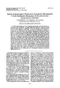
FIG. 1. N. meningitidis infection in Cp activity-deficient mice. Groups of control (circles) and hypoferremic, Cp activity-deficient (triangles) mice were injected with saline (open symbols) or 5 mg of iron dextran (closed symbols) immediately before injection of 104 CFU of N. meningitidis. Cp activity-deficient mice were also supplemented with exogenous Cp (N) immediately before infection with meningococci. Two mice from each group were sacrificed every 3 h, and bacterial counts in blood were determined by plate counts.
CERULOPLASMIN ACTIVITY IN MENINGOCOCCAL INFECTION
VOL. 45, 1984
225200L.
C
1751
0
°
150-
0
:.125.I
C._Cu
0
0.
co
E C.
50-
25 A
0 iA0
.0
L
A
18 2 6 12 Time during infection (hours)
FIG. 2. Variations of plasma Cp activity during N. meningitidis infection in normal and Cp activity-deficient mice. Control (0) and Cp activity-deficient (A) mice were infected intraperitoneally with 104 CFU of N. meningitidis. Control noninfected mice (0) were injected with physiological saline to estimate diurnal variation of Cp levels. Mice from each group were sacrificed every 3 h by cardiac puncture, and plasma samples were assayed for pPD oxidase activity. Values are expressed as a percentage of pPD oxidase levels determined for control noninfected mice at time zero.
IC 0 L.
135
values of ferroxidase activity as measured by the assay of Johnson et al. (12) (results not shown). Plasma ferroxidase activity (pPD oxidase activity) changed markedly during meningococcal infection (Fig. 2). There was an apparent decrease between 6 and 12 h of infection, followed by a rapid increase to levels approximately double those of control mice. Control (noninfected) mice displayed relatively constant levels of plasma ferroxidase activity. The marked increase in Cp activity during the convalescence phase was in keeping with the role of Cp as an acute-phase protein. It was unclear whether the drop in Cp activity which occurred during the onset of hypoferremia was related to iron metabolism. Influence of copper deficiency on plasma Cp and iron metabolism. The ferroxidase activity of Cp depends on the presence of an intramolecular chain of copper atoms. We therefore decided to investigate the effects of a lack of plasma Cp activity by producing copper deficiency in mice. Rapidly growing weanling mice placed on a copper-free diet quickly became Cp activity deficient (Fig. 3). Such mice were devoid of detectable plasma Cp activity by day 7 of treatment. The exponential loss of plasma activity (half-life of ca. 2 days) indicated a relatively high demand for exogenous copper or the lack of significant endogenous copper reserves in the mice or both. The copper-deficient mice continued to gain weight at control rates and appeared healthy even at 28 days of treatment. The only overt symptom noted was a change in fur color, with some loss of fur followed by regrowth of a more pale fur (black to brown). Key parameters of iron metabolism and iron balance were also monitored during copper deficiency. Total plasma transferrin levels (iron-binding capacity) remained constant (data not shown), but plasma transferrin iron levels fell gradually and linearly during copper deficiency (Fig. 3). Thus, mice maintained on a copper-free diet for 28 days had normal plasma transferrin levels but had only half as much transfer-
1002 A
75 -
a
cn
50 -
Cu'
25C0
C*
0I
0
I
I
I
I
I
25 20 15 10 5 diet Time on Cu-free (days)
30
FIG. 3. Plasma Cp activity and transferrin iron levels during the development of copper deficiency in mice. Weanling mice were placed on copper-deficient diet and given double-deionized water ad libitum. Plasma samples obtained by cardiac puncture were assayed for pPD oxidase activity (i) and transferrin iron levels (A). Values are expressed as a percentage of normal control values.
a
136
INFECT. IMMUN.
LETENDRE AND HOLBEIN 110-
O
z E ui
100-
A
A
.
0
A
A
I
III
IIA I
UL 0
A
I
90-
I
III III I
II III
U-
80-
I I I I
I
M
0
1. 1.
*
0
70-
0
(I) 0
0
z
60-
0
*11
IL
UD
I0
0%
0
120
240
MINUTES POST 59FeDRBC FIG. 4. Impaired release of hepatic 59Fe in Cp activity-deficient mice. Normal (0) and Cp activity-deficient (A) mice were injected intravenously with 59Fe-DRBC. Mice from each group were sacrificed every 30 min over 5 consecutive h. Liver-associated 59Fe was determined and expressed as a percentage of the total 59Fe taken up (maximal uptake at 30 min for both).
rin iron
as
controls. Interestingly, copper-deficient mice did
not become anemic as evidenced by normal hematocrit levels at day 28 of treatment (data not shown). Resistance of Cp activity-deficient mice to infection. After
28 days on the copper-free diet, mice lacked plasma ferroxidase activity and also had plasma transferrin iron levels that were only 45% that of control mice. This implicated a possible role of Cp in iron regulation in mice. We reasoned that an examination of meningococcal infection in Cp activity-deficient mice could provide further evidence of the importance of transferrin iron to meningococcal infection or the roles that Cp, as an acute-phase protein might play in host defense. Cp activity-deficient mice were much more resistant to meningococcal infection than normal mice (Fig. 1). There was only a slight and short-lived infection in these mice and no overt symptoms of infection could be seen. This suggested strongly that the hypoferremic state of Cp activitydeficient mice might be related to resistance. An absence of Cp ferroxidase activity would be expected to increase susceptibility if Cp was important to host defense as an acute-phase protein, i.e., in a function unrelated to iron metabolism. It should be noted in this regard that Cp activity-deficient mice exhibited no increase in plasma Cp activity during infection (Fig. 2), indicating a total lack of endogenous Cp or copper reserves. Susceptibility of Cp activity-deficient mice was returned readily to control levels by prior supplementation with exogenous Cp (Fig. 1). In addition, supplementation of Cp activity-deficient mice with exogenous iron also greatly stimulated infection. Both normal and Cp activity-deficient mice given exogenous iron displayed rapid and massive infections which resulted in death by 24 h of infection (Fig. 1 and data not shown). Both pieces of evidence suggested that it was altered iron regulation of the plasma transferrin iron
pool in Cp activity-deficient mice that resulted in resistance to meningococcal infection. Iron regulation in Cp activity-deficient mice. The transferrin iron pool in mice has been shown to be highly dynamic, with similar high rates of iron entry from the RES and exit to the bone marrow (13), and infection-induced hypoferremia has been shown to be due to an impaired entry of RESprocessed iron to the plasma transferrin iron pool (14). We have recently devised a model which allows study of the kinetics of processing of heme iron within the RES, the kinetics of incorporation of iron into RES stores, and the kinetics of the return of RES iron to the transferrin pool (14). We reasoned that studies of RES processing of iron might reveal the nature of the hypoferremia in Cp activity-deficient mice and the involvement of Cp activity in iron regulation. Normal and Cp activity-deficient mice injected with heatdenatured 59Fe-labeled erythrocytes accumulated similar amounts of 59Fe in their livers, with maximum uptake occurring by 30 min (data not shown) (14). However, the fate of this liver RES 59Fe (30 min total equal to 100%) was very different in control and Cp activity-deficient mice (Fig. 4). There was a rapid turnover of ca. 30% of the 59Fe in the case of control mice. This label has been shown to leave the RES quickly and enter the plasma transferrin iron pool of normal mice (14), whereas ca. 70% of the label remained RES associated and was mobilized more gradually. The initial rapid turnover of 59Fe was absent in Cp activity-deficient mice, and most of the 59Fe remained RES associated during the 5-h observation period. However, when Cp activitydeficient mice were injected with Cp 5 h after labeling with 59Fe-DRBC, there was a rapid increase in plasma 59Fe (Fig. 5). This indicated that exogenously supplied Cp could mobilize a portion of the RES-associated 9Fe readily in the Cp activity-deficient mice. This suggested the existence of a pool of iron in the RES of Cp activity-deficient mice that was available for release upon the availability of Cp activity. A pool of iron of similar size appeared to be absent in control mice, i.e., this pool had already "turned over" substantially in normal mice (Fig. 5). This evidence provided strong support for the hypothesis that the ferroxidase activity of Cp was involved in the movement of RES-processed iron to the plasma transferrin iron pool. Thus, Cp activity-deficient mice were impaired in the regulation (supply) of iron to the transferrin iron pool. This could relate directly to their resistance to meningococcal infection since iron limitation to meningococci would be much more prevalent under these conditions. DISCUSSION This study provides further evidence of the importance of transferrin to meningococcal infection and revealed that the ferroxidase activity of Cp is involved in the overall mechanism responsible for the regulation of the transferrin iron pool in mice. Transferrin iron has been shown to be important for the growth of N. meningitidis both in vitro (2, 9, 10, 15, 21) and in vivo (8, 9, 10). Normal mice control meningococcal infection successfully by the appearance of an infectioninduced hypoferremic response (8, 13). We have provided evidence that this response is due to altered movement of iron through the RES during infection (13, 14). The exact mechanism for hypoferremia remains obscure, but we have recently demonstrated an enhanced incorporation of iron into RES ferritin stores during infection-induced hypoferremia (14). The net result of this is to restrict the movement of RES-processed iron to the transferrin iron pool, thus limiting
CERULOPLASMIN ACTIVITY IN MENINGOCOCCAL INFECTION
VOL. 45, 1984
the availability of essential iron to meningococci in the host extracellular compartments where they replicate. Here we have shown that copper deficiency results in the disappearance of plasma Cp activity with a resulting hypoferremia in mice. This nutritionally induced hypoferremia also limits the availability of extracellular iron in the transferrin iron pool and restricts meningococcal infection in mice. This nutritionally induced hypoferremia also appears to result from an altered movement of RES-processed iron to the transferrin iron pool. However, this effect is reversible by supplementation with Cp. The ferroxidase activity of Cp has been implicated in the movement of iron from the RES to the transferrin iron pool in other animals (6), and several lines of evidence support this role (6, 17, 18, 20, 24). However, mice were shown to have relatively low levels of Cp, and it had been unclear whether Cp ferroxidase activity might be similarly involved in iron regulation. Our evidence indicates that Cp does play a role in the overall mechanism responsible for supplying iron to the transferrin iron pool in a manner similar to that in other animals, including humans.
150
A
i-
E100-
U. 0 0
0
LA0,
to
.5o
(A, -J
a.
137
Current models (6) suggest that Cp, functioning as a ferroxidase, acts on Fe2+ made available at the interface of the plasma and the RES compartments and oxidizes this iron to Fe3+, which is then picked up by plasma transferrin. This dictates the existence of an RES-associated iron pool supplied by catabolism of heme iron and available for Cp action. It is reasonable to suggest that this iron pool would be supplied either directly from heme catabolism or indirectly from ferritin and in a mobile form accessible by Cp. In Cp activity-deficient mice, all RES-processed heme iron remained intracellular, but a significant amount was released to the plasma upon resupply of Cp activity. On the other hand, a significant amount of RES-processed heme iron was quickly released to the plasma in normal mice. The results therefore suggest that processing of heme iron does occur in Cp activity-deficient mice, and the absence of Cp activity results in a buildup of an iron pool that is accessible to Cp activity when it becomes available. If Cp activity deficiency had resulted in all the processed heme iron entering intracellular ferritin stores, then one might expect an observable delay in RES iron release from Cp activity-deficient mice when Cp activity was resupplied. Thus, the mechanism for this hypoferremia appears somewhat distinct from that which we have determined in the case of infection-induced hypoferremia (14), in which Cp activity was present at normal or elevated levels but iron was incorporated into RES ferritin stores rather than being delivered to the transferrin pool. Our evidence has provided little support for the importance of Cp as an acute-phase protein which would act independent of its ferroxidase activity. One might expect a greater susceptibility to infection if Cp was important to host defense in this manner. The rapid increase of plasma ferroxidase activity during the convalescence phase of meningococcal infection of normal mice is noteworthy. We have recently shown that this requires de novo synthesis of Cp in the liver (D. Beaumier, M. Caldwell and B. E. Holbein, unpublished data). Based on our previous evidence of the kinetics of induction of the infection-induced hypoferremia (8, 13) and the results of this study, we suggest that Cp may be synthesized in response to the hypoferremia in normal mice. This implies a compensatory mechanism in which additional Cp, by virtue of its ferroxidase activity, would be synthesized to aid the mobilization of iron from the RES during the convalescence phase of infection. We are currently studying the mechanisms of infection and artificially induced hypoferremias and their regulation in mice since hypoferremia plays an important role in host defense during pathogenesis. ACKNOWLEDGMENTS The skillful technical assistance of Jane Donga is gratefully
acknowledged. We thank the Medical Research Council of Canada for its support in the form of a studentship to E.D.L. and operating grant MA-6772
0
0
30
60
90
120
MINUTES POST-INJECTION FIG. 5. Effect of exogenois Cp on plasma 59Fe levels in Cp activity-deficient mice. Cp activity-deficient mice were injected with 59Fe-DRBC and label incorporation was allowed to proceed for 5 h (see Fig. 4). Mice were then injected intravenously with saline (@) or Cp (A). Mice from each group were sacrificed every 30 min for 2 consecutive h and 59Fe radioactivity in plasma samples was determined.
to B.E.H.
LITERATURE CITED and E. P. Brown. 1977. The iron-binding function of 1. Aisen, P., transferrin in iron metabolism. Semin. Hematol. 14:31-53. 2. Archibald, F. S., and I. W. DeVoe. 1979. Removal of iron from human transferrin by Neisseria meningitidis. FEMS Microbiol.
Lett. 6:159-162. 3. Brener, D., I. W. DeVoe, and B. E. Holbein. 1981. Increased virulence of Neisseria meningitidis after in vitro iron-limited growth at low pH. Infect. Immun. 33:59-66.
138
LETENDRE AND HOLBEIN
4. Carver, F. J., D. L. Farb, and E. Frieden. 1982. The effect of albumin, ceruloplasmin, and other serum constituents on Fe(II)
oxidation. Biol. Trace Element Res. 4:1-19. 5. Cavill, I., and C. Ricketts. 1980. Human iron kinetics, p. 573604. In A. Jacobs and M. Worwood (ed.), Iron in biochemistry and medicine, vol. HI. Academic Press, Inc., New York. 6. Frieden, E., and H. S. Hsieh. 1976. The biological role of ceruloplasmin and its oxidase activity. Adv. Exp. Med. Biol. 74:505-529. 7. Goldstein, S. M., H. B. Kaplan, H. S. Edelson, and G. Weissman. 1979. Ceruloplasmin. A scavenger of superoxide anion radicals. J. Biol. Chem. 254:4040-4045. 8. Holbein, B. E. 1980. Iron-controlled infection with Neisseria meningitidis in mice. Infect. Immun. 29:886-891. 9. Holbein, B. E. 1981. Growth and surface binding of proteins by Neisseria meningitidis in normal human serum. Curr. Microbiol. 6:213-216. 10. Holbein, B. E. 1981. Enhancement of Neisseria meningitidis in mice by addition of iron bound to transferrin. Infect. Immun. 34:120-125. 11. Holbein, B. E., K. W. Jericho, and G. C. Likes. 1979. Neisseria meningitidis infection in mice, influence of iron, variations in virulence among strains and pathology. Infect. Immun. 24:545551. 12. Johnson, D. A., S. Osaki, and E. Frieden. 1967. A micro method for the determination of ferroxidase (ceruloplasmin) in human serums. J. Clin. Chem. 13:142-150. 13. Letendre, E. D., and B. E. Holbein. 1983. Turnover in the transferrin iron pool during the hypoferremic phase of experimental Neisseria meningitidis infection in mice. Infect. Immun. 39:50-59.
INFECT. IMMUN.
14. Letendre, E. D., and B. E. Holbein. 1984. Mechanism of impaired iron release by the reticuloendothelial system during the hypoferremic phase of experimental Neisseria meningitidis infection in mice. Infect. Immun. 44:320-325. 15. Mickelsen, P. A., and F. E. Sparling. 1981. Ability of Neisseria gonorrheae, Neisseria meningitidis, and commensal species to obtain iron from transferrin and iron compounds. Infect. Immun. 33:555-564. 16. Neilands, J. B. 1981. Microbial iron compounds. Annu. Rev. Biochem. 50:715-731. 17. Osaki, S., D. A. Johnson, and E. Frieden. 1971. The mobilization of iron from the perfused mammalian liver by a serum copper enzyme ferroxidase. J. Biol. Chem. 246:3018-3023. 18. Ragan, H. A., S. Nacht, G. R. Lee, C. R. Bishop, and G. E. Cartwright. 1969. Effect of ceruloplasmin on plasma iron in copper-deficient swine. Am. J. Physiol. 217:1320-1330. 19. Ravin, H. A. 1961. An improved colorimetric enzymatic assay of ceruloplasmin. J. Lab. Clin. Med. 58:161-168. 20. Roeser, H. P., G. R. Lee, S. Nacht, and G. E. Cartwright. 1970. The role of ceruloplasmin in iron metabolism. J. Clin. Invest.
49:2408-2417. 21. Simonson, C., D. Brener, and I. W. DeVoe. 1982. Expression of a high affinity mechanism for acquisition of transferrin iron by Neisseria meningitidis. Infect. Immun. 36:107-113. 22. Weinberg, E. D. 1974. Iron and susceptibility to infectious disease. Science 186;952. 23. Weinberg, E. D. 1978. Iron and infection. Microbiol. Rev. 42:45-66. 24. Williams, D. M., G. R. Lee, and G. E. Cartwright. 1974. Ferroxidase activity of rat ceruloplasmin. Am. J. Physiol. 227:1094-1097.