Hepatocyte growth factor (HGF) binds to MET and activates ... c-met proto-oncogene. ... For detailed protocols on conversion curves, kinase assays and inhibitor ...
ADP-Glo™ Kinase Assay Application Notes
TYROSINE KINASE SERIES: MET
MET Kinase Assay By Kevin Hsiao, M.S., Hicham Zegzouti, Ph.D., Jolanta Vidugiriene, Ph.D., and Said A. Goueli, Ph.D., Promega Corporation
Scientific Background: MET is a proto‐oncogene that encodes a transmembrane growth factor receptor which is a heterodimer of two disulphide linked chains of 50 kDa (alpha) and 145 kDa (beta). MET is widely expressed in the kidney, brain, lung, skin, and embryonic tissue (1). Hepatocyte growth factor (HGF) binds to MET and activates its tyrosine kinase activity. MET is overexpressed and activated in a variety of human cancers including pancreatic, colon, gastric, cervical and ovarian cancers and has been shown to be involved in tumor cell migration and invasion (2). 1. Giordano, S. et al: Biosynthesis of the protein encoded by the c‐met proto‐oncogene. Oncogene. 1989 Nov;4(11):1383‐8. 2. Iyer, A. et al: Structure, tissue‐specific expression, and transforming activity of the mouse met protooncogene. Cell Growth Differ. 1990 Feb; 1(2):87‐95.
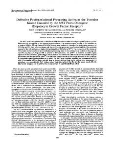
Figure 1. Principle of the ADP‐Glo™ Kinase Assay. The ATP remaining after completion of the kinase reaction is depleted prior to an ADP to ATP conversion step and quantitation of the newly synthesized ATP using luciferase/luciferin reaction.
ADP-Glo™ Kinase Assay Description ADP‐Glo™ Kinase Assay is a luminescent kinase assay that measures ADP formed from a kinase reaction; ADP is converted into ATP, which is converted into light by Ultra‐Glo™ Luciferase (Fig. 1). The luminescent signal positively correlates with ADP amount (Fig. 2) and kinase activity (Fig. 3A). The assay is well suited for measuring the effects chemical compounds have on the activity of a broad range of purified kinases—making it ideal for both primary screening as well as kinase selectivity profiling (Fig. 3B). The ADP‐Glo™ Kinase Assay can be used to monitor the activity of virtually any ADP‐generating enzyme (e.g., kinase or ATPase) using up to 1mM ATP.
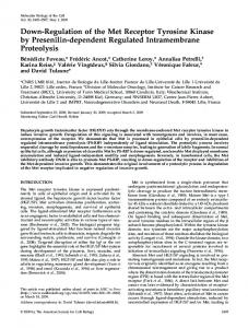
Figure 2. Linearity of the ADP‐Glo Kinase Assay. ATP‐to‐ADP conversion curve was prepared at 10µM ATP+ADP concentration range. This standard curve is used to calculate the amount of ADP formed in the kinase reaction. Z’ factors were determined using 192 replicates of each of the % conversions shown.
For detailed protocols on conversion curves, kinase assays and inhibitor screening, see The ADP‐Glo™ Kinase Assay Technical Manual #TM313, available at www.promega.com/tbs/tm313/tm313.html
Protocol
Add 5 μl of ADP‐Glo™ Reagent Incubate at room temperature for 40 minutes. Add 10 μl of Kinase Detection Reagent Incubate at room temperature for 30 minutes.
• • • •
• Dilute enzyme, substrate, ATP and inhibitors in Kinase Buffer. • Add to the wells of 384 low volume plate: 1 μl of inhibitor or (5% DMSO) 2 μl of enzyme (defined from table 1) 2 μl of substrate/ATP mix • Incubate at room temperature for 60 minutes.
• Record luminescence (Integration time 0.5‐1second).
Table 1. MET Enzyme Titration. Data are shown as relative light units (RLU) that directly correlate to the amount of ADP produced. The correlation between the % of ATP converted to ADP and corresponding signal to background ratio is indicated for each kinase amount.
MET, ng
100
50
25
12.5
6.25
3.13
1.56
0.78
0
Luminescence S/B % Conversion
54608 36.1 91.2
59274 39.2 99.3
45938 30.4 76.2
34821 23.0 57.0
24554 16.3 39.3
12571 8.3 18.6
5290 3.5 6.0
2252 1.5 0.8
1511 1 0
Figure 3. MET Kinase Assay Development: (A) MET enzyme was titrated using 10µM ATP and the luminescence signal generated from each of the amounts of the enzyme is shown. (B) Staurosporine dose response was created using 4ng of MET to determine the potency of the inhibitor (IC50).
Assay Components and Ordering Information:
Products ™
ADP-Glo Kinase Assay MET Kinase Enzyme System ADP-Glo + MET Kinase Enzyme System
Company
Cat.#
Promega Promega Promega
V9101 V3361 V9571
MET Kinase Buffer: 40mM Tris,7.5; 20mM MgCl2; 0.1mg/ml BSA; 50μM DTT.