REVIEW Korean J Intern Med 2015;30:1-5 http://dx.doi.org/10.3904/kjim.2015.30.1.1
Methods for the morphological and functional evaluation of microvascular damage in systemic sclerosis Barbara Ruaro1, Vanessa Smith2, Alberto Sulli1, Saskia Decuman2, Carmen Pizzorni1, and Maurizio Cutolo1
1
Research Laboratory and Academic Division of Clinical Rheumatology, Department of Internal Medicine, University of Genova, Genoa, Italy; 2 Department of Rheumatology, Ghent University Hospital, Gent, Belgium
Received : August 29, 2014 Accepted: November 10, 2014 Correspondence to Maurizio Cutolo, M.D. Research Laboratory and Academic Division of Clinical Rheumatology, Department of Internal Medicine, University of Genova, Viale Benedetto XV, n°6, Genova 16132, Italy Tel: +39-10-353-7994 Fax: +39-10-353-7994 E-mail:
[email protected]
Systemic sclerosis is a connective tissue disease characterized by alterations in microvascular structure and function. In these patients, numerous studies have demonstrated a relationship between capillary morphology and peripheral blood perfusion. Nailfold videocapillaroscopy reveals the peripheral microvascular morphology and thus allows classification and scoring of capillary abnormalities with respect to different microangiopathy patterns (early, active, and late). Laser Doppler f lowmetry and laser speckle contrast analysis can be used to estimate cutaneous blood flow through microvessels and to assess and quantify blood perfusion at peripheral sites. These two methods are also used to identify changes in digital blood perfusion after the infusion of vasodilators. Keywords: Systemic sclerosis; Microvascular damage; Nailfold videocapillaroscopy; Laser speckle contrast analysis; Peripheral blood perfusion
INTRODUCTION Systemic sclerosis (SSc) is an autoimmune connective tissue disease characterized by early impairment of the microvascular system, leading to functional and organic manifestations, including digital ulcers [1,2]. Progressive def iciency in the vasodilatory capacity of the vessels, along with intimal fibroproliferation, thrombosis of the digital arteries, and tissue ischemia have been postulated as mechanisms explaining the persistent vascular involvement [3,4]. Microvascular damage and dysfunction are the earliest morphological and functional markers of SSc and are correlated with prognosis and therapy planning [5]. Copyright © 2015 The Korean Association of Internal Medicine
The vascular changes occur during the early stage of SSc, with Raynaud phenomenon (RP) as the clinical expression of abnormal blood f low regulation due to altered microvascular structure and function [5,6]. Therefore, morphological and functional assessment of the peripheral microvasculature in SSc patients is a diagnostic necessity, with prognostic and therapeutic consequences [5]. Nailfold microangiopathy can be evaluated easily by nailfold videocapillaroscopy (NVC). Laser Doppler flowmetry (LDF) allows evaluation of the extent of perfusion in the peripheral microcirculation [7,8], while laser speckle contrast analysis (LASCA) is a relatively new method that permits quantitative assessment of the peripheral perfusion in larger areas
This is an Open Access article distributed under the terms of the Creative Commons Attribution Non-Commercial License (http://creativecommons.org/licenses/ by-nc/3.0/) which permits unrestricted noncommercial use, distribution, and reproduction in any medium, provided the original work is properly cited.
pISSN 1226-3303 eISSN 2005-6648 http://www.kjim.org
The Korean Journal of Internal Medicine Vol. 30, No. 1, January 2015
of different body sites [9-11]. These three methods for evaluating SSc patients are described below.
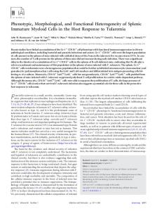
NAILFOLD VIDEOCAPILLAROSCOPY Nailfold microangiopathy can be evaluated easily using NVC. This non-invasive, well-accepted, safe method makes use of a magnif ication system (best at ×200 magnification) [12,13]. The video-capillaroscope has an optical/digital probe that can be moved over the surface of the fingernails from the second to the fifth digits [5,14,15]. NVC is used to evaluate the morphology of nailfold dermal papillary capillaries, thus allowing recognition of the qualitative, morphological patterns of microangiopathy (early, active, and late) (Fig. 1) [16]. The early SSc pattern is characterized by the presence of few giant capillaries, few capillary microhemorrhages, no evident capillary loss, and a relatively well-preserved capillary distribution. The active NVC SSc pattern is a marker of disease progression. It is characterized by frequent giant capillaries, frequent capillary microhemorrhages, moderate capillary loss, absent or mildly ramified capillaries, and a mild disorganization of the capillary architecture. In the late pattern, giant capillaries and microhemorrhages are nearly absent, but there is severe capillary loss, with evident extensive avascular areas, ramified or bushy capillaries, and se-
A
vere disorganization of the normal capillary array [16]. In patients with RP, NVC can be used to distinguish the primary from the secondary forms of the disease [5,6]. Under normal conditions or in the presence of primary RP, NVC shows a regular array of capillary loops along the nailfold bed, no abnormal enlargements, and no capillary loss [5,17,18]. In secondary RP, the morphological signs of giant capillaries, microhemorrhages, capillary loss, the presence of avascular areas, and angiogenesis are indicative of microvascular damage. These sequential capillaroscopic changes are typical of the microvascular involvement observed in more than 95% of SSc patients and are described by the term “SSc pattern” [5,19,20]. NVC also provides a semiquantitative assessment of microvascular damage that can be used to follow its progression, based on a semiquantitative rating scale (score 0 to 3) for the assessment of microvascular parameters [21]. The microangiopathy evolution score [14,15] is the sum of the three individual scores: capillary loss, disorganization of the microvascular array, and capillary ramifications. Qualitative and semiquantitative assessments also have been used to predict the clinical complications of SSc [15,21,22]. For example, patients with a reduced capillary density are at high risk of developing digital skin ulcers and pulmonary arterial hypertension [15,23]. Inclusion of the NVC patterns in the diagnostic procedure could increase the sensitivity of the classification criteria for SSc, such as the new European League Against Rheumatism and American College of Rheumatology criteria [24]. Moreover, NVC-based assessment of microvascular structure may be useful in the evaluation of drug effects during the treatment of SSc patients [25].
B PERIPHERAL BLOOD FLOW EVALUATION BY LDF
C
D
Figure 1. Nailfoild videocapillaroscopic (×200) patterns of microangiopathy: (A) normal versus (B) early, (C) active, and (D) late patterns. SSc, systemic sclerosis.
2
www.kjim.org
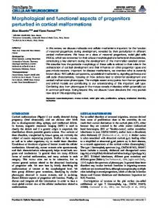
LDF is a user-friendly, well-accepted, and non-invasive assessment of microvascular flow at a single skin point. It provides an index of skin perfusion by measuring the Doppler shift induced by coherent light scattering caused by moving red blood cells [26]. An example of peripheral blood perfusion (BP) evaluated by LDF is shown in Fig. 2.
http://dx.doi.org/10.3904/kjim.2015.30.1.1
Ruaro B, et al. Microcirculation in systemic sclerosis
A
B Figure 2. Laser Doppler flowmetry: 1-minute analysis of blood perfusion in a healthy subject (A) and in a patient with systemic sclerosis (nailfold videocapillaroscopy late pattern) (B). Entity of the blood flow reported as perfusion units (PU).
The instrument comprises a heatable probe, an optic fiber connector, an instrument that detects both tissue perfusion and skin temperature, and a computer to evaluate the data. LDF measures local perfusion in a cubic millimeter of tissue (capillaries, arterioles, venules, shunts), which is recorded in perfusion units (PUs), for example at the level of the fingertips. Basal finger temperature (usually at the level of the fingertips, from the second to fifth digits of both hands) is measured first, after which the probe is heated to 36°C and capillary dilation capacity evaluated [8]. In the study by Binaghi et al. [27], a significant correlation was found between the digital flow parameters and capillaroscopy score in SSc patients. The results suggested the relevance of LDF in evaluating apparently primary RP during a standardized thermic test, because even in the absence of a definite scleroderma-like capillaroscopic pattern, flowmetry can be used to identify patients with secondary RP [27]. Some studies have demonstrated that SSc patients have lower blood flow compared with healthy subjects, and that among the patients evaluated by NVC, those with a late SSc microangiopathy pattern have a lower rate of blood f low on LDF compared with those with the early or active pattern [8,11,28,29]. SSc patients were also shown to have abnormal microvascular regulatory responses to heat stimulation, suggesting that the degree of endothelial dysfunction is related to the degree of peripheral microvascular involvement [8]. In addition, LDF can assess changes in peripheral BP during treatment with vasodilator drugs either within a few days or over a period of years [8,30]. However, one problem with the technique is the large site-to-site variation, which limits its efficacy in comparing blood
http://dx.doi.org/10.3904/kjim.2015.30.1.1
flow among sites and in monitoring changes over time [26]. A new technique, laser Doppler imaging, which evaluates flow over an anatomic area might overcome this problem [7].
PERIPHERAL BLOOD FLOW EVALUATION BY LASCA This relatively new technique quantifies BP over an area rather than only at a single point, and thus it may obviate the problems that occur when a single probe is used, especially site-to-site variability. LASCA is based on the principle that when an object is illuminated by laser light, the backscattered light will form a random interference pattern made up of dark and bright areas. This so-called speckle pattern is stationary, but when movement occurs, as in red blood cells within a tissue, the speckle pattern changes over time. These changes can be recorded by a detector camera [9,11]. An advantage of LASCA is that it allows commonly used settings (e.g., working distance, point density, frame rate resolution, and the width and height of the measurement area) to be saved for repeated use [11]. Moreover, LASCA does not require direct contact between the probe and the skin, which might in itself affect blood flow. It also evaluates the f luxes in different regions of interest (ROIs) and time regions of interest may also be created [11]. Using LASCA, fingertip BP, during or after recording (for example, at the fingertips and dorsum of the hand), is evaluated and the results reported in PUs. The different values can be represented graphically, and the BP ROI, whether depicted in color or as intensity images, are superimposed (Fig. 3) [11]. More recently, LASCA has been applied in research on RP and SSc [10,11]. One such study demonstrated that peripheral BP, evaluated by LDF and LASCA, correlates with the extent of the microangiopathy detected by NVC [11]. As noted above, when patients whose disease had been classified using NVC were then evaluated using LDF and LASCA, those with the late SSc microangiopathy pattern had a lower blood flow than that of patients with early or active SSc patterns [11]. A very recent study showed that BP, when monitored by LASCA, is significantly lower in SSc patients than
www.kjim.org
3
The Korean Journal of Internal Medicine Vol. 30, No. 1, January 2015
Conflict of interest No potential conflict of interest relevant to this article was reported.
REFERENCES 1. Gabrielli A, Avvedimento EV, Krieg T. Scleroderma. N
A
B
Figure 3. Imaging of cutaneous blood perfusion using laser speckle contrast analysis at the level of the palmar region in a healthy subject (A) and in a patient with systemic sclerosis (nailfold videocapillaroscopy late pattern) (B).
Engl J Med 2009;360:1989-2003. 2. Derk CT, Jimenez SA. Systemic sclerosis: current views of its pathogenesis. Autoimmun Rev 2003;2:181-191. 3. Cutolo M, Grassi W, Matucci Cerinic M. Raynaud’s phenomenon and the role of capillaroscopy. Arthritis Rheum 2003;48:3023-3030. 4. Cutolo M, Sulli A, Smith V. Assessing microvascular changes in systemic sclerosis diagnosis and manage-
in healthy subjects at the level of the fingertips, periungual areas, and palms of the hands. In addition, in SSc patients, there is a statistically significant negative correlation between the extent of nailfold microangiopathy and BP at the level of those same skin areas [31]. Similar combined investigations might optimize the follow-up of SSc patients [11]. Whether the combination of all three techniques (NVC, LDF, and LASCA) improves the classification of SSc, including distinguishing patients with SSc from those with primary RP and from healthy controls, has yet to be confirmed by the appropriate studies. However, thus far, when only a single microcirculatory tool is used, NVC seems to be the best discriminator [32,33].
ment. Nat Rev Rheumatol 2010;6:578-587. 5. Cutolo M, Pizzorni C, Sulli A. Nailfold video-capillaroscopy in systemic sclerosis. Z Rheumatol 2004;63:457462. 6. Smith V, Pizzorni C, De Keyser F, et al. Reliability of the qualitative and semiquantitative nailfold videocapillaroscopy assessment in a systemic sclerosis cohort: a two-centre study. Ann Rheum Dis 2010;69:1092-1096. 7.
Roustit M, Cracowski JL. Assessment of endothelial and neurovascular function in human skin microcirculation. Trends Pharmacol Sci 2013;34:373-384.
8. Cutolo M, Ferrone C, Pizzorni C, Soldano S, Seriolo B, Sulli A. Peripheral blood perfusion correlates with microvascular abnormalities in systemic sclerosis: a laser-Doppler and nailfold videocapillaroscopy study. J
CONCLUSIONS
Rheumatol 2010;37:1174-1180. 9. Draijer M, Hondebrink E, van Leeuwen T, Steenbergen W. Review of laser speckle contrast techniques for visu-
Peripheral microvascular morphology can be assessed using NVC, which also allows the classification and scoring of capillary abnormalities. NVC is the best method to distinguish patients with secondary RP from those with primary RP and from healthy subjects [32,33]. NVC, LDF, and LASCA together constitute an essential set of safe and noninvasive tools for the early diagnosis and reliable quantification of SSc, the response to therapy, and disease progression [22]. Extensive employment of these tools in rheumatology clinics is recommended accordingly [8,25,30,34].
alizing tissue perfusion. Lasers Med Sci 2009;24:639651. 10. Della Rossa A, Cazzato M, d’Ascanio A, et al. Alteration of microcirculation is a hallmark of very early systemic sclerosis patients: a laser speckle contrast analysis. Clin Exp Rheumatol 2013;31(2 Suppl 76):109-114. 11. Ruaro B, Sulli A, Alessandri E, Pizzorni C, Ferrari G, Cutolo M. Laser speckle contrast analysis: a new method to evaluate peripheral blood perfusion in systemic sclerosis patients. Ann Rheum Dis 2014;73:1181-1185. 12. Sulli A, Soldano S, Pizzorni C, et al. Raynaud’s phenomenon and plasma endothelin: correlations with capil-
4
www.kjim.org
http://dx.doi.org/10.3904/kjim.2015.30.1.1
Ruaro B, et al. Microcirculation in systemic sclerosis
laroscopic patterns in systemic sclerosis. J Rheumatol
sification criteria for systemic sclerosis: an American
2009;36:1235-1239.
College of Rheumatology/European League against
13. Kahaleh MB. Raynaud phenomenon and the vascular disease in scleroderma. Curr Opin Rheumatol 2004;16:718-722.
Rheumatism collaborative initiative. Arthritis Rheum 2013;65:2737-2747. 25. Cutolo M, Zampogna G, Vremis L, Smith V, Pizzorni C,
14. Smith V, Pizzorni C. The videocapillaroscopic tech-
Sulli A. Longterm effects of endothelin receptor antag-
nique. In: Cutolo M, Smith V, Sulli A, eds. Atlas of Cap-
onism on microvascular damage evaluated by nailfold
illaroscopy in Rheumatic Diseases. Milano: Elsevier Srl,
capillaroscopic analysis in systemic sclerosis. J Rheu-
2010:25-31. 15. Smith V, Cutolo M. When and how to perform the capillaroscopy. In: Cutolo M, Smith V, Sulli A, eds. Atlas of Capillaroscopy in Rheumatic Diseases. Milano: Elsevier Srl, 2010:33-45.
matol 2013;40:40-45. 26. Herrick AL, Hutchinson C. Vascular imaging. Best Pract Res Clin Rheumatol 2004;18:957-979. 27. Binaghi F, Cannas F, Mathieu A, Pitzus F. Correlations among capillaroscopic abnormalities, digital flow and
16. De Angelis R, Cutolo M, Salaffi F, Restrepo JP, Grassi
immunologic findings in patients with isolated Ray-
W. Quantitative and qualitative assessment of one rheu-
naud’s phenomenon: can laser Doppler flowmetry help
matology trainee’s experience with a self-teaching pro-
identify a secondary Raynaud phenomenon? Int Angiol
gramme in videocapillaroscopy. Clin Exp Rheumatol 2009;27:651-653. 17. De Angelis R, Grassi W, Cutolo M. A growing need for capillaroscopy in rheumatology. Arthritis Rheum 2009;61:405-410.
1992;11:186-194. 28. Wigley FM, Wise RA, Mikdashi J, Schaefer S, Spence RJ. The post-occlusive hyperemic response in patients with systemic sclerosis. Arthritis Rheum 1990;33:1620-1625. 29. Gunawardena H, Harris ND, Carmichael C, McHugh
18. Carpentier PH, Maricq HR. Microvasculature in sys-
NJ. Maximum blood flow and microvascular regulatory
temic sclerosis. Rheum Dis Clin North Am 1990;16:75-
responses in systemic sclerosis. Rheumatology (Oxford)
91.
2007;46:1079-1082.
19. Cutolo M, Sulli A, Secchi ME, Olivieri M, Pizzorni C.
30. Cutolo M, Ruaro B, Pizzorni C, et al. Longterm treat-
The contribution of capillaroscopy to the differential
ment with endothelin receptor antagonist bosentan and
diagnosis of connective autoimmune diseases. Best
iloprost improves fingertip blood perfusion in systemic
Pract Res Clin Rheumatol 2007;21:1093-1108.
sclerosis. J Rheumatol 2014;41:881-886.
20. Cutolo M, Pizzorni C, Secchi ME, Sulli A. Capillaroscopy. Best Pract Res Clin Rheumatol 2008;22:1093-1108. 21. Sulli A, Secchi ME, Pizzorni C, Cutolo M. Scoring the nailfold microvascular changes during the capillaroscopic analysis in systemic sclerosis patients. Ann Rheum Dis 2008;67:885-887. 22. Smith V, Riccieri V, Pizzorni C, et al. Nailfold capillaroscopy for prediction of novel future severe organ involvement in systemic sclerosis. J Rheumatol 2013;40:2023-2028.
31. Sulli A, Ruaro B, Cutolo M. Evaluation of blood perfusion by laser speckle contrast analysis in different areas of hands and face in patients with systemic sclerosis. Ann Rheum Dis 2014;73:2059-2061. 32. Murray AK, Moore TL, Manning JB, Taylor C, Griffiths CE, Herrick AL. Noninvasive imaging techniques in the assessment of scleroderma spectrum disorders. Arthritis Rheum 2009;61:1103-1111. 33. Rosato E, Rossi C, Molinaro I, Giovannetti A, Pisarri S, Salsano F. Laser Doppler perfusion imaging in system-
23. Hofstee HM, Voskuyl AE, Vonk Noordegraaf A, et al.
ic sclerosis impaired response to cold stimulation in-
Pulmonary arterial hypertension in systemic sclerosis
volves digits and hand dorsum. Rheumatology (Oxford)
is associated with profound impairment of microvascular endothelium-dependent vasodilatation. J Rheumatol 2012;39:100-105.
2011;50:1654-1658. 34. Cutolo M, Smith V, Sulli A. Training in capillaroscopy: a growing interest. J Rheumatol 2012;39:1113-1116.
24. van den Hoogen F, Khanna D, Fransen J, et al. 2013 Clas-
http://dx.doi.org/10.3904/kjim.2015.30.1.1
www.kjim.org
5