signals after interacting with processed antigen in conjunction with MHC gene prod- .... phosphate, pH 7.2, 0.4% glucose, and 1 MCi 1251 for 5 min and sepa-.
EXPRESSION OF A FUNCTIONAL CD3Ti ANTIGEN/MHC RECEPTOR IN THE ABSENCE OF SURFACE CD2 Analysis with Clonal Jurkat Cell Mutants BY PHILIPPE MOINGEON,1 ANDRES ALCOVER,1 LINDA K. CLAYTON,1 HSIU-CHING CHANG,1 CATHERINE TRANSY,1 AND ELLIS L. REINHERZ* From the Laboratory of Immunobiology, Dana-Farber Cancer Institute; and the Departments of *Medicine and 1Pathology, Harvard Medical School, Boston, Massachusetts 02115
Activation ofhuman T lymphocytes can be initiated through two major pathways : one of these is antigen specific, whereas the other is antigen independent. Antigenspecific activation is mediated via the TCR (CD3Ti) complex, which transducer signals after interacting with processed antigen in conjunction with MHC gene products. Similar activation can be induced experimentally by crosslinking the CD3 Ti complex with antibodies directed against individual TCR subunit proteins (1-3). An alternative antigen-independent pathway has been described based upon the in vitro observation that perturbation of the CD2 (T11) molecule with a combination of antibodies defining two different epitopes (anti-T112 and anti T113) is able to induce cell activation and proliferation (4). The physiological counterpart of antiT112+ 3 is still unknown; however, the cell adhesion structure LFA-3 appears to be one natural ligand for CD2 (5) that could provide part of the activation stimulus mediated by anti-T11 antibodies in vitro (6, 7). Interaction of membrane receptors with appropriate mitogenic stimuli for the antigen-specific and alternative pathways results in a virtually identical series of events involving phosphatidyl inositol turnover with subsequent production of 1, 4, 5 inositol trisphosphate (IP3)' and diacylglycerol (8, 9). IP3 is directly responsible for a rise in intracellular free calcium ([Ca2+ ]i) as a consequence of mobilization from intracellular stores (10) and/or transmembrane calcium flux (11, 12), while diacylglycerol is an endogenous activator of protein kinase C (13) . Previous studies have shown that both a rise in intracellular free calcium [Ca2+ ]i as well as phosphorylation events mediated through protein kinase C activation are necessary for IL-2 gene induction (14) . Given the similarities in early signal transduction via CD3Ti and CD2 structures and their coexpression on the surface ofmature T lymphocytes, the interdependence ofthe two pathways has been suggested, but remains to be fully investigated . Herein, we describe the production and characterization of CD2- CD3Ti+ variants derived This study was supported by NIH grants AI-19807, AI-21226, and CA-40134 . P Moingeon is supported by Ministere des Affaires Etrangeres (France) and the Philippe Foundation . A. Alcover was a recipient of a fellowship from Consejo S. Investigaciones Cientificas (Spain). C. Transy is supported by an Irvington House Institute fellowship. A. Alcover's present address is the Lab. Immunologie Moléculaire, Inst . Pasteur, Paris. i Abbreviation used in this paper: IP3, 1,4,5 inositol trisphosphate. J . Exp . MED. ® The Rockefeller University Press " 0022-1007/88/12/2077/14 $2 .00
Volume 168
December 1988
2077-2090
2077
2078
PRODUCTION AND CHARACTERIZATION OF CD2 - CD3Ti' VARIANTS
from the Jurkat CD2' CD3 Ti+ human T cell line. These mutants express normal levels of CD3 Ti-a/p molecules but lack any detectable surface CD2 molecules as judged by immunoprecipitation experiments and Scatchard analysis. Nevertheless, functional studies clearly establish that these cells can be activated via their CD3Ti receptor, retaining the capacity to mediate all the aforementioned events associated with the transduction of activation signals . Assuming that Jurkat cells are representative of their normal cycling cellular counterparts, we conclude that CD2 is not required to activate mature T lymphocytes through their TCRs. Materials and Methods Cells. Variants ofthe Jurkat T cell line (cloneJ77-6.8, kindly provided by Dr. K. Smith, Dartmouth Medical School, Hanover, NH) lacking surface expression of the CD2 molecule were derived by mutagenesis, immunoselection, and all sorting, as previously described (15). Briefly, 107 cells were irradiated (300 rad) in a y cell irradiator (model 1,000; Atomic Energy of Canada Ltd ., Ontario, Canada) using a 117 cesium source. After 5 d in culture, cells were incubated 1 h at room temp with a mixture of antiTll 1 (clone 3Pt2H9) and antiT112 (lold241C) ascites used at a 1 :100 final dilution . Rabbit sera (Pel-Freeze Biologicals, Rogers, AR) at a dilution of 1 :3 was added as a source of complement for 1 h at 37°C. The procedure was repeated four times and viable cells were isolated by centrifugation over a Ficoll-Hypaque density gradient (Pharmacia Fine Chemicals, Piscataway, NJ). CD3' Ti' CD2 - cells were sorted using an Epic V cell sorter (Coulter Electronics Inc., Hialeah, FL) and cloned at 1 cell/well . 19 CD3'Ti'CD2 - colonies were obtained, and three ofthese clones, termed Tll - a, T11 - b, and Tll - c, were randomly selected and extensively characterized . Indirect Immunofuorescence Assays. Phenotypic analyses were performed using indirect immunofluorescence assays with a fluorescein-conjugated goat anti-mouse IgG (Meloy Laboratories Inc., Springfield, VA) second antibody. 104 cells were analyzed in each sample and results were expressed as histograms displaying number of cells vs. fluorescence intensity on a log scale . Antibodies used in the study were anti-T3 (RW28C8, Leu-4), anti-Ti (9H5, directed against a clonotypic determinant present on Jurkat), antiTII1 (3Pt2H9), anti-T112 (lold241C), and ant-T113 (lmono2A6) . An irrelevant ascites (1HT4-4E5) was used as a control of fluorescence background . Antibodies were used at saturating dilutions in the assay (1 :200 final dilution) . Immunoprecipitations . 2 x 10' cells were surface labeled with 125 1 (IMS30, Amersham Corp., Arlington Heights, IL) for 15 min at room temperature using a standard lactoperoxidase method . Labeled cells were washed and cell lysates were prepared by resuspending the cell pellet in 400 pl of radioimmunoprecipitation assay (RIPA) buffer containing 1% Triton X-100, 0.15 M NaCl, 0.01 M NaH2PO4, 1 mM EDTA, 1 mM EGTA, 1 mM Naf, and protease inhibitors, and by agitating for 40 min at 4°C. Celllysates were then extensively precleared with formalin-fixed Staphylococcus aureus bacteria and protein A-Sepharose 4B beads (Pharmacia Fine Chemicals) bound to a nonreactive antibody (anti-T8) . Aliquots of precleared lysates were subsequently incubated with specific antibody (either antiT3 or anti-T11 1 antibodies) coated protein A-Sepharose beads for 6 h at 4°C. Immune precipitates were washed four times with RIPA buffer and submitted to SDS-PAGE analysis after treatment with 5% 2-ME (reducing conditions). Gels were dried and radiolabeled precipitates were visualized by autoradiography after 3 d of exposure. Binding Assays . Purified anti-T111 antibody (3Pt2H9) was labeled with 10 ul of immobilized lactoperoxidase/glucose oxidase (Enzymobeads ; Bio-Rad Laboratories, Richmond, CA) in 40 mM sodium phosphate, pH 7.2, 0.4% glucose, and 1 MCi 1251 for 5 min and separated over a 1 ml Biogel P-6 column (Bio-Rad Laboratories) . 0.1-6 x 10⁶ cpm labeled antibody (sp act 104 cpm/pmol) was added to 106 cells overlayed onto 0.2 ml of a 1.5:1 mixture of dibutyl phthalate :dioctyl phthalate (Aldrich Chemical Co., Milwaukee, WI). After 30 min at 4°C, tubes were centrifuged (8,500 g for 1 min), the tips of the tubes containing the cell pellets were cut, and the cell bound and free radioactivity was determined in a gamma counter. Nonspecific binding ofthe antibody was evaluated by performing the experiment in the pres-
MOINGEON
ET
AL .
2079
ence of an excess ofcold antibody (ascites used at a 1 :50 dilution) and was subtracted from total binding, with specific binding representing 95% of total binding . Northern Blot Analysis. Total RNA was extracted from 10' cells using the VRC method (Vanadyl Ribonucleoside Complex, New England Biolabs, Beverly, MA) . 20-25 Rg ofRNA per lane were run over a 1.3% agarose gel and blotted onto a Gene Screen Plus nylon (New England Nuclear, Boston, MA) membrane. Prehybridized filters were hybridized with a PBl (Barn HI fragment) probe (16) labeled with 32P using the random priming method to a sp act of 108 cpm/pg DNA. Filters were washed at 65°C with 2x SSC 0.5% SDS, then 0.5x SSC 0.5% SDS, and exposed for 18-36 h with intensifying screen (DuPont Co., Wilmington, DE) . The positions of28S and 18S RNA are given as a reference. For HC21 gene expression (H. C . Chang and E. L. Reinherz, submitted for publication), cells were stimulated either with antiT3, antiT112 and antiT113 antibodies, or .calcium ionophore in the presence of PMA (5 ng/ml final concentration), before extracting the RNA . Northern blots were hybridized with the 640-bp HC21-h probe and washed as above . Analysis of Phosphatidyl Inositol Turnover. Inositol metabolites were analyzed as previously described (17). Briefly, 107 cells were incubated in 1 ml of Hepes-buffered (pH 7.4) Hank's solution supplemented with 0.5% gelatin and 40 pCi of [3H]myo inositol (New England Nuclear) for 3 h at 37°C. Cells were diluted 10-fold in RPMI 1640 medium containing 10 1Y0 FCS and incubated overnight at 37°C, then washed and resuspended in 2 ml of 10% FCS, 10 mM Hepes buffer, pH 7.4, RPMI 1640 containing 10 mM LiCl (to inhibit the metabolism of inositol phosphates to inositol) . Cells were then triggered either with ascites (used at a 1 :200 final concentration) antiT3 (2Ad2A2), antiTi, anti-T11 2 and anti-T11 3, or antiT11 1 antibody (3Pt2H9, shown to bind to the CD2 molecule without inducing activation) (4) . After a 10-min incubation at 37 ° C, cells were rapidly pelleted and lysed with 0.75 ml of chloroform/methanol/HCI (at a ratio of 100:200 :2) before extraction of the inositol phosphates and separation over a Dowex column (Sigma Chemical Co., St. Louis, MO) (17). Inositol phosphates IP,, IP2, and IP3 were eluted together with 2 M ammonium formate, 0.1 M formic acid solution . Fractions of0.4 ml were collected and counted in the presence ofaquasol (New England Nuclear). Determination of Cytosolic Cat* . Cells were loaded with 2 pg/ml of the acetoxymethylester of indo-1 (Molecular Probes, Junction City, OR) in RPMI 1640 plus 2% FCS for 45 min at 37°C, and diluted fivefold in the same medium before running on an Epics V flow cytometer using an ultraviolet laser illumination (40 mW at 351-364 nm) provided by an argon ion laser (Innova 90-5; Coherent Laser Products, Palo Alto, CA) . After Cat* binding, the indo-1 dye exhibits changes in fluorescence emission wavelengths from 480 to 410 nm (18). The ratio of 410 :480 nm indo-1 fluorescence was recorded vs. time. The short wavelength fluorescence was detected through a 410-nm bandpass filter after being reflected by a blue reflective dichroid, and the longer wavelength fluorescence was detected through a 480-nm bandpass filter after passing through the same dichroic . Fluorescence ratio determination allows measurements of Cat* increase independent of variability of intracellular dye concentration. Samples were analyzed at room temperature by running the indo-l-loaded cells for 1 min to determine the baseline, then adding stimulating antibodies: antiT3 (2Ad2A2, IgM), antiTi (IgG2) at a 1 :100 final dilution, or antiT112 and antiT113 (IgG2 and IgG3, respectively) at a 1 :100 final dilution. In some experiments, the calcium ionophore A23187 (Sigma Chemical Co.) was added at a 1 pg/ml final concentration as a positive control. Samples were analyzed for 6 min and results are expressed as Cat* increase in ordinate (fluorescence ratio is given in arbitrary units) vs. time in min (abscissa) . One arbitrary unit represents -200 nM of Cat* rise (as evaluated in quantitative parallel experiments using the quin-2 fluorescent dye). IL-2 Production Assay. Evaluation ofIL-2 production was assayed as previously described by Marrack et al. (19). 5 x 10" cells were plated in 96-well round-bottomed plates (final volume, 0.2 ml) and incubated at 37°C with anti-T3 (2Ad2A2), anti-Ti at a 1 :200 final dilution or antiT112 and antiT113 antibodies at a 1 :100 final dilution in the presence of 5 ng/ml of PMA (Sigma Chemical Co.). Supernatants were harvested after 24 h and titrated in duplicate in serial twofold dilutions (ranging from 1 :4 to 1 :4,096) for their ability to support the growth of the IL-2-dependent cell line CTLL20 . The highest dilution ofculture supernatant
2080
PRODUCTION AND CHARACTERIZATION OF CD2- CD3Ti' VARIANTS
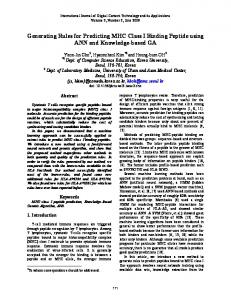
able to maintain 5 x 10³ CTLL20 viable cells (as evaluated in a trypan blue exclusion assay) was said to contain 1 U of IL-2. Results were expressed as arbitrary U/ml IL-2 secreted. We estimate one arbitrary unit to be equivalent to 0.25 U of rIL-2 from Biogen (Cambridge, MA). Cell Transfection. For reconstitution of CD2 expression on T11 - mutants, the full-length Tll cDNA (clone PB2) (16) was inserted into the Bam HI site of the pPink-2 vector (kindly provided by Dr. P Ohashi, Toronto, Canada), in which expression of the cDNA insert is under the control of the spleen focus-forming virus long terminal repeat (LTR), while the presence ofthe neomycin gene provides a selection marker for the antibiotic G418. Such generated plasmids were grown in Escherichia coli strain HB101 and amplified in the presence of chloramphenicol . Culture was converted to protoplasts and fused to the Tll - c cells in the presence of 40% polyethylene glycol 1500 as previously described (15). After 48 h, cells were plated at 30,000 cells/well in 96-well flat-bottomed microtiter plates in the presence of 2 mg/ml geneticin G418 (Gibco Laboratories, Grand Island, NY) . Resistant clones were selected for CD2 expression using an indirect immunofluorescence assay, and cells were maintained in culture in the presence of 0.5 mg/ml G418. Results Selection of CD3Ti+ CD2 - Variants of the J77 Cell Line. Variants of the jurkat cell line, clone J77-6.8 lacking the CD2 molecule, were obtained by mutagenesis, immunoselection, and cell sorting (see Materials and Methods). CD2 - colonies were screened by indirect immunofluorescence : 19 clones displaying the CD3 Ti + CD2 phenotype were obtained and three of these clones, termed Tll - a, Tll - b, and Tll - c, were randomly selected and used for further characterization . The phenotype of one of these CD2 - variants (T11 - b) is shown in Fig . 1. Antibodies directed to different epitopes of the CD2 molecule failed to react with the T11 - variants, while all three of these epitopes were found to be expressed on the J77 parental cell line. Furthermore, CD3 and Ti expression as judged by peak mean channel fluorescence intensity was clearly comparable onJ77 and Tll - clones, suggesting the presence of a similar number of TCR complexes with no discernable requirement for CD2 protein in CD3 Ti surface expression. An identical set of reactivities was observed for clones Tll - a and T11 - c (not shown). J77
Titb
anti-CD3
Phenotypic analysis ofJ77 and CD2 - cell lines. Reactivity ofdifferent mAbs with J77 and CD2 variant cells was assayed by indirect immunofluorescence using cytofluorimetric analysis . 104 cells were run per sample and the diagrams represent the number of cells (ordinate) vs . intensity offluorescence expressed in a logarithmic scale (abscissa). Reactivity of the different antibodies (thick line) is compared withbackground offluorescence obtained with an irrelevant antibody (thin line). FIGURE 1 .
anti Ti
anti J1 1 1
anti -T 11 2
anti T11 3 2 0 I Log f0 Relative Fluorescence
MOINGEON ET AL .
208 1
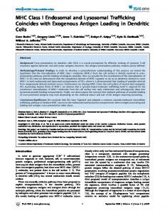
CD2 Molecules Are Undetectable on the Surface of CD2- Variants. As an alternative approach for the identification of CD2 molecules, immunoprecipitation experiments were performed on the J77 parental line, and on T11 - a and T11 - c clones . To this end, lysates from surface-labeled cells were immunoprecipitated either with purified anti T11 i or anti T3 antibody linked to protein A-Sepharose beads and immunoprecipitates washed and analyzed by SDS-PAGE . As shown in Fig. 2 a, antiT111 immunoprecipitates a 50-kD protein from J77 (lane C) . In contrast, even overexposure of the autoradiogram failed to reveal any corresponding band from T11 cells (lanes A and B) . Anti-T3 antibody precipitates the CD3 subunits on the three cell lines (lanes D-F) that appear as two bands of -27 and N21-kD from theJurkat cell line . Parallel immunoprecipitation conducted with the antiTi antibody on T11 variants and 177 cells established that a Ti molecule with similar biochemical characteristics was also present on these cells (our unpublished data). As a more quantitative means of assessing numbers of CD2 molecules on theJurkat variants, a radioisotope binding assay was developed using 1251-labeled anti-T11 1 antibody. As shown in Fig. 2 b, a high and saturable specific binding was observed on J77 cells. Scatchard analysis of the data indicated the presence of "14,000 sites per cell (not shown). By contrast, no specific binding was detected on T11 - c. Similar data was observed with T11 - a and T11 - b clones . Given the sensitivity of this binding method, we estimate that the T11 - mutants must express < 100 Tll molecules on their cell surface. Using an identical methodology with 1251-labeled antiT3 and anti-Ti antibodies, we determined that J77 and T11 - c each expressed
Absence of detectable CD2 protein on variant cell lines. (a)J77 and Tll- a and T11 - c cells were iodinated before lysis and immunoprecipitation with anti-T11 1 (lanes A-C) or antiT3 antibodies (lanes D-F) . Immunoprecipitates were analyzed in 12 .5% polyacrylamide gel in the presence of 5% 2-ME . Lanes A and D, Tll - a cells; lanes B and E, Tll - c cells; lanes C and F, J77 cells. (b) Binding assays were conducted with 12'1 anti-T11 I as described in Materials and Methods. Specific binding was obtained by subtracting nonspecific binding from total binding. Results are expressed as mean of a triplicate determination (SD