CE Update Submitted 11.12.09 | Revisions Received 12.23.09, 1.7.10, and 3.9.10 | Accepted 3.22.10
Microarray-Based Gene Expression Profiling for Molecular Classification of Breast Cancer and Identification of New Targets for Therapy
Rupninder Sandhu, MBBS,1,3,4 Joel S. Parker, MS,2,4 Wendell D. Jones, PhD,5 Chad A. Livasy, MD,1,3,4 William B. Coleman, PhD1,3,4 (1Department of Pathology and Laboratory Medicine, 2Department of Genetics, 3Program in Translational Medicine, 4UNC Lineberger Comprehensive Cancer Center, University of North Carolina, School of Medicine, Chapel Hill, NC 5Expression Analysis, Durham, NC ) DOI: 10.1309/LMLIK0VIE3CJK0WD
Abstract
Basal-like breast cancers represent approximately 15%–20% of all breast carcinomas, are aggressive, have variable responses to chemotherapy, and associated with poor clinical outcome. The molecular mechanisms governing the biological behavior of basal-like breast cancers are not well understood. Hence, it is difficult to predict which chemotherapeutics
are most likely to be effective and to determine appropriate management strategies for individual patients. Transcriptomic analysis may allow further stratification of basal-like breast cancers enabling prediction of 1) cancer recurrence after surgery; 2) likelihood of metastatic spread; 3) probable tissue sites for metastatic spread; and 4) responses to specific therapies and treatment modalities. Furthermore, careful
After reading this article, readers should be able to describe the basic features of the various molecular subtypes of breast cancer, with special emphasis on traits characterizing basal-like breast cancer. Readers should also understand the basic technical attributes of gene expression microarrays, and be able to discuss the advantages and disadvantages of using microarray-based gene expression profiling for making decisions
Breast cancer is a disease that is diverse in natural history, response to treatment, and patient outcomes. It remains the most common non-cutaneous female malignancy with an estimated 192,370 new cases in 2009 in the United States.1 Breast cancer-associated mortality is second only to lung cancer in the United States, with an estimated 40,170 deaths in 2009.1 Breast cancer is also the most commonly diagnosed female malignancy worldwide. According to the American Cancer Society, approximately 1.2 million women worldwide are diagnosed with breast cancer each year. Breast cancer is not a single disease, rather it represents a diverse spectrum of diseases including several distinct biological entities and subtypes. These subtypes are associated with specific morphological characteristics and
examination of microarray-based gene expression profiles may identify new molecular targets (or pathways) for the development of targeted therapeutics. Targeted therapies may prove to be more efficacious in basal-like breast cancer treatment than the cytotoxic chemotherapeutics currently employed. Keywords: AP breast, molecular diagnostics, genetics
related to the clinical management of basal-like breast cancers. In particular, readers should understand the molecular assays already in clinical use for breast cancer. Molecular Diagnostics exam 81002 questions and corresponding answer form are located after this CE Update on page 373.
different clinical outcomes.2-10 The molecular signatures of these breast cancer subtypes reflect not only the distinct biological features of these malignant neoplasms, but also predict their clinical behavior and responses to chemotherapy,11-14 with certain subtypes having better outcomes than others. To some extent, the observed variation in disease outcome among breast cancer patients reflects the successful identification of therapeutic targets for some subtypes and the development of effective targeted therapies. The molecular mechanisms contributing to the biological/ clinical behavior of basal-like breast cancer are not well understood. These aggressive cancers constitute 15%–20% of all breast carcinomas. They resist chemotherapy and are associated
Corresponding Author
Abbreviations
William B. Coleman, PhD
[email protected]
ER, estrogen receptor; PR, progesterone receptor; HER1 and HER2, human epidermal growth receptors; OS, overall survival; DFS, disease-free survival; PARP, Poly(ADP-ribosyl)ation polymerase; SSP, single sample predictor
364
LABMEDICINE ■ Volume 41 Number 6 ■ June 2010
labmedicine.com
CE Update
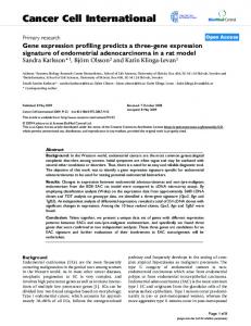
Figure 1_Differential expression of estrogen receptor (ER), progesterone receptor (PR), and human epidermal growth receptor 2 (HER2), among different subtypes of breast cancer. Breast cancer is classified into various subtypes based on differential immunohistochemical staining for ER, PR, HER2, HER1 (not shown), and cytokeratins (not shown). Panels A-D, luminal A breast cancer; Panels E-H, luminal B breast cancer; Panels I-L, HER2+ breast cancer; Panels M-P, basal-like breast cancer. Panels A, E, I, and M show H&E staining for each breast cancer subtype. Panels B, F, J, and N show immunostaining for ER and the results (ER+ or ER-) are indicated. Panels C, G, K, and O show immunostaining for PR and the results (PR+ or PR-) are indicated. Panels D, H, L, and P show immunostaining for HER2 and the results (HER2+ indicative of HER2 amplification or HER2-) are given.
with poor clinical outcomes, contributing disproportionately to breast cancer-related mortality.15 In this article, we discuss the possible use of microarray-based gene expression profiling to further examine basal-like breast cancers and to identify gene expression signatures that predict their clinical behaviors and responses to treatment. Transcriptomic analysis of basallike breast cancer may enable further subclassification within this molecular subtype, creating biological subgroups reflecting differences in 1) cancer recurrence after surgery; 2) likelihood of metastatic spread; 3) probable tissue sites for metastatic spread; and 4) responses to specific therapies and treatment modalities. Furthermore, careful examination of microarraybased gene expression profiles may identify new molecular targets (or pathways) for development of targeted therapeutics directed against biological subgroups of basal-like breast cancer. Targeted therapies may prove to be more efficacious in basal-like breast cancer treatment than the cytotoxic chemotherapeutics currently employed. Application of personalized and targeted therapies will improve long-term outcomes for patients with basal-like breast cancer and will lessen the burden associated with over-treatment of these patients. labmedicine.com
Breast Cancer: A Heterogeneous Entity Breast cancer is a diverse disease with a number of morphologic subtypes. Invasive ductal carcinoma is the most common morphological subtype, representing 80% of the invasive breast cancers. Invasive lobular carcinoma is the next most common subtype, representing approximately 10% of invasive breast cancers. The less common subtypes of the invasive breast cancers include mucinous, cribriform, micropapillary, papillary, tubular, medullary, metaplastic, and inflammatory carcinomas. Representative examples of invasive ductal carcinomas are shown in Figure 1. Routine subclassification of invasive ductal carcinomas is accomplished by immunostaining tumor tissues for estrogen receptor (ER), progesterone receptor (PR), human epidermal growth receptors (HER1 and HER2), and various cytokeratins. The differential expression of ER, PR, and HER2 in different subtypes of breast cancer based upon immunohistochemical staining is shown in Figure 1. The differential expression of these protein biomarkers is used as an immunohistochemical surrogate for gene expression analysis to determine molecular subtype. Approximately 70%–75% of invasive breast cancers June 2010 ■ Volume 41 Number 6 ■ LABMEDICINE
365
CE Update express the ER positive cancers (ER+). Collectively, the ER+ malignant neoplasms are classified as luminal cancers. These cancers are further subclassified into luminal A and luminal B subtypes based on their HER2 status and proliferation rate. The majority of ER+ tumors also express PR. The presence of normal PR levels suggests an intact ER signal transduction pathway in the breast cancer cells, and PR expression typically follows the ER expression pattern. The ER- breast cancers are subclassified as HER2+ and basal-like based on the HER2 overexpression/gene amplification, basal cytokeratin expression, and EGFR (HER1) expression. An immunohistochemical staining proxy based on 5 biomarkers classifies breast cancers into the major subtypes (shown schematically in Figure 2): 1) ER+ are subclassified into luminal A (ER+, PR+, HER2-) and luminal B (ER+, PR+, HER2+); 2) ER negative cancers (ER-) are subclassified into triple-negative breast cancer (ER-, PR-, HER2-) and human epidermal growth factor receptor 2-positive (ER-, PR-, HER2+); and 3) unclassified cancers (negative for all 5 markers).2-4,16 Basal-like breast cancers are distinguished from other triple-negative breast cancers (ER-, PR-, HER2-) by expression of cytokeratin 5/6 and/or EGFR. There is no international consensus on the criteria used to define cancers as basal-like in formalin-fixed, paraffin-embedded tissues. Therefore, the term basal-like is not yet routinely used in clinical practice. Rather, the basal-like breast cancers are contained in the triplenegative classification. Breast cancers, like most epithelial cancers, are associated with better treatment and survival outcomes when diagnosed at an early stage. However, outcomes of early stage breast cancers differ depending upon the molecular subtype (Figure 3). In general, the ER+ breast cancer subtypes (luminal A and luminal B) exhibit a good prognosis and excellent long-term survival (approximately 80%–85% 5-year survival), while the ER- subtypes (HER2-positive and basal-like) are difficult to treat and are associated with poor prognosis (approximately 50%–60% 5-year survival). The ability of patients with ER+ breast cancers to survive their disease reflects the availability of effective targeted therapy in the form of anti-estrogen treatment (eg, tamoxifen). However, among the ER+ breast cancers, the luminal B neoplasms are associated with a significantly worse prognosis than luminal A subtype4 (Figure 3). This difference in outcome is partly due to variations in response of ER+ subtypes (luminal A and luminal B) to anti-estrogenic treatment.17 Targeted therapy of HER2 overexpressing breast cancers, (luminal B or HER2positive [ER-] neoplasms) with trastuzumab (herceptin), either concurrent or sequential with adjuvant chemotherapy has improved survival for these breast cancer subtypes.18 Basal-like breast cancers are characterized by autonomy of growth that is independent of expression of hormone receptors. Since these cancers lack the appropriate targets for the drugs like tamoxifen (targeting ER) and trastuzumab (targeting HER2), patients with these cancers do not derive benefit from these drugs. Basal-like breast cancers are associated with overall poor prognosis and shorter long-term survival. The poor clinical outcomes associated with basal-like breast cancer reflect the fact that these cancers show variable response to chemotherapy or recur following therapy. Lack of identification of “druggable” targets in basal-like breast cancers and poor prognosis makes the identification of molecular signatures and therapeutic targets in these cancers to be of utmost significance. No widely available targeted therapies for this breast cancer subtype have been developed to date, although phase II studies of Poly(ADP-ribosyl)ation polymerase (PARP) inhibitors have shown promising results.19 366
LABMEDICINE ■ Volume 41 Number 6 ■ June 2010
Figure 2_Schematic illustrating various breast cancer subtypes. The blue and pink rectangles group the subtypes based on the expression of ER/PR, positive in the blue (Luminal A and Luminal B) and negative (HER2+ and basal-like) in the pink. The central grey rectangle (with black outline) indicates the presence of HER2 amplification in Luminal B and HER2+ subtypes.
Figure 3_Survival plot of 294 breast cancer patients. A Kaplan-Meier survival plot of overall survival corresponding to 294 breast cancers from the publicly available UNC database is shown grouped by molecular subtype. The P-value was calculated using the Log-rank test. Details of these 294 samples along with clinical annotation can be found at https://genome.unc.edu/pubsup/breastGEO/.
Basal-Like Breast Cancer Discovery of Basal–Like Breast Cancers The basal breast cancer subtype was first described in studies based on immunohistochemistry.20-23 These cancers are designated basal-like because they exhibit some cellular characteristics associated with the basal myoepithelial cell layer, such as expression of cytokeratins 5/6, 14, or 17, vimentin, and laminin, but these tumors are clearly not derived from myoepithelial cells.24-26 The basal-like breast cancer subtype was rediscovered following the application of microarray-based gene expression labmedicine.com
CE Update profiling to breast cancer classification.2-7,9 That the basal-like breast cancers were identified independently by 2 different methodologies indicates strongly that these cancers represent a distinct biological entity. Basal-like breast cancers are best classified through gene expression profiling.2-7,9 However, in routine clinical practice, immunohistochemistry has become the surrogate for the gene expression analysis for diagnosis of basallike breast cancers (Figure 1). Correctly classifying these cancers significantly impacts clinical decisions and research efforts. In the clinic, there is a need to correctly identify breast cancer subtypes for prognostication purposes in relation to individual patients and for decision-making related to appropriate treatment course. On the other hand, in the research environment, correct breast cancer subclassification is essential to ensure investigations expand our understanding of the biological basis for the behavior and characteristics of these cancers. Association With Risk Factors The development of basal-like breast cancer is associated with distinct risk factors, including early-onset menarche, younger age at first full-term pregnancy, high parity combined with lack of breast feeding, and abdominal adiposity (based upon waist-hip ratio).27 These breast cancers are overrepresented among patients of certain age and ethnic groups, and are frequently associated with certain genetic mutations. Specifically, basal-like breast cancer is overrepresented among premenopausal, African-American women.11 However, these cancers can and do affect women of every age and ethnic group.27 The differences in distribution of basal-like breast cancer by age and race can be partially attributed to variations in the distribution of the risk factors described and to other risk factors (eg, waist-hip ratio, use of lactation suppressants, and overexpression of leptin receptor).27 In addition, basal-like breast cancer occurs more frequently among hereditary breast cancer patients harboring germ-line BRCA1 mutation.28 Foulkes and colleagues showed that 17/72 triple-negative breast cancers harbored a BRCA1 mutation, and 88% (15/17) of these expressed the basal-like phenotype.29 Likewise, Sorlie and colleagues observed that 100% (18/18) of breast cancers from patients carrying BRCA1 mutations clustered within the basallike subgroup.4 However, the other molecular subtypes of breast cancer can be associated with BRCA1 mutations as well. Morphological Features Morphologically, basal-like breast cancers are characterized by the presence of central necrotic zones, pushing borders, and conspicuous lymphocytic infiltrate.30-34 The presence of metaplastic elements3,30-32 and medullary/atypical medullary features31,32,35 are more prevalent in basal-like breast carcinomas than in other types of breast cancer. Recent studies have shown that more than 90% of metaplastic breast carcinomas,3 as well as the majority of medullary carcinomas,35,36 exhibit a basal-like phenotype. Basal-like breast cancers are aggressive, with high rates of cellular proliferation, high histological grade, and extremely poor clinical outcomes.3,4 These factors combine to account for the disproportionate contribution of basal-like breast cancer to breast cancer mortality. It has been suggested the high level of cellular proliferation observed in these neoplasms might account for the over-representation of basal-like breast cancers among the so-called interval breast cancers (the cancers arising between annual mammograms). labmedicine.com
Clinical Behavior of Basal-Like Breast Cancers Currently, there is no consensus on the immunohistochemical criteria for the diagnostic classification of basal-like breast cancers. Studies have shown the profile constructed using ER-/ PR-, HER2-, CK5/6+, and/or EGFR+ is 100% specific but only 55% to 76% sensitive.37 Breast cancers that are ER-/PR-/HER2are broadly classified as triple-negative neoplasms (Figure 1). The triple-negative breast cancers include most (or all) basal-like breast cancers.38 Interpreting the percentage of positive cells and intensity of immunohistochemical staining is subjective. Variability in immunostaining techniques and procedures is a concern as well. Hence, standardization and/or automation of immunostaining procedures and interpretation to remove technical and subjective variation will benefit this analysis in the clinical laboratory. The low sensitivity associated with the classification of basal-like breast cancers using immunohistochemical staining may indicate that these cancers are much more heterogeneous than previously thought. Gene expression profiling-based molecular classification of breast cancers predicts the general clinical behavior of breast cancers corresponding to the different molecular subtypes. Microarray studies show the basal-like breast cancers express a common gene expression signature, and these cancers are associated with an extremely bad prognosis.3 Among the patient cohort examined in the initial study of this type, 100% of the patients with basal-like subtype succumbed to their disease within 4 years of diagnosis.3 Basal-like breast cancers respond to preoperative (neoadjuvant) chemotherapy.39,40 However, despite the observation of pathologic complete response in many patients, these individuals exhibit poor long-term survival. The poor survival outcomes among these patients, despite response to chemotherapy, may be related to a higher likelihood of relapse in individuals where pathologic complete response was not achieved.40 The malignant neoplasms constituting the basal-like breast cancer subtype are not biologically homogeneous. For example, in 1 study unsupervised hierarchical clustering within 43 cytokeratin-14 positive (basal-like phenotype) tumors revealed 4 clusters, and 1 of these displayed a worse prognosis than the other 3, strongly suggesting intra-subtype heterogeneity.41 Variable prognosis within the basal-like subtype has also been reported by other groups.42,43 Rakha and colleagues divided the basal-like breast cancers into those with a dominant basal pattern (>50% of cells are positive for cytokeratin 5/6 and 14) and the remaining basal cancers (