Available online at www.sciencedirect.com
ScienceDirect Agriculture and Agricultural Science Procedia 6 (2015) 324 – 331
“ST26733”, International Conference "Agriculture for Life, Life for Agriculture"
Microbial population dynamics in presence of lactococcal bacteriophage during ripening of traditional raw milk Romanian cheese Mara Georgescua*, Mimi Dobreaa, Dragos Georgescub a
University of Agronomic Sciences and Veterinary Medicine of Bucharest, 59 Mărăúti Blvd, District 1, Bucharest 011464, Romania b Carol Davila University of Medicine and Pharmacy of Bucharest, Eroii Sanitari 8 Blvd, Bucharest, 050474, Romania
Abstract A massive Lactococcus lactis ssp.lactis c2 and e72 phage contamination of raw milk burduf cheese was revealed in all analyzed samples. Contamination with c2 phage specific for Lactococcus lactis ssp.lactis C2 is lower than the contamination induced by e72 phage. The contamination level with E.coli and coliforms in the core of burduf cheese was initially high, while the initial Staphylococcus spp. contamination was not high enough to pose a threat through toxin production. During the ripening period of the analyzed cheese, there was a high statistical significant decrease in E. coli, coliforms and staphylococci counts. This decrease was highly unexpected according to the scientific literature that indicates low lactococcal counts and low microbiological quality in bacteriophage contamination cases. The presence of wild bacteriophages, known to reduce the number of lactic acid bacteria, preventing the inhibition effect of the latter on contaminating bacteria growth, was proven not to induce a poor hygiene level of the raw milk burduf cheese, even if the initial contamination was considered to be high. This study is an example of natural, persistent and statistical significant lactococcal resistance to contaminating bacteriophages. The results are also scientifically significant due to the atypical time resistance of the microbiological balance between the contaminating bacteriophages and cheese bacterial microflora with positive consequences over cheese quality. © 2015 2015 The Authors. Published © Published by byElsevier ElsevierB.V. B.V. This is an open access article under the CC BY-NC-ND license (http://creativecommons.org/licenses/by-nc-nd/4.0/). Peer-review under responsibility of the University of Agronomic Sciences and Veterinary Medicine Bucharest.
Peer-review under responsibility of the University of Agronomic Sciences and Veterinary Medicine Bucharest Keywords: lactococcal bacteriophage; contaminating microorganism; microbiological quality; raw milk cheese.
* Corresponding author. Tel.: +4-0751-109-241; fax: + 4-021-318-25-67. E-mail address:
[email protected]
2210-7843 © 2015 The Authors. Published by Elsevier B.V. This is an open access article under the CC BY-NC-ND license (http://creativecommons.org/licenses/by-nc-nd/4.0/). Peer-review under responsibility of the University of Agronomic Sciences and Veterinary Medicine Bucharest doi:10.1016/j.aaspro.2015.08.086
Mara Georgescu et al. / Agriculture and Agricultural Science Procedia 6 (2015) 324 – 331
1. Introduction Bacteriophage contamination still challenges the dairy industry leading to economical loss and compromising the safety of the final product by interference with the starter bacteria (Hicks et al., 2000; Hicks, 2001; Hicks, 2004; Atamer et al., 2010; Garneau and Moineau, 2011; Verreault et al., 2011; Mahony et al., 2012). Advanced research is being conducted in order to enhance lactococcal resistance to phage contamination or to select naturally resistant strains (Van Pijkeren and Britton, 2012; Garneau et al., 2012). Yet, recent studies proved that phage eventually overcome resistance and so far no long term protection against phage has been achieved (Mahony et al., 2012). A massive phage contamination of raw milk cheese could prevent the self-sterilizing natural process, thus compromising the hygiene level of traditional milk products made of unpasteurized milk, as no heat treatment is applied (Madera et al., 2004). Therefore, the present study aimed to investigate phage contamination of a traditional Romanian raw milk cheese assortment and evaluate the correlation between bacteriophage and the fluctuations of certain contaminating bacteria during ageing. 2. Materials and methods The investigation trials implied the use of burduf cheese, a popular traditional Romanian cheese, manufactured from a mixture of raw sheep’s and cow’s milk and wrapped in natural membrane, by different local producers. Research was conducted at the University of Kentucky, Lexington, KY, USA. Ten samples of cheese up to 250g each, were transported in refrigeration bags, pending further storage and analysis at the premises were the research was undertaken (Department of Animal Sciences, University of Kentucky, 410 W. P. Garrigus Bldg., Lexington, KY 40546, USA). Investigations were made at 20, 64 and 92 days of ripening, within three analysis sets, I-III, respectively. The samples were tested for lactococcal bacteripohage, E.coli, coliforms and Staphylococcus spp. contamination; also, the Lactococcus spp. counts were determined. 2.1. Bacteriophage isolation was performed using the double layer technique and pure cultures of lactic acid bacteria (LAB), as described by Hicks (2001). The method consists in preparation of a high viscosity bottom agar and a low viscosity top agar. The LAB culture and a fraction of filtrated and diluted sample are inoculated in the sterilized top agar. This mixture is dispensed into Petri dishes over the bottom agar, incubated and examined for bacteriophage plaques. 2.1.1. Sample preparation and obtaining of serial dilutions. The natural membrane of burduf cheese was removed and samples of the outer and inner cheese layers were harvested using autoclaved cheese samplers. The samples were suspended in monobasic phosphate (phosphate buffer with pH adjusted to 7) and 4 serial dilutions were prepared (D1-D4). 2.1.2. Bottom and top agar preparation was performed according to the method described by Douglas et al. (1974), using BactoTM-agar (Becton, Dickinson and Company, Oxford, England), M17 powder (Oxoid Ltd., Hampshire, England) and lactose (BD DifcoTM, Becton, Dickinson and Company, Oxford, England). 2.1.3. Isolation and enumeration of phage were performed using the method described by Hicks (2001). Lactococcus lactis ssp.lactis C2 and E72 active cultures (1 x 10⁹ cfu mL-1) were obtained from University of Kentucky (Lexington, KY, USA). 2.2. Isolation and enumeration of E.coli, coliforms and Staphylococcus spp. E.coli and coliforms enumeration was performed using 3MTM PetrifilmTM E.coli/Coliform Count Plate (PetrifilmTM EC, St. Paul, Minnesota, USA). Plating, incubation and interpretation (counting on standard colony counter) were performed according to NMKL Method (147.1993). Staphylococcus aureus enumeration was performed using 3MTM PetrifilmTM Staph Express Count System (St. Paul, Minnesota, USA) and Petrifilm Staph Express disk (AOAC Official Method of Analysis 2003.08, for dairy foods) (www.eoma.aoac.org). 2.3. Lactococcus spp. isolation and enumeration was performed using M17 agar (Oxoid Ltd., Hampshire, England) (Terzaghi and Sandine, 1975) containing 0.5% lactose (LM17). Dilutions of the samples were directly plated on LM17. The cultures were incubated at 30ºC for 48 h. 2.4. Data analysis was based on the method “within-subject t-test for a mean” (DeCoster, 2006). After gathering all data regarding E. coli, coliforms and staphylococci counts in the core and outer layers of the analyzed cheese
325
326
Mara Georgescu et al. / Agriculture and Agricultural Science Procedia 6 (2015) 324 – 331
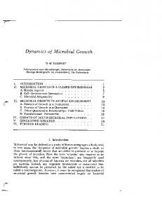
samples, microbial populations decrease was calculated for the ripening time considered. Thus the decrease I (DI) was calculated by subtraction of log cfu g-1 values of the second set of values (64 days of ripening) from the log cfu g-1 values of the first set of data (20 days of ripening). Similarly, decrease II (first set-third set) was then calculated. Data analysis aimed to assess the statistical significance of the decrease in bacterial counts from the first set of determinations by comparison with the second and the third set. The research hypothesis for this study stated that there is a significant decrease in E. coli, coliforms and staphylococci counts during the ripening period. This would mean that the average decrease of the microbial counts would be greater than zero (one tailed hypothesis). The null hypothesis stated that there is no significant decrease in microbial counts (there is no improvement in microbiological quality of cheeses), thus the average decrease values of microbial counts should tend to be equal to zero. Calculating the standard deviation of the microbial counts decreases from the mean decrease for each microorganism was performed according to DeCoster (2006). The t-test statistic was calculated in order to determine whether the mean microbiological decrease is different from the constant value considered to be zero (according to formulae described by DeCoster, 2006). The P values were obtained using standard calculators (www. easycalculation. com; www.statstodo.com; www. statisticsmentor.com), and the significance threshold was considered p< .05. 3. Results and discussions 3.1. Lactococcus lactis ssp.lactis c2 and e72 isolation and enumeration. After 19 hours of incubation, all phages were enumerated, the counts being surprisingly high (Lactococcus lactis ssp.lactis c2 average contamination of 7.322 log pfu g-1) (Figure 1). According to scientific literature, curd and cheese are generally tested for bacteriophage only if acid production has failed during cheese manufacture. Therefore, considering that all cheese samples indicated regular sensory characteristics, no phage contamination was expected. Yet, despite this presumption, bacteriophage Lactococcus lactis ssp.lactis c2 and e72 contamination levels of burduf cheese proved to be considerably high. Both Lactococcus lactis ssp.lactis c2 phages concentration of 1.3x10⁶ pfu g-1 cheese and e72 contamination level of 1.7x10⁷ pfu g-1 are considered to be high concentrations, given the fact that the threshold for phage titres in fermentation vessels is 106- 107 pfu mL-1 (at this concentration, lactic acid production is significantly reduced and the organoleptic properties of the product are altered) (McGarth and Douwe van Sinderen (a), 2007). The analyzed samples of raw milk cheese appeared to contain a higher number of e72 phage in comparison with c2 phage. All samples revealed higher concentrations of e72, the average contamination level being approximated at 8.985 log pfu g-1 cheese (Figure 2). On some plates, white bacterial colonies grew in the middle of the phage plaques. The white bacteria colonies consist of mutant lactic acid bacteria, that are resistant to the c2/e72 phage responsible for lysis. Due to the different morphology of phage plaques formed on the same enumeration plate, noticeable for e72 and c2 bacteriophage, the presence of more than one phage strain may be suspected.
Fig. 1. Enumeration of c2 phage in sample 1 (STD – standard; D1-D4 – dilution 1-4)
Mara Georgescu et al. / Agriculture and Agricultural Science Procedia 6 (2015) 324 – 331
Fig. 2. Comparison lysis intensity for Lactococcus lactis ssp.lactis c2 (B, D, F, H, dilutions 1-4) and e 72 phages (A, C, E, G – dilutions 1-4)
3.2. E. coli, coliforms and S. aureus isolation and enumeration in burduf cheese. After 10 days of ripening, the first set of investigations (I) concerning the degree of contamination with E.coli, coliforms and S.aureus of burduf cheese revealed that the initial contamination of the cheese before ripening can be considered high (Figure 3). S. aureus was mostly present in the outer layer of the cheese chunks, with an average of 2.52 log cfu g-1 (not enough to produce toxins that would induce food poisoning). Only four samples were found to have staphylococci in the core of the chunk. The average E.coli and coliforms contamination was higher in the core of the cheese chunk: 3.84 log cfu g-1 (E. coli) and 4.29 log cfu g-1 respectively (coliforms), compared to the outer layer of burduf cheese: 3.06 log cfu g-1 (E. coli) and 3.56 log cfu g-1 respectively (coliforms). Microbial population decrease is more intense in the core of the cheese samples, for all samples. The presence of S. aureus in the outer layer of the cheese chunk and its absence in the core of the product suggest that contamination of burduf cheese with this bacteria occurred most probably after milking, during processing. Yet, higher concentrations of E. coli and coliforms in the core of the cheese chunk indicate either a poor milking hygiene, or an infectious mastitis. E. coli, coliforms and staphylococcal count dynamics during ripening, indicated bacterial decrease both in the core and in the outer layer of analyzed samples (Figure 3).
Figure 3. Bacterial contaminants counts decrease and fluctuation of Lactococcus spp. counts during ripening (I, II, III / set of analysis)
Decrease values of bacterial counts were calculated for the three chosen intervals, as explained previously (Materials and Methods section), both for the core and the outer layer of the cheese samples. The values of the microbial count decrease varied within the range 0.1- 5.56 log cfu g-1. Moreover, all values were positive, proving there were no rising shifts of microbial counts between the considered time intervals in any of the samples. The statistical significance of the microbial decrease was calculated for each of the considered contaminant. After running the results through the data analysis using the method of “within-subjects t test”, we concluded that the null hypothesis (stating that there is no significant decrease in microbial counts, meaning that there is no improvement in microbiological quality of cheeses) must be rejected, and that the research hypothesis is true. Therefore, according to this study, there is a high statistically significant (p values ranging from less than 0.001, to p = 0.004) decrease in
327
328
Mara Georgescu et al. / Agriculture and Agricultural Science Procedia 6 (2015) 324 – 331
E. coli, coliforms and staphylococci counts for the cheese samples analyzed during the ripening period (table 1). Even though there are numerous available articles highlighting the potential benefits for food industry of benefic bacteriophages such as coliphages, staphylococcal phages etc., which proved to successfully improve food safety of dairy products (Sillankorva et al., 2012a; Goodridge and Bisha, 2011), however, there is no sufficient information related to the direct or indirect effect on food safety of lactococcal phage present in finished dairy products. Nevertheless, the effects of lactococcal phage on lactococcal starter bacteria are well known, usually phage contamination being associated with low Lactococcus spp. counts; high phage levels (107 pfu mL-1 and above) in the curd lead to the absence of starter lactococci as a result of intense lysis of starter by phage (Madera et al., 2004; Neve et al., 2005; Lortal and Chapot-Chartier, 2005). On the other hand, the antagonistic effect of lactic acid bacteria on contaminant germs and some pathogens growth is extremely well documented in the literature of the past decades (Hugas, 1998; Stiles, 1996; Parada et al., 2007; Portella et al., 2009; Yesillik et al., 2011; Kousta et al., 2010). Therefore, by destroying the lactic acid bacteria, which are responsible for limitation of pathogen growth in dairy products, the presence of lactococcal phage in finished dairy foods would obviously be associated with a poor bacteriological quality. As a consequence, considering the rich content of wild bacteriophage of analyzed samples and the possible presence of more strains for each type of phage, an obvious decrease in the hygiene and safety levels of the final product was expected (McGarth and Douwe van Sinderen (b), 2007), especially because the analyzed cheese sample is a raw milk cheese. Sanitary quality of sheep milk is often poor when obtained by local producers in rural areas. Moreover, considering that traditional cheeses are manufactured using unpasteurized milk, the high initial contamination level of milk would be associated with a compromised product with respect to its safety for consumption. Phage infection of the lactic acid bacteria is known to contribute on one hand to a reduction of the cheese yield and on the other hand to preventing bacteria competition that would limit contaminant microorganisms’ growth. Despite the poor hygiene level of the cheese before ripening and the considerable high concentration of bacteriophage, the investigation trials performed in order to monitor bacteria level throughout the ripening period, indicated a significant decrease of bacteria. Also, due to the fact that the initial S.aureus contamination level was not high enough to allow toxin formation, the loss of this microorganism by the end of the ripening period proved that the product became safe for consumption after two months of ageing. Considering these findings, it is obvious that the self-sterilization process of the cheese without any artificial interventions, associated with a natural ripening, was not disturbed by the presence of phage. Table 1. Data analysis regarding the decrease of E.coli, coliforms and Staphylococcus spp. counts in the core and the outer layer of the cheese samples
E.coli
Core Outer layer
Coliforms
Core Outer layer
Staphylococci
Core Outer layer
D1* D2** D1 D2 D1 D2 D1 D2 D1 D2 D1 D2
N
Average
Standard deviation (sd)
One-tailed t test value
P value
10 10 10 10 10 10 10 10 4 4 10 10
( ) 2.266 3.737 1.091 2.24 2.065 4.192 1.343 2.542 1.4875 1.4875 1.536 2.227
0.609028 0.71997 0.718122 0.850059 0.847431 1.20244 1.06801 1.098977 0.168597 0.168597 1.001978 1.012017
11.764 16.4123 4.803 8.3322 7.705 11.0235 3.9761 7.3138 27.8977 27.8977 4.8472 6.9581
< 0.001 < 0.001 0.0003 < 0.001 < 0.001 < 0.001 0.0013 < 0.001 < 0.001 < 0.001 0.0041 0.0011
*D1=Decrease 1: I-II; **D2=Decrease 2: I-III.
3.3. Lactococcus spp. isolation and enumeration. The results of Lactococcus spp. enumeration indicated a massive presence of lactococci in all cheese samples (7.38 – 11.07 log cfu g-1), as revealed in figure 4. The average lactococcal content was higher in the outer layer than in the core of the cheese samples, throughout the examination period (average 9.92 log CFU g -1 in the outer layer versus 9.02 in the core of the samples for the first set of analysis; set II: 10.62 versus 9.88 average log CFU g -1; set III: 9.65 versus 8.62 average log CFU g -1 in the outer layer and core, respectively).
Mara Georgescu et al. / Agriculture and Agricultural Science Procedia 6 (2015) 324 – 331
329
Fig. 4. Enumeration of Lactobacillus spp. on Difco™ Rogosa SL Agar: A-C – dillutions 1-3, sample 1; D-F – dillutions 1-3,sample 2.
This study shows a rising shift with an average of 0.865 log cfu g-1 in the core and 0.699 log cfu g-1 in the outer layer of analyzed samples, followed by a slight decrease (average of 1.261 log cfu g-1 for both layers) within the next 4 weeks of ripening. The level of Lactococcus spp. at the end of the ripening period was in average 8.625 log cfu g-1 in the core and 9.357 log cfu g-1 in the outer layer. There may be a connection between the fact that the rising shift of lactococci is more intense in the core of the cheese samples on one hand, and the fact that it was noticed that bacterial decrease is also more intense in the core, on the other hand. Nevertheless, a higher lactococci level in the outer layer of cheese samples at the end of the ripening is contradictory with the bacterial dynamics, which indicated less pronounced decrease. Another interesting connection may be seen between the rising shift of Lactococcus spp. population during the first 54 days of ripening, on one hand, and the fact that bacterial decrease is more intense during that same interval, on the other hand. Also, the decrease of lactococci population during the remaining ripening period may be associated with the less intense bacterial decrease. Regarding the Lactococcus genus dynamics during traditional or artisanal cheese ripening, most authors reported a rising shift during the first 7-8 days of ripening, followed by an average decrease of up to 1 log cfu g-1 by the end of the first ripening month (Vasek et al., 2013; Pesic-Mikulec and Jovanovic, 2005; Mas et al., 2002). Unlike the usual findings reported by literature, this study shows first a rising shift both in the core and in the outer layer of analyzed samples, during the first month of the ripening, followed by a slight decrease within the next 30 days of ripening. It is noticeable that the lactococcal count at the end of the ripening time considered (92 days) was not much lower than the initial values, unlike the findings reported by the scientific literature. Also, these results are highly unexpected considering the considerably high lactococcal bacteriophage contamination. The decrease of contaminating bacteria number in the background of high phage concentration may be associated with a natural phage resistance of natural lactic acid bacteria present in cheese. This theory is also supported by the appearance after only 16 hours of incubation of mutant bacteria in the culture used for phage enumeration. The natural lactic acid bacteria could be adapted and resistant to the wild strains of phage that contaminate the sample of traditional cheese that was subjected to research. Overcoming of the phage problem remains a challenge for the dairy science and technology, as documented by the numerous articles approaching this issue. Scientific literature provides significant volume of data related to different methods lactococcal phages inactivation (Lavigne et al., 2009; Mills et al., 2010; Guglielmotti et al., 2011). In addition to physical and chemical barriers encountered in dairy industry facilities, phage-resistant starter cultures are often mentioned (Weimer et al., 1993; Viscardi et al., 2003). Also, there are many recent studies revealing phage resistance in lactococcal starters, such as the work of Roces et al. (2012), or the studies of natural anti-phage systems of Garneau et al. (2012) and Deveau et al. (2010). Moreover, in order to achieve enhanced lactococci resistance, different genome engineering strategies have been recently developed, thus being possible to generate point mutations for resistance in this type of bacteria without antibiotics or other means of selection (Van Pijkeren and Britton, 2012; Van Pijkeren et al., 2012). Therefore, in the light of these continuous preoccupations, the findings in this article may pave the way to a better understanding of natural phage resistance.
330
Mara Georgescu et al. / Agriculture and Agricultural Science Procedia 6 (2015) 324 – 331
Even though natural phage resistance was noticed as early as 1976, and at that time the first attempts of using phage resistant mutants in dairy industry were communicated in studies such as the one published by Limsowtin and Terzaghi (1976) and despite the numerous forms of natural and induced resistance to lactococcal phages, there is a well known phage capability of overcoming these resistance systems, thus complete protection against them on a long term is known not to be achievable (Boucher et al., 2000). A recent example of such scientific findings is the article of Mahony et al. (2012), which reveals that protection against lactococcal phages is limited and while the 936-type phages appear to be sensitive to the majority of lactococcal anti-phage strategies, phage mutations occur rapidly and with relative ease, to overcome resistance and thus persisting in the dairy facilities environment, leading to phage related problems reoccurring. It is generally agreed throughout the scientific literature regarding phage related quality and safety issues in the dairy industry (Garneau and Moineau, 2011), that despite all efforts to find or create new anti-phage mechanisms, the industrial use of phage resistant bacteria will eventually lead to the emergence of phage mutants, able to overcome resistance systems. Nevertheless, the findings in this study are in contradiction with the scientific literature conclusion because, in addition to the natural resistance, the environment in which the cheeses were manufactured was used for this purpose for generations, long enough to allow the eventual overcoming of a resistance system. This is the reason why the results of this study are scientifically significant, because while extensive literature examples show increased difficulty in overcoming bacteriophage problems, a particular traditional raw milk cheese, proved significant and consistent bacteriophage resistance of lactococcal flora and good microbial quality. 4. Conclusions The contamination of raw milk cheese samples with e72 and c2 phage proved to be massive. Even though the initial contamination level of all cheese samples was important, during the ripening period there was a highly statistical significant (P from< 0.001, to = 0.004) decrease in E. coli, coliforms and Staphylococcus spp. counts. In addition, the massive phage contamination did not influence the lactococci levels of cheese, which continued to remain high throughout ripening. Microbial decrease and important lactococci levels are highly unexpected findings according to the scientific literature, which indicates low lactococcal counts and low microbiological quality in bacteriophage contamination cases. The presence of wild bacteriophages, known to reduce the number of lactic acid bacteria, preventing the inhibition effect of the latter on contaminating bacteria growth, was proven not to induce a poor hygiene level of the raw milk burduf cheese, even if the initial contamination was considered to be high. This study could be an example of natural, persistent and significant lactococcal resistance to contaminating bacteriophages, which is worth studying for further understanding of mechanisms that lead to the development of such a time resistant microbiological balance with positive consequences over cheese quality. Acknowledgements This research was supported by USAID – USDA scholarship, within FEP, through the University of Kentucky, Department of Animal and Food Sciences, Kentucky, USA. Our gratitude goes to Professor C.L. Hicks, for his guidance and persistent help and to Professor P. Jelen, for his valuable and knowledgeable advice. References Atamer, Z., Dietrichb, J., Neveb, H., Hellerb, K.J., Hinrichsa, J., 2010. Influence of the suspension media on the thermal treatment of mesophilic lactococcal bacteriophages. International Dairy Journal 20 408–414. Boucher, I., Emond, E., Dion, E., Montpetit, D., Moineau, S., 2000. Microbiological and molecular impacts of AbiK on the lytic cycle of Lactococcus lactis phages of the 936 and P335 species. Microbiology. 2000 Feb; 146 ( Pt 2):445-53. Caeser, A., Portella, F., Karp, S., Newton, Scheidt, G., Woiciechwski, A.L., Parada, J.L., Soccol, C.R., 2009. Modelling antagonic effect of lactic acid eacteria supernatants on some pathogenic bacteria. Brazilian Archives of Biology and Technology, [online]. 2009, vol.52, pp. 29-36. DeCoster, J., 2006. Testing Group Differences using T-tests, ANOVA, and Nonparametric Measures. Retrieved 04.15.2013, from http://www.stat-help.com/notes.html. Deveau, H., Garneau, J.E., Moineau, S., 2010. CRISPR/Cas system and its role in phage-bacteria interactions. Annual Review of Microbiology, 2010;64:475–493. Douglas, J., Qanber-Agha, A., Phillips, V., 1974. Medium for the propagation and assay of lactic and other phages. Lab. Pract. 23:3-4. Garneau, J.E., Moineau, S., 2011. Bacteriophages of lactic acid bacteria and their impact on milk fermentations. Microbial Cell Factories, 10 (Suppl 1):S20.
Mara Georgescu et al. / Agriculture and Agricultural Science Procedia 6 (2015) 324 – 331 Garneau, J.E., Dupuis, M.E., Villion, M., Romero, D.A., Barrangou, R., Boyaval, P., Fremaux, C., Horvath, P., Magadán, A.H., Moineau, S., 2010. The CRISPR/Cas bacterial immune system cleaves bacteriophage and plasmid DNA. Nature. 2010; 468:67–71. Goodridge, L.D., Bisha, B., 2011. Phage-based biocontrol strategies to reduce foodborne pathogens in foods, Bacteriophage. 2011 May;1(3):130137. Guglielmotti, D.M., Mercanti, D.J, Reinheimer, J.A, Quiberoni, A.L., 2011. Efficiency of physical and chemical treatments on the inactivation of dairy bacteriophages, Frontiers in Microbiology, 2011;2:282. Hicks, C.L., Clark-Safko, P.A., Surjawan, I., O’Leary, J., 2004. Use of bacteriophage-derived peptides to delay phage infections, Elsevier, Food Research International 37, 115-122; Hicks, C.L., inventor, 2001. Cheese making with bacteriophage resistant bacteria, University of Kentucky, assignee, Pat. No. 6297042, Oct. 2; Hicks, C.L., Onuorah, C.E., Surjawan, I., 2000. Use of hydrolysed whey peptide to inhibit culture agglutination, Journal of Dairy Science, 83, 1196-1202; Hugas, M., 1998. Bacteriocinogenic lactic acid bacteria for biopreservation of meat and meat products. Meat Science, 49, 139-150. Kousta, M., Mataragas, M., Skandamis, P. Drosinos, E.H., 2010. Prevalence and sources of cheese contamination with pathogens at farm and processing levels, Food Control, 21 (2010) 805–815. Lavigne, R., Darius, P., Summer, E.J., Seto, D., Mahadevan, P., Nilsson, A.S, Ackermann, H.W., Kropinski, A.M., 2009. Classification of Myoviridae bacteriophages using protein sequence similarity, BMC Microbiology, 2009 Oct 26; 9:224. Limsowtin, G.K.V., Terzaghi, B.E., 1976. Phage resistant mutants: their selection and use in cheese factories. New Zealand journal of dairy science and technology, 1976;11:251–6. Lortal, S., Chapot-Chartier, M.P., 2005. Role, mechanisms and control of lactic acid bacteria lysis in cheese. International Dairy Journal. 2005; 15:857–71. Madera, C., Monjardín, C., Suárez, J.E., 2004. Milk contamination and resistance to processing conditions determine the fate of Lactococcus lactis bacteriophages in dairies. Applied and Environmental Microbiology, 2004 Dec; 70(12):7365-7371. Mahony, J., Murphy, J., Van Sinderen, D., 2012. Lactococcal 936-type phages and dairy fermentation problems: from detection to evolution and prevention, Frontiers in Microbiology, 2012; 3: 335. Mas, M., Moriche, J., Gonzales, J., 2002. Ibores goat's milk cheese: microbiological and physicochemical changes throughout ripening. Lait, v. 82, p. 579-587, 2002. McAuliffe, O., Ross, P.R., Fitzgerald, G.F., 2007. The new phage biology: from genomics to applications (Chapter 1), p. 29-30, in McGarth, S. and Van Sinderen, D. (Eds.) Bacteriophage genetics and molecular biology, Caister Academic Press, 2007, Norfolk UK. Mill, S., Griffin, C., Coffey, A., Meijer, W.C, Hafkamp, B., Ross, R.P., 2010. CRISPR analysis of bacteriophage-insensitive mutants (BIMs) of industrial Streptococcus thermophilus--implications for starter design., Journal of Applied Microbiology, 2010 Mar; 108(3):945-55. Neve, H., Dietrich, J., Helle, K.J., 2005. A short note on long-term stability of Lactococcus lactis bacteriophages in cheese brine. Kieler Milchwirtschaftliche Forschungsberichte. 2005; 57:191–200. Parada, J.L., Caron, C.R., Medeiros, A.B.P., Soccol, C.R., 2007. Bacteriocins from lactic acid bacteria: purification properties and use as biopreservatives. Brazilian Archives of Biology and Technology, 50, 521-542. Pesic-Mikulec, D., Jovanovic, L., 2005. Microbiological study of fresh white cheese (a Serbian craft variety). Applied Ecology and Environmental Research, v. 4, p. 129-134, 2005. Roces, C., Courtin, P., Kulakauskas, S., Rodríguez, A., Chapot-Chartier, M.P., Martínez, B., 2012. Isolation of Lactococcus lactis Mutants Simultaneously Resistant to the Cell Wall-Active Bacteriocin Lcn972, Lysozyme, Nisin, and Bacteriophage c2, Applied and Environmental Microbiology, 2012 June; 78(12): 4157–4163. Sillankorva, S.M., Oliveira, H., Azeredo, J., 2012. Bacteriophages and Their Role in Food Safety. International Journal of Microbiology, 2012: 863945. Stiles, M.E., 1996, Biopreservation by lactic acid bacteria. Antonie Leeuwenhoek, 70, 331-345. Terzaghi, B.E, Sandine, W.E., 1975. Improved medium for lactic streptococci and their bacteriophages Applied and Environmental Microbiology, 29:807-813. Van Pijkeren, J.P., Britton, R.A., 2012. High efficiency recombineering in lactic acid bacteria, Nucleic Acids Research, 2012 May; 40(10): e76. Van Pijkeren, J.P., Neoh, K.M., Sirias, D., Findley, A.S., Britton, R.A., 2012. Exploring optimization parameters to increase ssDNA recombineering in Lactococcus lactis and Lactobacillus reuteri. Bioengineered. 2012 Jul-Aug; 3(4):209-17. Vasek, O.M., Mazza, S.M., Giori, G.S., 2013. Physicochemical and microbiological evaluation of corrientes artisanal cheese during ripening. Food Science and Technology (Campinas), 33(1), 151-160. Verreault, D., Gendron, L., Rousseau, G.M., Veillette, M., Massé, D., Lindsley, W.G, Moineau, S., Duchaine, C., 2011. Detection of airborne lactococcal bacteriophages in cheese manufacturing plants. Applied and Environmental Microbiology, 77(2):491-7. Viscardi, M., Capparelli, R., Di Matteo, R., Carminati, D., Giraffa, G., Iannelli, D., 2003. Selection of bacteriophage-resistant mutants of Streptococcus thermophilus. Journal of Microbiological Methods, 2003;55:109–19. Weimer, B.C., Blake, M., Hillier, A.J., Davidson, B.E., 1993. Studies on the isolation of phage-resistant derivatives of Lactococcus lactis subsp. cremoris FG2 with phage sk1. Australian Journal of Dairy Technoogy, 1993;48:59–61. Yesillik, S., Yldirim, N., Dikici, A., Yildiz, A., Yesillik, S., 2011. Antibacterial Effects of Some Fermented Commercial and Homemade Dairy Products and 0.9% Lactic Acid against Selected Foodborne Pathogens, Asian Journal of Animal and Veterinary Advances 6(2): 189-195, 2011. www.easycalculation.com/statistics/p-value-t-test.php. www.easycalculation.com/statistics/t-distribution-critical-value-table.php. www.statisticsmentor.com/tables/table_t.htm. www.statstodo.com/TTest_Tab.php. www.nmkl.org/Engelsk/methods.htm www.eoma.aoac.org/methods
331