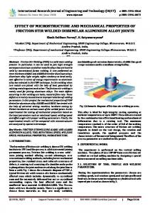
by the doughnut-like bright contrast area surrounding the. Fig. 3 Temperature change at the joint interface during FSSW. Melting temperatures of plated layer of ...
Materials Transactions, Vol. 52, No. 7 (2011) pp. 1418 to 1425 #2011 The Japan Institute of Light Metals
Microstructure and Joint Strength of Friction Stir Spot Welded 6022 Aluminum Alloy Sheets and Plated Steel Sheets*1 Keyan Feng1; *2 , Mitsuhiro Watanabe2 and Shinji Kumai1 1 2
Department of Materials Science and Engineering, Tokyo Institute of Technology, Yokohama 226-8502, Japan Department of Metallurgy and Ceramics Science, Tokyo Institute of Technology, Tokyo 152-8552, Japan
Friction stir spot welding (FSSW) was performed for joining of an aluminum alloy sheet to a steel sheet. A 6022 aluminum alloy sheet, a non-plated steel and four kinds of plated steel sheets were prepared. They were plated by pure zinc (GI), zinc alloy (ZAM), Al-Si alloy (AS) and zinc alloy including Fe (GA). The melting temperature of each plated layer was 420, 330, 640 and 880� C. The aluminum alloy sheet was overlapped on the steel sheet. A rotating tool was inserted from the aluminum alloy sheet side and the probe tip was kept at the position of 0.2 mm above the lapped interface for 3 s. Temperature change at the welding interface was measured during FSSW by using thermocouples which were located at the joint interface below the rotating tool. The maximum operating temperature was 430� C. It was found that interface morphology, strength and joining area of the joint varied depending on whether melting temperature of the plate layer was higher or lower than the maximum operating temperature. Large joint strength and joining area were obtained for the steel sheet with the low melting temperature of plated layer. In this case, the original plated layer was removed from the interface during FSSW and aluminum/steel interface with a thin intermediate layer was observed. [doi:10.2320/matertrans.L-M2011811] (Received October 5, 2010; Accepted March 2, 2011; Published June 15, 2011) Keywords: friction stir spot welding, microstructure, joint strength, aluminum alloy, plated steel
1.
Introduction
Solid-state welding methods for joining of dissimilar metals are attracting a lot of interests.1–3) This is because they can avoid mechanical property degradation of original metals due to melting and re-solidification and they can be applicable to metal combinations with a large melting temperature difference. Friction stir spot welding (FSSW), which is one of the solid-state welding methods, has been applied for joining of dissimilar materials. In particular, it has been applied for the combination of aluminum alloy sheets to steel sheets from the strong industrial interests.4–7) A number of studies have been made so far for joining of the aluminum alloy sheets to the steel sheets by the friction stir welding methods (FSW) and FSSW. These works suggested the existence of the plated layer significantly improved their weldability.8–10) The previous reports, however, did not mention the reason why the plate metals could improve the weldability and the behavior of the plated layers during FSW and FSSW operation. In the present study, we focus on the relationship between the operation temperature and the melting temperature of the plate metals or alloys on the steel sheets. An aluminum alloy sheet and several kinds of plated steel sheets were FSSWed. Microstructures of the joint interface, joint strength and fracture behavior of the joints were investigated and the role of plated layers was discussed. 2.
Experimental Procedure
2.1 Materials A 1.1 mm-thick 6022 aluminum alloy sheet (called Al *1The
Paper Contains Partial Overlap with the ICAA12 Proceedings by USB under the Permission of the Editorial Committee. *2Graduate Student, Tokyo Institute of Technology
alloy, hereafter), four kinds of 1.2 mm-thick plated steel sheets and a 1.2 mm-thick non-plated low carbon steel sheet were prepared. These materials were supplied from the Mitsubishi Motors Corporation. The four kinds of plated layers were as follows: (1) Zinc alloy (Zn-6%Al-3%Mg (in mass%)) (called ZAM), (2) pure Zinc (called GI), (3) Al-Si alloy (called AS) and (4) Zinc alloy including Fe (called GA). They have different melting temperatures, 330, 420, 640 and 880� C for ZAM, GI, AS and GA, respectively. The microstructure of plated layer for each steel sheet is shown in Figs. 1(a)–(d). The thicknesses of plated layers were almost the same and about 10 mm. It should be mentioned that Al-Fe intermetallic compound layer (IMC) with about 3 mm-thick was observed at the interface of steel and Al-Si alloy for AS plated sheet, an indicated by a black arrow in Fig. 1(c). 2.2 Welding tool A conventional vertical milling machine was used for FSSW. The welding tool was made of SKH51 steel. The shoulder diameter and probe diameter of the tool were 10 mm and 5 mm, respectively. The probe length was 0.8 mm. Vertical grooves were introduced to the probe surface. 2.3 Welding procedure Prior to welding, the Al alloy sheet surface and the nonplated low carbon steel sheet surface were polished by a waterproof abrasive paper (# 4000) and cleaned with acetone and dried. No special surface treatment was made. The Al alloy sheet was overlapped on the steel sheet and they are tightly clamped by the fixture. The tool with a rotational speed of 3000 rpm was inserted from the Al alloy sheet side at a rate of 4 mm/min. The probe tip was kept at the position of 0.2 mm above the lapped interface for 3 s, and the rotating tool was retracted from the Al alloy sheet. 2.4 Microstructure observation The joints were sectioned along the normal direction to the
Microstructure and Joint Strength of Friction Stir Spot Welded 6022 Aluminum Alloy Sheets and Plated Steel Sheets
Plated layer
Steel
Plated layer
Steel (a)
(b)
Plated layer
Steel
1419
Plated layer
IMC layer
Steel (d)
(c)
Fig. 1 Plated layers on the plated steel sheets. (a) ZAM, (b) GI, (c) AS, (d) GA.
lapped sheets for the microstructure observation of the joint interface. The polished specimens were observed using both an optical microscope (OM) and a scanning electron microscope (SEM). 2.5 Temperature measurement during FSSW Temperature change at the welding interface was measured during FSSW with a series of thermocouples. The thermocouples were set at the interface of Al alloy sheet and steel sheet below the probe center and at the location 2.5 mm away from the center (probe surface). 2.6 Joint strength evaluation Joint strength was evaluated using a modified cross tension test (called M-CTT, hereafter). Figure 2 shows how to produce the test specimen. Two Al alloy sheets were overlapped on the steel sheet and the three sheets were welded together. The sandwiched middle Al alloy sheet was welded to both the upper Al alloy and the steel sheet. The upper Al alloy and the steel sheet were fixed to the loading apparatus using the bolts which were inserted to the holes of the sheet. In the present specimen configuration, the upper sheet acts as a part of the specimen grip system. Deformation of the upper Al alloy sheet during loading is available to apply a vertical tensile load to the joint interface between the middle Al alloy sheet and the steel sheet. This is quite important to examine the strength of the Al alloy sheet/steel sheet joint interface. The tests were performed using an Instron-type tensile testing machine under a crosshead speed of 0.5 mm/ min at room temperature. In some cases, M-CTT was interrupted in order to obtain the joint specimen in the course of fracture and to examine the fracture process.
Al alloy Al alloy
Steel
Fig. 2 Schematic illustration of the FSSW method and the modified cross tension test (M-CTT) for the lap joint.
3.
Results
3.1 Temperature change during FSSW Figure 3 shows the temperature change at the joint interface during the FSSW. Melting temperatures of plated layer of ZAM, GI, AS and GA were shown by dashed lines in Fig. 3, respectively. Those are temperature-time curves for beneath of the probe-center and for outer-region of the probe. The temperature increased gradually as increasing the plunging depth. The temperature became almost constant when the probe tip reached the target position (0.2 mm above the lapped interface). The temperature was stable for 3 s of the dwell time. The maximum temperature was 430� C for the probe-center and it was 350� C for the outer-region of the probe. The important finding here is the maximum temperature at the joint interface is higher than the melting temperature of the two plated layers, that is, ZAM and GI can be melted at the joint interface during the welding.
1420
K. Feng, M. Watanabe and S. Kumai
Al alloy Steel
Fig. 5 Macroscopic view of the cross section of joint interface (optical micrograph).
(a)
Joining area
Al alloy Plated steel
Fig. 3 Temperature change at the joint interface during FSSW. Melting temperatures of plated layer of ZAM (330� C), GI (420� C), AS (640� C) and GA (880� C) are shown by dashed lines.
(b)
c
d
c
Joining area
Al alloy
1.8
Plated steel
c
Fracture load, P / kN
1.6 1.4
Fig. 6 Schematic illustration of the joint interface. (a) ZAM and GI, (b) AS and GA.
1.2 1 0.8 0.6 0.4 0.2 0 ZAM
GI
AS
GA
Non-plated steel
Fig. 4 Fracture loads of four kinds of plated steel sheet joints and Nonplated steel sheet joint.
3.2 Joint strength Figure 4 shows the fracture load of joints. Under the present welding condition (3 s of the dwell time), lap joining of Al alloy to non-plated steel was not achieved sufficiently. In contrast, joining was successful for all the plated steel sheets. The obtained fracture load was low for AS and GA. The joints of ZAM and GI showed relatively high joint load values. 3.3 Joint interface morphology Figure 5 shows the optical micrograph of the cross-section of joint interface for ZAM. The upper side is the Al alloy sheet and the lower side is the steel sheet. The Al alloy sheet was given a hollow by insertion of the welding tool with the probe. About 0.2 mm-thick Al alloy region observed on the steel sheet corresponds to the area beneath the probe tip. The macroscopic cross-sectional view like this was common for all the joints. Careful observation, however, found that joining area was not common for all the joints. Figures 6(a) and (b) are schematic illustrations showing the difference in joining area. Joining was achieved in the limited area close to the periphery of the probe for ZAM and GI, as indicated by ‘‘c’’ in Fig. 6(a). There was a gap between the sheets in the region ‘‘d’’ in Fig. 6(a). While, the joining area encompassed through the lapped-interface under the probe, in the area ‘‘c’’, as shown in Fig. 6(b) for AS and GA. The interface microstructures of all joints were shown in Figs. 7(a)–(f).
Figures 7(a)–(f) show the back-scattered electron images (BEIs) in the areas at the joint interface corresponding to ‘‘c’’ and ‘‘d’’ in Figs. 6(a) and (b). Bright and dark contrasts correspond to steel and Al alloy, respectively. The intermediate layers with a medium contrast are clearly observed at the joint interface for all joints. The thickness of intermediate layer was about 1 mm for ZAM and GI joints, as shown in Figs. 7(a) and (b) in the areas at the joint interface ‘‘c’’ in Fig. 6(a). These intermediate layers were different from the original plated layers of ZAM and GI, as shown in Figs. 1(a) and (b). Cavities (un-bonded area) were often observed under the probe center region for ZAM, GI joint, as indicated by white arrows in Figs. 7(c) and (d). The joint interface of AS is shown in Fig. 7(e). No intermediate layer was observed. The important finding is that the IMC layer at the original Al-Si alloy/plated steel interface remained after FSSW. Figure 7(f) is the joint interface of GA. The thick layer was clearly observed between Al alloy and steel sheets. The thickness of layer was about 20 mm and thicker than the original plated layer as shown in Fig. 1(d). In addition, many voids and cavities were observed at the bottom side of the layer. Formation mechanism of the layer will be reported precisely in the next paper. 3.4
Macroscopic appearance of the fracture surface after M-CTT Figures 8(a)–(d) are optical micrographs of the fracture surface on the steel sheet side after M-CTT. In the picture, Dp represents the probe diameter and Ds does the shoulder diameter. As shown in Fig. 8(a), for ZAM, a double doughnut-like area is a slightly larger than the probe diameter, Dp . We can also find the dark contrast area at the central region of the round fracture surface. The fracture surface of GI (Fig. 8(b)) is also characterized by the doughnut-like bright contrast area surrounding the
Microstructure and Joint Strength of Friction Stir Spot Welded 6022 Aluminum Alloy Sheets and Plated Steel Sheets
Al alloy
Al alloy
ZAM steel
GI steel (a)
Al alloy
1421
(b)
Cavity
ZAM steel
Cavity
Al alloy
GI steel (c)
(d)
Al alloy
Al alloy
AS steel
GA steel (e)
(f)
Fig. 7 Back-scattered electron images (BEIs) of the joint interface. (a) ‘‘c’’ in Fig. 6(a) for ZAM, (b) ‘‘c’’ in Fig. 6(a) for GI, (c) ‘‘d’’ in Fig. 6(a) for ZAM, (d) ‘‘d’’ in Fig. 6(a) for GI, (e) ‘‘c’’ in Fig. 6(b) for AS, (f) ‘‘c’’ in Fig. 6(b) for GA.
dark contrast region at the center of fracture surface. The size of the bright contrast area is a slightly larger than Dp in this case, too. In contrast, the fracture surface of AS is round pancakelike and the diameter of the bright contrast area is also a slightly larger than Dp , as shown in Fig. 8(c). As for GA (Fig. 8(d)), the round pancake-like area is also observed, but the contrast of this area is relatively dark and complicated. The diameter of this area is slightly larger than Dp , too. 3.5
Microscopic view of the fracture surface and fracture path at the joint interface In order to investigate fracture surface more precisely, SEM observation was performed for each region marked with frames in Fig. 8: a1 to a4 in ZAM, b1 to b4 in GI, c1 in AS and d1 in GA. Figures 9(a)–(j) show SEM images for these regions. Figures 9(a)–(d) correspond to areas ‘‘a1 ’’–‘‘a4 ’’ for ZAM. The area ‘‘a1 ’’ shows the flat featureless surface with many small voids and sharp cracks. The area ‘‘a2 ’’ is covered with relatively flat fracture surface with dark contrast. A bright contrast network-like pattern is also observed. The area ‘‘a3 ’’ exhibits dimple fracture morphology. Rough and compli-
cated appearance is observed for the area ‘‘a4 ’’. However, the doughnut-like area including the area ‘‘a4 ’’ is not fracture surface but the surface of solidified ZAM plate metals which once melted and being squashed out from the probe center region during FSSW. These features are common for GI, as shown in Figs. 9(e)– (h) corresponding to the areas ‘‘b1 ’’–‘‘b4 ’’ in Fig. 8(b), respectively. Figure 9(i) is the fracture surface of AS for the area ‘‘c1 ’’. This is characterized by the relatively flat appearance with many cracks forming ‘‘mosaic’’ structure. Figure 9(j) correspond to the area ‘‘d1 ’’ in GA exhibits more complicated fracture surface covered with the flat area with sharp cracks and the relatively rough area. 3.6
Measurement of substantial joining area in the FSSW joints Based on both optical micrographs of fracture surface and cross sectional view of the joint interface, the substantial joining area was estimated for the joints after M-CCT. Relationship between the obtained fracture load and the estimated joining area is summarized as shown in Fig. 10. Fracture load increased as increasing the joining area in all joints.
1422
K. Feng, M. Watanabe and S. Kumai
(a)
(b) a4
b4
a3
b3
a2
Dp
Dp
Ds
Ds
(c)
(d)
d1
c1
Dp
Dp Ds
Fig. 8
b2
b1
a1
Ds
Macroscopic view of the fracture surface of the plated steel side (optical micrograph). (a) ZAM (b) GI (c) AS (d) GA.
3.7 Fracture path at the joint interface during M-CTT Fracture path at the joint interface was examined with the help of the interrupted M-CTT. The loading was interrupted at the maximum load point and the unbroken, but partially cracked specimens were obtained. Figures 11(a)–(d) show cross sectional views of the cracked joint interfaces observed in the unbroken specimens. Figure 11(a) is BEI in the area corresponding to the area ‘‘a2 ’’ in Fig. 8(a) and Fig. 9(b). A crack is clearly observed along the interface between the steel surface and the intermediate layer. See Fig. 7(a) for comparison. Figure 11(b) is for the area ‘‘a3 ’’ in Fig. 8(a) and Fig. 9(c). In this case, we can see a large cavity in the Al matrix close to the joint interface. This suggests that the crack propagated in the Al alloy matrix. Please note that the intermediate layer on the steel sheet is thin in this area and no fracture took place at this interface. Note that the fracture manners are common for GI. Figure 11(c) is for the area ‘‘c1 ’’ in Fig. 8(c) and Fig. 9(i) for AS. It was found that the crack propagated in the original IMC layer in Al-Si plate alloy. See Fig. 1(c) and Fig. 7(e) for comparison. Figure 11(d) shows the fractured area corresponding to the area ‘‘d1 ’’ in Fig. 8(d) and Fig. 9(j) for GA. We can see that the fracture took place in the layer between Al alloy and steel sheets. See Fig. 7(f) for comparison.
4. 4.1
Discussion
Effect of melting temperature of the plated layer on the Al alloy/steel joint interface in FSSW The joint interface morphology as shown in Figs. 5 to 7 can be classified into two patterns. One is the case that no joining takes place below the probe center but joining occurs at the lapped interface close to the periphery of the probe. The other one is the case that joining occurs in the area under the probe almost evenly. The former is for ZAM and GI, in which melting temperature of the plated layer is lower than operating temperature of the present FSSW, as shown in Fig. 3. The latter is for AS and GA, which have the plated layer with high melting temperature. Such difference in joining area is considered to cause the characteristic macroscopic difference in fracture surface morphology: doughnutlike and pancake-like one. Let us discuss the formation mechanism of the joint interface. When the melting temperature of the plated layer is lower than the operating temperature, the plated layer will melt and the molten plated layer can be squeezed outward. This facilitates direct contact between the aluminum plate surface and the refreshed steel surface. At the probe-center region, a thin liquid film of the plated layer possibly stays and hinders the direct contact of aluminum and steel surface.
Microstructure and Joint Strength of Friction Stir Spot Welded 6022 Aluminum Alloy Sheets and Plated Steel Sheets
(a)
(b)
(c)
(d)
(e)
(f)
(g)
(h)
(i)
(j)
Fig. 9 Secondary electron images (SEIs) of fracture surface. (a) ‘‘a1 ’’ in Fig. 8(a) for ZAM, (b) ‘‘a2 ’’ in Fig. 8(a) for ZAM, (c) ‘‘a3 ’’ in Fig. 8(a) for ZAM, (d) ‘‘a4 ’’ in Fig. 8(a) for ZAM, (e) ‘‘b1 ’’ in Fig. 8(b) for GI, (f) ‘‘b2 ’’ in Fig. 8(b) for GI, (g) ‘‘b3 ’’ in Fig. 8(b) for GI, (h) ‘‘b4 ’’ in Fig. 8(b) for GI, (i) ‘‘c1 ’’ in Fig. 8(c) for AS, (j) ‘‘d1 ’’ in Fig. 8(d) for GA.
1423
1424
K. Feng, M. Watanabe and S. Kumai
plastic flow and both mechanical and metallurgical alloying must be achieved by FSSW. In the present welding condition, the probe tip is away from the IMC layer and there is almost no chance the rotating tool breaks the IMC layer. Consequently, joining between 6022 aluminum alloy sheet and Al-Si alloy plate takes place, but the IMC layer remains almost intact after FSSW in spite that some small broken pieces were observed in the Al alloy matrix.
1.8 1.6
ZAM
GI
AS
GA
Fracture load, P / kN
1.4 1.2 1 0.8 0.6 0.4 0.2
4.2
0 0
20
60
40
Joining area, A / mm
80
2
Fig. 10 Relationship between joining area and fracture load.
Since the temperature decrease occurs by pulling out of the probe, the isolated liquid film will solidify and this will form shrinkage cavity. This may result in the gap formation as shown in Fig. 7(c) for ZAM and Fig. 7(d) for GI. At the outer region close to the periphery of the rotating probe, corresponding to the area ‘‘c’’ in Fig. 6, the aluminum matrix above the lapped surface must be subjected to a relatively severe plastic flow condition. This is considered to promote solute diffusion between aluminum and steel. It should be mentioned that thickness of the intermediate layer decreases as increasing the distance from the probe center. The effect of this thickness difference on fracture behavior will be discussed later. The joining area spreads over entirely under the probe for AS, as shown in Fig. 6(b). In this case, no intermediate layer was observed as shown in Fig. 7(e). The original IMC layer formed on the steel sheet in Al-Si alloy plated layer was clearly remained in the welded joint interface. The Al-Si alloy will be mixed to the 6022 aluminum alloy sheet by
Relationship among joint interface structure, fracture surface and joint strength Figure 9(a) shows SEM image of the area ‘‘a1 ’’ in Fig. 8(a) for ZAM. Flat featureless surface with many small voids and sharp cracks is observed. As shown in Fig. 7(c), the cross sectional view of this area revealed that there is a cavity between Al alloy sheet and the intermediate layer on the steel sheet. Therefore, this is not the fracture surface produced by M-CTT but the image of original interfacial morphology of this region after FSSW. Careful observation found that this is the surface of intermediate layer formed during FSSW. This is partially covered with a thin film which was the once-melted and re-solidified Zn alloy plate. Figure 9(b) is for the area ‘‘a2 ’’ in Fig. 8(a). The cross sectional view of the joint interface for this area is shown in Fig. 7(a). The thin and continuous intermediate layer is formed at the joint interface. Figure 9(c) is for the area ‘‘a3 ’’ in Fig. 8(a). This thickness difference is considered to affect fracture manner. It is considered that when the intermediate layer is thick, the crack propagates along the interface between the steel surface and the intermediate layer and/or in the intermediate layer, as shown in Fig. 11(a). It is generally recognized that fracture and/or peeling of the intermediate layer from the matrix seldom occurs when the intermediate
Al alloy
Al alloy
Al alloy ZAM steel
ZAM steel (a)
(b)
Al alloy
Al alloy
AS steel
GA steel (c)
(d)
Fig. 11 Cross-sectional views of the cracked joint interface after the interrupted M-CTT. (a) ZAM, (b) ZAM, (c) AS, (d) GA.
Microstructure and Joint Strength of Friction Stir Spot Welded 6022 Aluminum Alloy Sheets and Plated Steel Sheets
layer is very thin. In this case, fracture tends to take place in the matrix with weaker strength. This corresponds to the appearance of dimple fracture shown by Fig. 11(b), which is for the area ‘‘a3 ’’ in Fig. 8(a) and Fig. 9(c). The crack propagates in the Al alloy matrix. As described in 4.1, at the joint interface in ZAM, thickness of the intermediate layer decreases as increasing the distance from the probe center. Therefore, we can say that transition of fracture manner from the type ‘‘a2 ’’ to the type ‘‘a3 ’’ is reasonable. This interpretation can be applicable to the case of GI. In contrast, fracture of AS occurred mainly in the original IMC layers in the plated Al-Si alloy. This was well demonstrated by the interrupted M-CTT as shown in Fig. 11(c). Relatively flat fracture surface with many cracks, which is like a forming ‘‘mosaic’’ structure, represents the broken surface of IMC layer. As shown in Fig. 11(d), the thick layer at the joint interface provides a preferential crack growth path in GA. Fracture surface of GA as shown in Fig. 9(j) consists of two different fracture surfaces, flat and rough, this reflects the crack growth path very well. Figure 10 indicates that relatively large joining area is obtained for ZAM and GI. This is because the direct bonding between 6022 aluminum alloy and steel was promoted by the appearance of refreshed steel surface due to melting of the plate layer. Fracture strength was controlled by the strength of original IMC layer and that of intermediate layer for AS and GA, respectively. 5.
Summary Lap joining of aluminum alloy sheet/four kinds of plated
1425
steels was carried out using the friction stir spot welding method. Sound joining was achieved for all plated steel sheets. The interface morphology, strength and joining area of the joint varied depending on whether melting temperature of the plate layer was higher or lower than the maximum operating temperature. Large joint strength and joining area were obtained for the steel sheet with the low melting temperature of plated layer. In this case, the original plated layer was removed from the interface during FSSW and aluminum/steel interface with a thin intermediate layer was observed for ZAM and GI joints. REFERENCES 1) S. Kumai, H. Sato, K. Suzuki, T. Ookawa, K. Lee and M. Watanabe: J. JILM 57 (2007) 529–535. 2) K. Lee, S. Kumai, T. Arai and T. Aizawa: Mater. Sci. Eng. A 471 (2007) 95–101. 3) S. Kumai, M. Watanabe and K. Feng: Mater. Sci. Forum 654–656 (2010) 596–601. 4) K. Feng, M. Watanabe and S. Kumai: Mater. Sci. Forum 654–656 (2010) 970–973. 5) S. Bozzia, A. L. Helbert-Ettera, T. Baudinb, B. Criquic and J. G. Kerbiguetc: Mater. Sci. Eng. A 527 (2010) 4505–4509. 6) K. Feng, M. Watanabe and S. Kumai: Proc. 117th Conf. JILM, (2009) pp. 57–58. 7) M. Watanabe, K. Feng, Y. Nakamura and S. Kumai: Proc. 12th Int. Conf. on Aluminum Alloys, (The Japan Inst. Light Metals, 2010) pp. 984–987. 8) Y. C. Chen, T. Komazaki, T. Tsumura and K. Nakata: Mater. Sci. Technol. 24 (2008) 33–39. 9) K. Miyagawa, M. Tsubaki, T. Yasui and M. Fukumoto: Q. J. JWS 26 (2008) 131–136. 10) A. Elrefaey, M. Takahashi and K. Ikeuchi: Q. J. JWS 23 (2005) 186– 193.