hydrazine, containing 30% (v/v) water and 1% (w/v) hydrazine sulfate, at 100 âC for 4 h. After repeated evaporation in the presence of toluene the mixture was ...
THEJOURNALOF BIOLOGICAL CHEMISTRY 0 1993 by The American Society for Biochemistry and Molecular Biology, Inc
Vol. 268, No. 32, Issue of November 15, pp. 23898-23905,1993 Printed in U.S.A.
Minimal Sequence in Heparin/Heparan Sulfate Required for Binding of Basic Fibroblast Growth Factor* (Received for publication, March 9, 1993, and in revised form, June 11, 1993)
Marco MaccaranaSg,Benito Casufl, and Ulf Lindahl(1 From the Department of Medical and Physiological Chemistry, Box 575, the Biomedical Center, University of Uppsalu, S-751 23 Uppsalu, Sweden, Sltalfarmaco S.p.A., Viale Fulvio Testi, 330, 20126 Milan, Italy, and the VG. Ronzoni Institute for Chemical and Biochemical Research, Via G. Colombo 81, 20133 Milan, Italy
Experiments based on interaction in free solution between basic fibroblast growth factor (FGF-2) and saccharides related to heparinbeparan sulfateshowed that the growth factor binds to heparin and to selectively glucosaminyl6-O-desulfated heparin but poorly to iduronosyl 2-O-desulfated heparin. 2-O-sulfate groups thus are essential to the interaction, whereas 6-O-sulfates are not required nordo they interfere with FGF-2 binding. Comparison of various bound/ nonbound oligosaccharides implicated a minimal pentasaccharide sequence for FGF-2 binding, with the structure: -hexuronic acid-glucosamine N-sulfatehexuronic acid-glucosamine N-sulfate-iduronic acid 2-O-sulfate- (reducing terminus to the right). Such (overlapping) sequences are abundant in heparin, albeit heavily obscured by irrelevant O-sulfate groups, and occur also in heparan sulfate,with or without additional O-sulfates.
Most of the biological activities known to be associated with proteoglycans are due to interactions between the negatively charged glycosaminoglycan chains and various proteins (Jackson et al., 1991; Kjell6n and Lindahl, 1991). Such interactions range from highly specific, “lock-and-key” type binding, as described for the antithrombin-binding region in heparin (Lindahl et al., 1984), to relatively nonspecific, co-operative electrostatic association. A group of proteins that has attracted particular interest in recent years is the heparinbinding growth factors (Burgess and Maciag, 1989; Klagsbrun, 1990),of which the best characterized members are theacidic and basic (FGF-2)’ fibroblast growth factors. Neither the mode of binding between these proteins and heparin or heparan sulfate (HS) nor the functional implications of the interactions have been well understood. Recent studies have indicated an important role for HS proteoglycans in mediating functional responses to fibroblast growth factors. HS proteoglycan binds FGF-2 at the cell surface andinthe extracellular matrix, thus providing a storage form of the growth factor, which is relatively protected * This study was supported by Swedish Medical Research Council Grant 2309, Italfarmaco S.p.A. (Milan), and Polysackaridforskning AB (Uppsala). The costs of publication of this article were defrayed in part by the payment of page charges. This article must therefore be hereby marked “advertisement” in accordance with 18 U.S.C. Section 1734 solelyto indicate this fact. J Present address: Dept. of Medical and Physiological Chemistry, University of Uppsala, Sweden. 1) To whom correspondence should be addressed. Tel.: 46-18174196; Fax: 46-18-174209. The abbreviations used are: FGF-2, basic fibroblast growth factor; HS, heparan sulfate; HPLC, high pressure liquid chromatography.
against proteolytic degradation (Saksela et al., 1988). The factor may be released in active form either by addition of heparin or through mobilization by enzymatic cleavage of the HS carrier (Naparstek et al., 1984; Ishai-Michaeli et al., 1990; Vlodavsky et al., 1991a). Furthermore, cells that are lacking cell surface HS proteoglycan or produce undersulfated HS are unable to respond to added FGF-2, unless supplemented with exogenous HS or heparin. It has been proposed that the polysaccharides induce a conformational change in FGF-2, which is a prerequisite to efficient binding of the growth factor to its high affinity receptor at the cell surface (Yayon et al., 1991; Rapraeger et al., 1991). The present study has been aimed at defining the minimal saccharide structure required for binding of heparin/HS to FGF-2. EXPERIMENTALPROCEDURES
Polysaccharides-Heparin from pig intestinal mucosa (stage 14, Inolex Pharmaceutical Division, Park Forest South, IL) was purified as described (Lindahl et al., 1965). Two samples of selectively 0desulfated heparin were given by A. Naggi and G. Grazioli, Institute G. Ronzoni, Milan, Italy. One sample was generated by preferential 6-O-desulfation (treatment with dimethyl sulfoxide, 10% water at 110 “C for 5 h) (Nagasawa et al., 1977) along with N-desulfation of thestarting material, followed by re-N-sulfation; compositional analysis of the product (see below) indicated non-0-sulfated -HexAGlcNX- (39%) and-IdoA(2-OSOo)-GlcNX- (46%)( X = (N)-sulfate or (N)-acetyl)asthe predominant disaccharide units (Fig. 5A). Because -70% of the disaccharide units of the unmodified heparin would contain IdoA(2-OSOo)residues (Kusche et al., 1990), about one-third of the 2-O-sulfate groups must have been lost along with the 6-O-sulfates. The other sample was obtained by selective 2-0desulfation under alkaline conditions (Jaseja et al., 1989) and contained 21% non-0-sulfated -HexA-GlcNX- and 70% -1doAGlcNX(6-OS03)- disaccharide units (Fig. 5 8 ) . A low sulfated HS preparation (-0.6 sulfate group/disaccharide unit), isolated from human aorta (Iverius, 1971), was provided by W. Murphy, University of Monash, Melbourne, Australia. HS from bovine kidney was given by K. Sugahara, Women’s Collegeof Pharmacy, Kobe, Japan. The preparations of chondroitin sulfate (bovine cartilage) and dermatan sulfate (pig intestinal mucosa) were as described (Lycke et al., 1991). Oligosaccharides-Even numbered heparin oligosaccharides were generated by partial deaminative cleavage of the polysaccharide with nitrous acid (pH 1.5; cleavage at N-sulfated GlcN units) (Shively and Conrad, 1976a), essentially as described (Pejler et al., 1988), and the resulting 2,5-anhydro-~-mannoseresidues were reduced with NaB3H4 (The Radiochemical Center, Amersham, U.K.). The labeled oligosaccharides (-0.6 X lo6 dpm 3H/nmol) were separated by repeated gel chromatography on Sephadex G-50 (superfine, 1 X 190 cm column, eluted with 1 M NaCl) into even numbered species, from 4- to 14saccharides (designated H-4 to H-14), homogeneous with regard to molecular size (Lane et al., 1984); in addition, a labeled fraction estimated to contain largely 20- to 24-saccharides was recovered. The selectively 6-O-desulfated heparin was treated in a similar way to yield 4- to 12-saccharides (-2 X lo6 dpm 3H/nmol) and a fraction of larger oligosaccharides. The isolated and desalted oligosaccharides were subjected to mild acid treatment (25 mM HzSO,, 80 “C, 30 m i d ,
23898
Binding Site
for FGF-2 in HeparinlHeparan Sulfate
followed by gel chromatography, to eliminate sequences containing products of “anomalous” deaminative ring contraction (Shively and Conrad, 1976a, 1976b). Odd numbered heparin oligosaccharides were obtained as follows. A sample (3 X lo6 dpm) of the fraction containing 20- to 24-meric (even numbered), [3H]aManR-labeledoligosaccharides, isolated after deaminative cleavage (see above), was incubated with 4.2 mIU (2.5 Sigma units) of heparinase I (Sigma) from Fluuobacteriumheparinurn in 200 pl of 5 mM phosphate buffer, 200 mM NaCl, 0.5 mg/ml bovine serum albumin, pH 7.0. After 10 min a t 30 “C the digest was heated at 100 “C for 1 min and centrifuged, and the supernatant was fractionated bygel chromatography on Sephadex G-50, as indicated above. The hexa- and octasaccharide fractions, indicated by distinct peaks corresponding to 16 and 14%,respectively, of the 3H applied to the column, were recovered and desalted by passage through PD-10 columns (Pharmacia). The nonreducing terminal,4,5-unsaturated hexuronic acid units were eliminated by treatment with 40 p1 (final volume) of 10 mM mercuric acetate in 130 mM sodium acetate, pH 5.0, for 30 min a t room temperature (Ludwigs et al., 1987; Kusche et al., 1988). The samples were adjusted to 0.5 ml with 5 M NaCl and were then desalted by successive passage through PD-10 columns equilibrated with 1 M NaCl and water; the high salt separation step was found to be necessary to ascertain removal of mercuric ions. The predicted penta- and heptasaccharide structures of the products (H5 and H-7) were confirmed by analytical gel chromatography on Sephadex G-50, against even numbered oligosaccharide standards (data not shown). Generation of oligosaccharides (HS-4 toHS-14) from HS (human aorta) followed a different strategy, involving cleavage at sites occupied by N-acetylated GlcN units. A sample (5 mg) of HS was Ndeacetylated (Guo and Conrad, 1989) by treatment with 1 mlof hydrazine, containing 30% (v/v) water and 1% (w/v) hydrazine sulfate, a t 100 “C for 4 h. After repeated evaporation in the presence of toluene the mixture was passed through Sephadex G-15, lyophilized, and deaminated with nitrous acid (pH 3.9; cleavage at Nunsubstituted GlcN units; Shively and Conrad, 1976a). The resulting oligosaccharides were radiolabeled by reduction with NaB3H4 and separated by gel chromatography as described in the legend to Fig. 2. The various oligosaccharides, reisolated after mild acid treatment as described above, had a specific radioactivity of -0.6 X IO6 dpm 3H/ nmol. Octasaccharide was converted into heptasaccharide (HS-7) by digestion with B-D-glucuronidase as described in the legend to Table 11. Molar concentrations of oligosaccharides were determined on the basis of hexuronic acid contents, as measured by the carbazole reaction (Bitter andMuir, 1962). Interaction between Saccharides and FGF-2”Human recombinant FGF-2, purified from yeast cells (Zymogenetics, Inc.) as described by Olwin and Hauschka (1990),was given byDr. B. B. Olwin (University of Wisconsin-Madison, Madison, Wisconsin). For analytical scale experiments FGF-2 was incubated at room temperature for 2 h with the appropriate saccharide samples (see the text) in 300 pl (unless otherwise stated) of50 mM Tris-HC1, pH 7.4, containing NaCl at various concentrations and 0.5 mg of bovine serum albumin/ml. The growth factor, along with any bound carbohydrate, was recovered by quick passage of the samples through nitrocellulose filters (Sartorius, pore size 0.45 p; 25 mm diameter), which had been placed onto a 10well vacuum-assisted manifold filtration apparatus. The filters were prewashed with 2 X 5 ml of 50 mM Tris-HC1, 130 mM NaCl, pH 7.4, before application of the samples, which were immediately followed by another 2 X 5 mlof the same buffer; each washing step was completed within 5 sec. Protein-bound radioactivity was determined after submersion of the filters in 2 ml of 3 M NaCl for 30 min.; 1 ml of the eluate was mixed with 0.5 ml of H 2 0 and 3.5 ml of OptiPhase scintillation mixture (Pharmacia LKB Biotechnology Inc.) and counted ina Beckman LS 6000IC scintillation spectrometer. No residual radioactivity could be detected on the filters. In preparative experiments, incubation mixtures of various volumes were passed through larger filters (Sartorius; 38 mm diameter); the number of filters used for each incubation was adjusted so that the amount of bovine serum albumin (0.1-0.5 mg/ml of incubation mixture) never exceeded 100 pg/cm2 of filter. Nonbound material was recovered after washing (2 X 20 ml) as described above. Proteinbound saccharides, eluted from the filters with 3 ml of 3 M NaCI, were concentrated and desalted by passage through columns (PD-10; Kabi Pharmacia) of Sephadex G-25. Analytical Methods-Compositional analysis of heparin or HS oligosaccharides involved degradation of samples with nitrous acid
23899
(pH 1.5) followed by reduction of the resulting di- and tetrasaccharides with NaB3H4.The procedure of Kusche et al. (1990) was scaled down so that 20-100 X IO3 dpm of end group-labeled oligosaccharide (-30-170 pmol, corresponding to -80-400 ng of a heparin-derived octasaccharide) was deaminated and reduced with 25-50 pCi NaB3H4. The resulting HexA-[1-%]aManR disaccharide fractions wereisolated and analyzed further by anion-exchange HPLC (Bienkowski and Conrad, 1985), as described in more detail in the legend to Fig. 5. Due to the poor separation of the non-0-sulfated disaccharides in this procedure, G l ~ A - [ l - ~ H ] a Mand a n ~I d ~ A - [ l - ~ H ] a M were a n ~ resolved by descending paper chromatography (ethyl acetate/acetic acid/H20, 3:l:l) andquantified by liquid scintillation counting of the paper strips. The proportion of labeled H ~ X A - G ~ C N A C - G ~ C A [ ~ - ~ H ] aManR tetrasaccharides in relation to total disaccharides was determined by gel chromatography (Sephadex G-15) following mild acid treatmentas described above. Di- and tetrasaccharide sequences occupying the reducing terminal position were identified in separate experiments, utilizing the 3H label introduced upon initial labeling of the 2,5-anhydromannose end group in the intactoligosaccharide.For compositional analysis of full sized glycosaminoglycans (aorta HS and partially 0-desulfated heparins) the compounds were N-deacetylated by hydrazinolysis (4 h) as described above and were then subjected to deamination first at pH1.5 (cleavageat N-sulfated GlcN units) and then a t pH 3.9 (cleavage at N-unsubstituted GlcN units) according to Riesenfeld et al. (1982), followedby reduction of the disaccharide products with NaB3H4. High-voltage paper electrophoresis (40 V/cm) was conducted on Whatman No. 3MM paper in 1.6 M formic acid (pH 1.7). Additional separation methods (gel chromatography and anion-exchange HPLC of oligosaccharides) are described in the legends to the figures. RESULTS
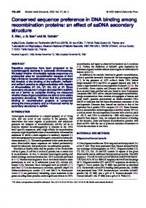
Binding to FGF-2 of PHIHeparin alongwithUnlabeled Glycosaminoglycans-The ability of selected polysaccharide preparations to displace 3H-labeled heparin from FGF-2 was investigated, using a nitrocellulose filter disc procedure (see “Experimental Procedures”). The labeled ligand consisted of fragments >18 monosaccharide units, recovered after partial deaminative cleavage of pig mucosal heparin and reduction of the products with NaB3H4.Binding of this fragment to FGF2 was efficiently precluded in the presence of intact, unlabeled heparin, 50% inhibition of binding occurring at a heparin concentration of -0.2 pg/ml (30 ngaddedper 150 pl of incubation mixture) (Fig. 1).The displacing ability of selectively 6-0-desulfated heparin was only slightly lower than
Unlabeled polysaccharide added(pg)
FIG. 1. Competitive binding of ‘H-labeled heparin and unlabeled polysaccharides to FGF-2. Incubation mixtures containing 150 pl of50 mM Tris-HC1 (pH 7.4), 130 mM NaC1,0.42pgof FGF-2 (160 nM), 15 X lo3dpm of 3H-labeledheparin fragments (-20to 24-saccharides; -170 nM), and various amounts of unlabeled polysaccharides (1 pgof heparin corresponding to -500 nM concentration) were passed through nitrocellulose filters as described in “EXperimental Procedures.” Controls without added cold polysaccharide showed -8,000 dpm bound to FGF-2, thus retained by the filter; blanks without added FGF-2 gave