□ CLINICAL RESEARCH □
Korean J Thorac Cardiovasc Surg 2016;49:421-426
https://doi.org/10.5090/kjtcs.2016.49.6.421
ISSN: 2233-601X (Print) ISSN: 2093-6516 (Online)
Minimally Invasive Cardiac Surgery versus Conventional Median Sternotomy for Atrial Septal Defect Closure Joon Chul Jung, M.D., Kyung-Hwan Kim, M.D., Ph.D. Department of Thoracic and Cardiovascular Surgery, Seoul National University Hospital
Background: Median sternotomy is the standard approach for atrial septal defect (ASD) closure. However, minimally invasive cardiac surgery (MICS) has been introduced at many centers in adult/grown-up congenital heart patients. We retrospectively reviewed the results of right anterolateral thoracotomy compared with conventional median sternotomy (CMS) for ASD closure at Seoul National University Hospital. Methods: We retrospectively analyzed 60 adult patients who underwent isolated ASD closure from January 2004 to December 2013 (42 in the CMS group, 18 in the MICS group). Preoperative, operative, and postoperative data were collected and compared between the 2 groups. Results: The MICS group was younger (44.6 years vs. 32.4 years, p=0.002) and included more females (66.7% vs. 94.4%, p=0.025) than the CMS group. Operation time (188.4 minutes vs. 286.7 minutes, p<0.001), cardiopulmonary bypass time (72.7 minutes vs. 125.8 minutes, p<0.001), and aortic cross-clamp time (25.5 minutes vs. 45.6 minutes, p<0.001) were significantly longer in the MICS group. However, there were no significant differences in morbidity and mortality between groups. Only chest tube drainage in the first 24 hours (627.1 mL vs. 306.1 mL, p<0.001) exhibited a significant difference. Conclusion: MICS via right anterolateral thoracotomy is an alternative choice for ASD closure. The results demonstrated similar morbidity and mortality between groups, and favored MICS in chest tube drainage in the first 24 hours. Key words: 1. 2. 3. 4.
Sternotomy Minimally invasive surgery Congenital heart disease Atrial heart septal defects
Introduction Atrial septal defect (ASD) is a common congenital heart disease with an incidence of 900 per million in the United States [1], and surgical results of ASD closure are favorable [2]. Most patients are diagnosed in childhood, but some patients are diagnosed as adults. Seoul National University Hospital has a separate children’s hospital, but ASD patients over 15 years old generally visit and undergo surgery at the
adult cardiac center. Median full sternotomy is the standard approach for surgical ASD closure in children and adults. The ASD closure procedure is relatively simple compared to other complex congenital and adult heart diseases, and more people are concerned about cosmetic outcomes. Therefore, minimally invasive cardiac surgery (MICS) for ASD closure has been introduced at many centers. Our center has used right anterolateral thoracotomy
Received: December 23, 2015, Revised: April 10, 2016, Accepted: April 26, 2016, Published online: December 5, 2016 Corresponding author: Kyung-Hwan Kim, Department of Thoracic and Cardiovascular Surgery, Seoul National University Hospital, Seoul National University College of Medicine, 101 Daehak-ro, Jongno-gu, Seoul 03080, Korea (Tel) 82-2-2072-3971 (Fax) 82-2-765-7117 (E-mail)
[email protected] © The Korean Society for Thoracic and Cardiovascular Surgery. 2016. All right reserved. This is an open access article distributed under the terms of the Creative Commons Attribution Non-Commercial License (http://creativecommons.org/ licenses/by-nc/4.0) which permits unrestricted non-commercial use, distribution, and reproduction in any medium, provided the original work is properly cited.
− 421 −
Joon Chul Jung, et al
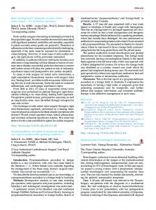
Fig. 1. (A) Femoral artery and vein cannulations are performed. (B) Right internal jugular vein cannulation and right anterolateral thoracotomy are performed.
for ASD closure in adult patients for the past 10 years. This study retrospectively reviewed the results of right anterolateral thoracotomy for ASD closure and compared the outcomes with conventional median sternotomy (CMS).
Methods A total of 125 consecutive patients over 15 years old underwent ASD closure at our institution by a single surgeon from January 2004 to December 2013. Patients who underwent concomitant procedures, such as the Maze procedure, mitral valve annuloplasty, and tricuspid valve annuloplasty, were excluded. Sixty patients with isolated ASD closure constituted the final study population. The age of the patients was between 16 and 73 years. CMS was performed in 42 patients, and MICS was performed in 18 patients. Preoperative, operative, and postoperative data were collected from patients’ medical records and compared between the 2 groups. This study was approved by the institutional review board of Seoul National University Hospital (1507-007-683). In the CMS group, standard median sternotomy was performed, and the usual aortic and bi-caval cannulations were used. Cardioplegia was infused in an antegrade manner via the aortic root cannula, and the ascending aorta was cross-clamped. The right atrium was opened, and the ASD was closed with glutaraldehyde-fixed auto-pericardium. Patients in the MICS group were positioned with their right side up by inserting towels under the right back. A small incision (approximately 2–3 cm) was made in the inguinal region for femoral artery and
vein development. A purse string suture was applied at the common femoral vein, and a venous cannula was inserted and the tip positioned at the inferior vena cava (IVC) level under trans-esophageal echocardiography guidance. Another venous cannula was inserted via the internal jugular vein and positioned at the superior vena cava (SVC) level. Internal jugular vein cannulation was aided by ultrasonography-guided puncture in the operative field. Two venous cannulae were connected with a Y-connector. A purse string suture was applied at the common femoral artery, and an arterial cannula was inserted (Fig. 1A). A small anterolateral thoracotomy (approximately 6–8 cm) was made at the sub-mammary line, and the pleural cavity was entered through the fourth intercostal space (Fig. 1B). The pericardium was opened and retracted using a suture that passed through the chest wall via separate incision. The SVC and IVC were snared down with umbilical tape and a slider after cardiopulmonary bypass (CPB) was initiated. Aortic cross-clamp (ACC) was achieved using a Chitwood DeBakey Clamp (Scanlan International Inc., St. Paul, MN, USA) through a separate incision at the second intercostal space. Cardioplegia was infused in an antegrade manner via the aortic root cannula. The right atrium was opened, and the ASD was closed using glutaraldehyde-fixed auto-pericardium. During the entire procedure, CO2 gas was infused into the chest wall to reduce the risk of air embolism. No thoracoscopy or thoracoscopic devices were used. The right atrium was closed, the ACC was released, and CPB weaning and decannulation procedures were performed using standard techniques. Thoracotomy and femoral wounds were closed with special concern for
− 422 −
Right Anterolateral Thoracotomy for Atrial Septal Defect Closure Table 1. Preoperative patient characteristics Conventional median sternotomy (n=42)
Characteristic Sex (female)
28 (66.7)
Age (yr) 2 Body mass index (kg/m ) Ejection fraction (%)
Minimally invasive cardiac surgery (n=18) 17 (94.4)
44.6±13.8 21.9±2.5 58.4±6.8
p-value 0.025
32.4±11.3 21.7±3.1 60.1±5.4
0.002 0.787 0.309
Hypertension Diabetes mellitus Coronary artery disease
8 (19.0) 1 (2.4) 0
1 (5.6) 0 0
0.255 >0.99
Chronic obstructive pulmonary disease Congestive heart failure Transient ischemic attack
1 (2.4) 0 0
0 0 0
>0.99
Stroke Peripheral vascular disease Renal failure
0 0 2 (4.8)
0 0 0
>0.99
Arrhythmia
1 (2.4)
0
>0.99
Values are presented as number (%) or mean±standard deviation.
Table 2. Operative details Variable Operation time (min) Cardiopulmonary bypass time (min) Aortic cross-clamp time time (min) o
Lowest rectal temperature ( C) o Lowest nasopharyngeal temperature ( C) Intraoperative transfusion Intraoperatively infused blood products (mL)
Conventional median sternotomy (n=42)
Minimally invasive cardiac surgery (n=18)
p-value
188.4±37.8 72.7±21.5 25.5±9.8
286.7±30.8 125.8±17.8 45.6±9.4
<0.001 <0.001 <0.001
32.0±1.6 30.0±2.1 9 (21.4)
27.8±1.8 25.4±2.0 1 (5.6)
<0.001 <0.001 0.256
128.6±312.4
13.9±59.0
0.114
Values are presented as mean±standard deviation or number (%).
cosmetics. Categorical variables are described using frequency and percentage. Continuous variables are described as the means and standard deviation. Comparisons of categorical variables between 2 groups were performed using the chi-square or the Fisher exact test. Comparisons of continuous variables were performed using the Student t-test or Mann-Whitney U-test after pretests for normal distribution. Statistical significance was accepted when the p-value was <0.05.
Results The MICS group was younger (44.6±13.8 years vs. 32.4±11.3 years, p=0.002) and included more females (28 [66.7%] vs. 17 [94.4%], p=0.025) than the CMS croup. However, preoperative ejection fraction and
underlying diseases were not significantly different between the 2 groups (Table 1). The operation time (188.4±37.8 minutes vs. 286.7± 30.8 minutes, p<0.001), CPB time (72.7±21.5 minutes vs. 125.8±17.8 minutes, p<0.001), and ACC time (25.5±9.8 minutes vs. 45.6±9.4 minutes, p<0.001) were significantly longer in the MICS group. The rectal o o o o temperature (32.0 C±1.6 C vs. 27.8 C±1.8 C, p<0.001) and nasopharyngeal temperature (30.0oC±2.1oC vs. 25.4oC±2.0oC, p<0.001) were significantly lower in the MICS group. There were no significant differences in intraoperative transfusion (Table 2). There were no significant differences in morbidities and mortalities between groups. Mechanical ventilation time (14.6±12.2 hours vs. 12.2±7.1 hours, p=0.566), intensive care unit length of stay (LOS) (37.5±37.1 hours vs. 27.4±11.7 hours, p=0.675), and hospital LOS
− 423 −
Joon Chul Jung, et al Table 3. Postoperative results Variable
Conventional median sternotomy (n=42)
Conversion to sternotomy
Minimally invasive cardiac surgery (n=18)
p-value
0
In-hospital mortality Reoperation for bleeding Cardiac arrest
1 (2.4) 2 (4.8) 0
0 0 0
Neurological complication Respiratory failure Postoperative intra-aortic balloon pump insertion
0 0 0
0 0 0
Myocardial infarction Atrial fibrillation Renal failure
0 8 (19.0) 1 (2.4)
0 1 (5.6) 0
Wound complication Complication of femoral cannulation Mechanical ventilation time (hr)
1 (2.4) 14.6±12.2
0 0 12.2±7.1
>0.99 >0.99
0.255 >0.99 >0.99 0.566
Intensive care unit LOS (hr) Hospital LOS (day) Chest tube drainage in first 24 hours (mL)
37.5±37.1 7.3±10.9 627.1±742.6
27.4±11.7 5.9±1.9 306.1±354.2
0.675 0.746 <0.001
Blood product requirement Postoperatively infused red blood cell (mL) Postoperatively infused total blood products (mL)
8 (19.0) 196.4±598.6 367.9±1,226.5
4 (22.2) 83.3±257.2 183.9±559.6
0.740 0.471 0.889
Residual defect Early re-intervention Late re-intervention
0 0 0
0 0 0
Values are presented as number (%) or mean±standard deviation. LOS, length of stay.
(7.3±10.9 days vs. 5.9±1.9 days, p=0.746) were shorter in the MICS group, but these differences were not statistically significant. Postoperative transfusion was also not significantly different. Only chest tube drainage in the first 24 hours (627.1±742.6 mL vs. 306.1±354.2 mL, p<0.001) was significantly different. There were no residual shunts or early or late re-interventions in the 2 groups (Table 3).
Discussion Several methods of MICS for ASD closure have been reported, including partial sternotomy, right parasternal mini-incision, right anterolateral thoracotomy, right posterolateral thoracotomy, video-assisted mini-thoracotomy, robot-assisted surgery, and total thoracoscopic surgery without robotic assistance [3-11]. MICS exhibits a cosmetic advantage. However, MICS has the disadvantages of a restricted operative field and technical difficulties in peripheral cannulation and aortic cross-clamping. Therefore, MICS should ex-
hibit similar outcomes as conventional median sternotomy. Several studies of MICS for ASD closure have been performed, and most results have revealed comparable outcomes between the 2 groups [12-15]. Right anterolateral thoracotomy has the advantage of providing a direct field of vision to the right atrium, which may be incised and approached for ASD. Therefore, the skin incision is minimized and adjusted to the size required for ASD closure. The incision may be entirely hidden in the innerwear line. Our results revealed that the MICS group experienced significantly longer operation times, CPB times, and ACC times, primarily because femoral vessel development and peripheral cannulation are time-consuming procedures. The restricted operative field also results in longer CPB and ACC times. Therefore, we used lower target body temperatures in the MICS group for major organ protection during the longer procedure. There were no significant differences in postoperative morbidities and mortalities. Re-intervention due to residual defects is an important issue,
− 424 −
Right Anterolateral Thoracotomy for Atrial Septal Defect Closure
but there were no residual defects or re-interventions in either group. Because ASD closure embraces a very low level of overall risk, we think that the absence of a marked difference paradoxically reflects the stable implementation of ASD closure using MICS. There was no conversion to full sternotomy in the MICS group, which means there was no failure of the minimally invasive procedure. There were also no femoral cannulation site complications, such as lymphocele, wound infection, or pseudoaneurysm, in the MICS group. These results suggest that there were no additional complications following the peripheral cannulation procedure. There was one case of hospital mortality in the CMS group. A 64-year-old woman with chronic renal failure underwent ASD closure and intraoperative hemodialysis catheter insertion via the femoral vein. Vascular injury and intra-abdominal bleeding occurred during catheter insertion. Hematoma evacuation and bleeding control were performed on postoperative day 5. However, ischemic colitis occurred, and the patient died after a long duration of conservative management. Preoperative characteristics revealed that the MICS group was younger and included more female patients than the CMS group, which reflects the fact that young females are more concerned about cosmetics. They especially dislike median sternotomy, which creates scars that are easily visible in the neckline. Naturally, MICS was performed to satisfy the demand for smaller invisible incision. Finally, patients in the MICS group were satisfied with their small incision and favorable postoperative outcomes. The limitations of this study are its retrospective nature and small population. To overcome selection bias, propensity score matching was applied. But the matching left only 11 patients in each group, therefore further comparison could not be made. Because we do not bring patients with excellent outcomes back for follow-up in our outpatient clinic several years after surgery, we cannot collect data on cosmetic satisfaction from all patients. Therefore, objective and subjective parameters, such as a visual analogue pain scale, a satisfaction questionnaire, or an exercise test, are absent in this study. Finally, data related to ASD severity, such as Qp/Qs, pulmonary artery systolic pressure, and ASD size, were not compared between groups, because cardiac catheterization was not performed in every patient, and ASD size
was not measured in every patient. In conclusion, MICS via right anterolateral thoracotomy is an alternative choice for ASD closure. The results demonstrated similar morbidity and mortality between groups, and favored MICS in chest tube drainage in the first 24 hours.
Conflict of interest No potential conflicts of interest relevant to this article are reported.
References 1. Hoffman JI, Kaplan S. The incidence of congenital heart disease. J Am Coll Cardiol 2002;39:1890-900. 2. Murphy JG, Gersh BJ, McGoon MD, et al. Long-term outcome after surgical repair of isolated atrial septal defect: follow-up at 27 to 32 years. N Engl J Med 1990;323:1645-50. 3. Black MD, Freedom RM. Minimally invasive repair of atrial septal defects. Ann Thorac Surg 1998;65:765-7. 4. Cremer JT, Boning A, Anssar MB, et al. Different approaches for minimally invasive closure of atrial septal defects. Ann Thorac Surg 1999;67:1648-52. 5. Grinda JM, Folliguet TA, Dervanian P, Mace L, Legault B, Neveux JY. Right anterolateral thoracotomy for repair of atrial septal defect. Ann Thorac Surg 1996;62:175-8. 6. Massetti M, Babatasi G, Rossi A, et al. Operation for atrial septal defect through a right anterolateral thoracotomy: current outcome. Ann Thorac Surg 1996;62:1100-3. 7. Yoshimura N, Yamaguchi M, Oshima Y, Oka S, Ootaki Y, Yoshida M. Repair of atrial septal defect through a right posterolateral thoracotomy: a cosmetic approach for female patients. Ann Thorac Surg 2001;72:2103-5. 8. Min HK, Yang JH, Jun TG, et al. Closure of atrial septal defects through a video-assisted mini-thoracotomy. Korean J Thorac Cardiovasc Surg 2008;41:568-72. 9. Bonaros N, Schachner T, Oehlinger A, et al. Robotically assisted totally endoscopic atrial septal defect repair: insights from operative times, learning curves, and clinical outcome. Ann Thorac Surg 2006;82:687-93. 10. Ma ZS, Dong MF, Yin QY, Feng ZY, Wang LX. Totally thoracoscopic repair of atrial septal defect without robotic assistance: a single-center experience. J Thorac Cardiovasc Surg 2011;141:1380-3. 11. Kim JE, Jung SH, Kim GS, et al. Surgical outcomes of congenital atrial septal defect using da VinciTM Surgical Robot System. Korean J Thorac Cardiovasc Surg 2013;46:93-7. 12. Ryan WH, Cheirif J, Dewey TM, Prince SL, Mack MJ. Safety and efficacy of minimally invasive atrial septal defect closure. Ann Thorac Surg 2003;75:1532-4. 13. Poyrazoglu HH, Avsar MK, Demir S, Karakaya Z, Guler T, Tor F. Atrial septal defect closure: comparison of vertical
− 425 −
Joon Chul Jung, et al axillary minithoracotomy and median sternotomy. Korean J Thorac Cardiovasc Surg 2013;46:340-5. 14. Chu MW, Losenno KL, Fox SA, et al. Clinical outcomes of minimally invasive endoscopic and conventional sternotomy approaches for atrial septal defect repair. Can J Surg
2014;57:E75-81. 15. Sabate Rotes A, Burkhart HM, Suri RM, et al. Minimally invasive video-assisted surgical closure of atrial septal defects: a safe approach. World J Pediatr Congenit Heart Surg 2014;5:527-33.
− 426 −