chain cytoplasmic domain amino acids may be required to achieve maximal ...... state of any integrin in any cell, using cheap and readily avail- able reagents (ie.
Vol. 269, No. 31, Issue ofAugust 5, pp. 19859-19867, 1994 Printed in U.S.A.
THE JOURNAL OF BIOLOGICAL CHEMISTRY 0 1994 by The American Society for Biochemistry and Molecular Biology, Inc
Minimum a Chain Cytoplasmic Tail Sequence Needed to Support Integrin-mediated Adhesion* (Received for publication, April 12, 1994, and in revised form, May 27, 1994)
Paul D. Kassner, Satoshi Kawaguchi, and Martin
E. HemlerS
From the Dana-Farber Cancer Institute, Haruard Medical School, Boston, Massachusetts 02115
of t h e a4(191, a’ Previously, we found that deletion of the integrin a4 altering ligand binding affinity (18). Deletion and 2 subunit cytoplasmic domains,just after the con- (20), a n d a6 (22) cytoplasmic domains causes loss of both conserved GFFKR motif, causes a loss of adhesive activity stitutive adhesive activity and phorbol ester-stimulated activmediated by VLA-4 or VLA-2, respectively (Kassner, P. D., ity mediated by VLA-4, VLA-2, or VLA-6 in several different and Hemler, M. E. (1993)J.Exp. Med. 178,649-60; Kawagu- cellular environments. Exchange of t h e a’ a n d a4 cytoplasmic chi, S., and Hemler, M. E. (1993)J.BioZ. Chem. 268,16279- sequences with the a’, a4,a n d a5 sequences does not alter the 12685).Here, we showfor a4and 2 chains (expressed in levels of integrin-mediated adhesion (19, 20, 271, suggesting MIPlOl and Chinese hamster ovary cells) that adding that these domains playa similar positive rolein the regulation only 3-4 amino acids after the GFFKR motifrestores sub- of integrin activity. Because thea’, a4,a n d a5 cytoplasmic dostantial adhesive activity and that5-7 amino acids con- mains have no obvious shared sequence motif, ithas not been fers maximal adhesiveactivity (to VCA”1, CS1 peptide, clear how they could make similar contributions to cell adheor collagen, respectively). Point mutations within the sion. In contrast to results seenwith a’, a4,a n d a6,deletion of most critical 5 a4residues had no effecton a4adhesive t activity, nor did exchange of the a4tail with that of a2. h e aL(241, a5(281, and a’ (29) cytoplasmic domains appears to Thus, onlya short and relatively nonspecificstretch of a have no impact on integrin-mediated adhesion. Furthermore, chain cytoplasmic domain amino acids may be required deletion of allb (18)causes an increase in adhesion mediated by hasitbeen to achieve maximalintegrin adhesive activity.Also, com- t h e a11bp3integrin. Thus, from these diverse results, prehensive divalent cation titration assays revealed (i) difficult to derive general concepts applicable to most integrins. that Using a4 a n d a’ as model a chains, we have determined that deletion of (Y chain cytoplasmic domains caused a marked decrease in the efficiency of divalent cation uti- relatively short cytoplasmic tail sequences suffice to allow the lization during cell adhesion assays and (ii) that cyto- positive regulation of cell adhesion. Also, we have found that plasmic domain deletion effects could be either sup- deletion of integrin a chain cytoplasmic domains causesa propressed or accentuated depending on the type and nounced decrease in the efficiency of divalent cation utilization amount of divalent cation and the cellular environment a n d that a chain tails need an active cellular environment in utilized. Notably, integrin cy chain tail deletions did not which to exert theireffects. appear to alter the intrinsic ability to interact with ligand becausedeletion effects were minimalin the presMATERIALSANDMETHODS ence of metabolic energy inhibitors and were absent durAntibodies and Purified Ligands-Monoclonal antibodies used in ing cell-free ligand binding assays. this study were mouse anti-human a’ mAb’ TS2/7 (301, a’ mAb 12F1 (311, a3mAb IIF5 (32), a4mAb B5G10 (33) and mAb HP1/2 (341,a5mAb PUJ-5,’ a6mAb 135-13C (351,and PI mAb TS2/16(30).Also, we used rat anti-human PI mAb 13 (36); mouse anti-hamster p1mAb 7E2 (37); rat Integrin-mediated adhesion is involved in a wide variety of anti-human, -mouse, and -hamster “active”p1 mAb 9EG7 (38); mouse biological processes including hematopoietic differentiation (1negative control mAb J2A2 (39); and rat negative control mAb 187.1 31, leukocyte trafficking (4-6), wound healing (71, tumor cell (40). Recombinant soluble VCAM-1 (SVCAM-1)with seven Ig domains metastasis (81, and muscle development (9). The adhesive ac- (41) was a gift from Dr. Roy Lobb (Biogen Inc., Cambridge, MA). The tivity of integrin heterodimers is highly controlled as exempli- fibronectin-derived CS1 peptide conjugated to BSA (CS1-BSA) was a fied during T-cell activation (10-121, keratinocyte differentia- gift from Dr. Tadashi Shimo-Oka (Iwaki Glass Co., Tokyo, Japan). Rat collagen type I was purchased from Collaborative BiomedicalProducts tion (131, and platelet activation( 1 P 1 6 ) . Regulation of integrin (Bedford, MA). function proceeds by an unknown mechanism, termed “insideConstruction of a Mutant Chains-Cytoplasmic deletion mutants out” signaling,that may cause conformational changes altering (see Table I) were created by introduction of termination codons into the a4 cDNA by PCR. The following antisense oligonucleotides (each introthe affinity of integrin for ligand (17). The cytoplasmic domains of both a a n d p chains are candidates for translating signals ducing termination codons and an XbaI site) were used for mutation from cytosolic elements into conformational changes within theand amplification of the cytoplasmic domain:X4CO (also called a4-974; TGTAGTCTAGATTTCTATTATCTTTT),a‘-980 (CCTCTAGACTTATTAextracellular domains. TAGGATAGATTTGTA),a‘-984 (CCTCTAGACTCATCAGTTTTCTTCTThe cytoplasmic domains of both a (18-22) and p (23-26) TGTAG), a4-990 (CTTCTAGAG‘M‘M‘ATTAACTCCAACTGTCTC),and chains influence integrin-mediated adhesion, presumably by a4-997(AGTCTAGATTATTAATTGCTTTTACTGTTG).Each oligonucleotide was paired with the pBluescript T3 primer (ATTAACCCTCAC* This work was supported by National Institutes of Health Grant TAAG), and pBluescript (Stratagene, La Jolla, CA) containing the a4 GM46526 and by the Dana-Farber Cancer Institute-Sandoz Drug Discovery Program. The costs of publication of this article were defrayedin The abbreviations used are: mAb, monoclonal antibody; sVCAM-1, part by the payment of page charges. This article must therefore be soluble vascular cell adhesion molecule; BSA, bovine serum albumin; hereby marked “aduertisement”in accordance with 18 U.S.C. Section PCR, polymerase chain reaction; CHO, Chinese hamster ovary; MFI, 1734 solelyto indicate this fact. mean fluorescence intensity; PAGE, polyacrylamide gelelectrophoresis; $ To whom correspondence should be addressed: Dana-Farber Cancer BCECF-AM, 2’,6‘-bis(2-carboxyethyl)-5(6)-carboxyfluorescein acetoxInst., Rm. M613, 44 Binney St., Boston, MA 02115. Tel.: 617-632-3410; ymethyl ester. Fax: 617-632-2662. C. Pujades and M. E. Hemler, unpublished data.
19859
19860
Integrin a Chain Cytoplasmic Domain Deletion Effects
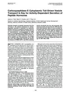
cytoplasmic tailcDNA(HindII13043-E~oR13805) was usedas a template for Adhesion Assays-Adhesion assays were carried out essentially as amplification. TheHindII13043site was introduced intoa4 as previously described (48). Briefly, cells were labeled by incubation with thefluodescribed (27). PCR products were digested with HindIII and XbaI andrescent dye BCECF-AM (Molecular Probes,Inc., Eugene, OR), and then then subcloned into pBluescriptand sequenced, pBluescript containing 5 x lo4 cells i n RPMI 1640 medium with0.1% BSA were added to each PCR products was digested with SalI and HindIII and then ligated to well of 96-well microtiter plates (Flow Laboratories, Inc. or Life Techthe remainder of the a4 cDNA (SalIPBS~"'~'i"ke'-Kpn12527 and Kpn12527- nologies, Inc.) that had been coated with protein ligands and blocked HindII13043).The ( ~ ~ - 9 7a4-977, 6, a4-979, Y976A, Y976F, K977A, and with 0.1% heat-denatured BSA. For assays analyzing cation usage, S978Aconstructs were created in pBluescript using the method of Deng fluorescently labeledcells were washed with phosphate-buffered saline and Nickoloff (42). The unique B ~ s H I I " ~ s i t ea4iwas n mutated with the containing 5 mM EDTA and then with phosphate-buffered saline and selection oligonucleotide (CACTGAGAGCCCGCTGCTC), and the finally were resuspended in 25 mM Tris-HC1, pH 7.5, 150 mM NaCl following oligonucleotides were used to introduce specific mutations: containing 0.1% BSA and 2 m~ glucose (TBSBG buffer). Then, 5 x lo4 a4-976(AAGACAATACTAATCTATCCTAC),a4-977 (ACAATACAAATA- of these cells were plated into each microtiter well already containing GATCCTACAAG), a4-979 (CAAATCTATCTAACAAGAAGAAA), Y976A divalent cation a t twice the final concentration in TBSBG buffer. After (TAAAAGACAAGCCAAATCTATC),Y976F (TAAAAGACAATTCAAAT- 30 min of incubation a t 37 "C, unbound cells were removed (three CTATC), K977A(AAGACAATACGCATCTATCCTAC), and S978A(ACA- washes with phosphate-buffered saline usinga multichannel pipetter). ATACAAAGCTATCCTACAAG).Sequencing of plasmids confirmedthat Cells remaining attached to the plate were analyzed using Cytofluor the only the intended mutations were introduced. 2300 measurement system(Millipore Corp., Bedford, MA). Background Constructs were excised from pBluescript with SalI-XbaI digestion binding (assessed using BSA-coated wells) was typically 4 % of the and subcloned into pSR~xNeo.~ Chimeric a4 containing the cytoplasmic total and was subtracted from experimental values. For every data domain of ' a (X4C2) wascreatedasdescribed (19). Full-length ' a point, results are reported as the means S.D. of triplicate determina(X2C2) and ' a with cytoplasmic deletion (X2CO) were created as detions. For assays in which blocking antibodies were used, cells were scribed (20). Deletion mutants of ' a were created by PCR with the incubated withblocking or control antibodies (1pg/ml) for 30 min prior introduction of a termination codon followed by an XbaI cloning site to the timeof plating. For assays in which metabolic energy inhibitors using one of,the following oligonucleotides: a'-1136 (TCTGGTCTAGAT- were used, 0.1% NaN, and 2 mM 2-deoxyglucose were added to cells TACTACTTTTCATATTTTC),a'-1139 (TCAATTCTAGATTACTA'ITTG- during cell labeling withBCECF-AM and thenwere added againat the GTCATCTTTT), or a2-1146 (CTACTTCTAGATTACTACTCATCMTCT- start of the assay. From divalent cation titration curves, values for CAT) pairedwith the common oligonucleotide (CAAGCCTTAAGT- maximal and half-maximal adhesion were estimated and used to deterGAAAGCCAAGAAA). During this procedure, an irrelevant Glu+ Lys mine the cation concentration at which half-maximal adhesion was mutation was introduced at amino acid 890 within the extracellular attained (ED,, value). domain of ' a for the a'-1139 and 01'-1146 truncation mutants. This Binding of Solubilized V U - 2 to Collagen Beads-Interaction of a'& change did not influence a2 function because these mutants had adhe- with collagen beads was carried out essentially as described (49, 501, sive properties identical to those of wild-type ' a (see Fig. S A ) . pBluewith slight modifications. Briefly, cells (2 x 10⁶/lane) were iodinated script containing ' from a was digested withAf2112784 and XbaIPBSply'1nker and using the lactoperoxidase method (with divalent cations excluded ligated with thePCR products that hadbeen similarly digested. The ' a the reaction). Following washing, cells were lysed in 0.1 M octyl thiocDNAs containing premature truncations were excised from pBlueglucopyranoside (Calbiochem), 0.1 M n-octyl glucopyranoside (Sigma)in script using SalI and XbaI and subcloned into the pECE expression Tris-buffered saline containing protease inhibitors(0.15 mg/ml phenylvector (43). The pMDR901 plasmid encoding the dhfr gene (44) was methylsulfonyl fluoride,5 pg/ml leupeptin, and5 pg/ml aprotinin). Following clarification by centrifugation, Mn2+ was addedat various conkindly provided by Dr. M. Rosa (Biogen Inc.). Dansfection, Selection, and Cell Culture-MIP101 carcinoma cells centrations that were maintained during preclearing with mAb J2A2 were transfected using Lipofectin reagent (Life Technologies, Inc.), se- and protein A-Sepharose (1 h, 4 "C) and subsequent incubationof cell lected in 2.0 mg/ml G418, and then enrichedfor cell-surface expression lysates with collagen-Sepharose beads for 18 h a t 4 "C. Beads were using anti-a4mAb B5G10 and goat anti-mouse Ig-coated immunomag- washed six times with 1 ml of Tris-buffered saline containing 0.025 M netic beads (Dynal, Inc.). The PMWK melanoma and K562 erythroleu- octyl thioglucopyranoside, 0.025M n-octyl glucopyranoside, and the apkemia cell lines transfected withX4CO and X4C4 (19) as well as the RD propriate concentration ofMn". Nonreducing SDS sample buffer was added to washed beads,boiled for 3 min, and loaded ontoa 7% polyacrhabdomyosarcoma and K562 cell lines transfected with X2CO and X2C2 (20) were previously described. Stably transfected MIP101, K562, rylamide gel for SDS-PAGE. The quantity of 'a was determined by PMWK, and RD cell lines were cultured in RPMI 1640 medium supple- scanning of the autoradiograph usinga n Ultrascan XL enhanced laser mented with 10% fetal bovine serum, L-glutamine, and antibiotics. Chi- densitometer (PharmaciaBiotech Inc.). nese hamster ovary (CHO) cells (lackingthe dhfr gene) were cotransRESULTS fected by electroporationwith pMDR901 togetherwithexpression vector containing ' a or a4 mutants and then selected as previously Analysis of Adhesion Mediated by a* Cytoplasmic Deletion described (45). Cells were enriched for cell-surface expression by fluo- MutantsinMIPlOl Cells-To identify specific residuesinrescence-activated cell sorting using mAb 12F1 followed by fluorescein of adhesion by the a4cytoplasvolved in the positive regulation isothiocyanate-conjugatedgoat anti-mouse Ig (Calbiochem) (a2transmic tail, wild-type a4 and multiple truncated a4 constructs (Tafectants) or phycoerythrin-conjugated mAb HP1/2 (a4transfectants). ble I,upper) wereexpressed intheMIPlOl cell line at Stable CHO cell lines were maintained in alpha minimal essential medium without ribonucleosides and deoxyribonucleosides containing moderate t o high levels as determinedby flow cytometry (Fig. 10% dialyzed fetal bovine serum. 1).In cell adhesion assays, MIPlOl cells transfected with the Flow Cytometry and Immunoprecipitation-Indirect immunofluoresa4-974 construct (also called X4CO) displayed no attachment to cence and flow cytometry were performed using a FACScan machine CS1-BSA at anycoating concentrationtested (Fig. 2 A ) . Also, no (Becton Dickinson Co., San Jose, CA) as described (46). Cell-surface expression levels were estimated using computer-generated mean flu- attachment to sVCA"1 was observed at low coating concentrations, and only limited adhesion occurred at the highest orescence intensity (MFI) values. MFI values due to autofluorescence were obtained using matched negative control antibodies and were concentrations of sVCA"1 (Fig. 2B), as previously demonsubtracted toyield specific MFI values. The percentage of p1expressing strated (19). In contrast, the a4-980, ( ~ ~ - 9 8a4-990, 4, and a4-997 the mAb 9EG7 epitope was calculated from (MF19EG7/MFI~l) x 100. The transfectants showed pronounced adhesion to CS1-BSA (Fig. percentage of associatedwith a4 wasdetermined from (MFI,,,,d 2 . 4 ) and sVCA"1 (Fig. 2B) comparable t o that seen with the MFI,,, x 100 in the case of K562 cells and from (MFIB5Gl@IFIP1) x X4C4 transfectants. Complete inhibition by anti-a4mAb HP1/2 100 in the case of CHO cells. indicated that adhesion was a4-dependent (data not shown). Following CHO cell-surface labeling using sulfosuccinimidyl "ioAlthough eachof the constructs was notexpressed at exactly tinamido)hexanoate (Pierce ChemicalCo.), cells were lysed and immunoprecipitated as described (471, and then proteins were treated with the same level in MIPlOl cells (Fig. l),this variation does not N-glycosidase F (New England Biolabs Inc., Beverly, MA). Samples alter the essential conclusions derived from Fig. 2. For exwere then separated by 7% SDS-PAGE, transferred t o nitrocellulose, ample, ( ~ ~ - 9and 8 0 X4C4 showed substantially greateradhesion blotted with peroxidase-conjugated ExtrAvidin (Sigma), and visualized at than X4CO (a4-974), eventhough they wereexpressed using the Amersham ECL kit. H. Band, manuscript in preparation.
slightly lower levels. Because the a4-990 and a4-984 proteins were expressed at 1.4-1.6-fold higher levels compared with the
Integrin a Chain Cytoplasmic Domain
Deletion Effects
19861
TABLEI List of d and a* mutants Cell line 'a constructs
Cytoplasmic sequence tail
x 4 c 4 (wt)" X4CO (974) 976 977 979 980 984 990 997
KAGFFKRQYKSILQEENRRDSWSYINSKSNDD KAGFFKR KAGFFKRQY KAGFFKRQYK KAGFFKRQYKSI KAGFFKRQYKSIL KAGFFKRQYKSILQEEN KAGFFKRQYKSILQEENRRDSWS KAGFFKRQYKSILQEENRRDSWSYINSKSN
Y976A Y976F K977A S978A X4C2
KAGFFKRQAKSILQEENRRDSWSYINSKSNDD KAGFFKRQFKSILQEENRRDSWSYINSKSNDD KAGFFKRQYASILQEENRRDSWSYINSKSNDD KAGFFKRQYKAILQEENRRDSWSYINSKSNDD KAGFFKRKYEKMTKNPDEIDETTELSS
a2constructs
X2x2 (wt) X2CO (1132) 1136 1139 1146
KLGFFKRKYEKMTKNPDEIDETTELSS KLGFFKR KLGFFKRKYEK KLGFFKRKYEKMTK KLGFFKRKYEKMTKNPDEIDE
RD
MIP
CHO
+
+
+
+
+
+
+ + + +
+ +
+ + + + + +
+
+ + + + +
wt, wild-type.
more adhesive than CHO-X4CO cells (Fig. 3 A ) . However, this difference was not nearly as dramatic as that observed in a4transfected MIPlOl or PMWK cells (see Fig. 2 and Ref. 20). No adhesion by mock-transfected CHO-Neo cells was observed (Fig. 3 A ) , indicating that adhesion mediated by both CHO-a4 transfectants is entirely a4-dependent. In a search for conditions that might alter therelative adhesive capabilities of CHO-X4C4 and CHO-X$CO cells, we examined the effects of different levels of Mg2' (Fig. 3B). In the presence of 10 mM M e , CHO-X4CO and CHO-X4C4 showed similar levels of adhesion at three of the four doses of sVCA"1 tested. In contrast, in the presence of 0.3 mM M e , adhesion of CHO-X4CO cells was considerably diminished in comparison with CHO-X4C4 cells, as also seen inFig. 2 and elsewhere (19) with other cell lines. Consistent with these results, we have generally found that titration of divalent cation (at a constant dose of ligand) yields a more sensitive and reproducible indication of relative integrin activity compared with results obLog Fluorescence Intensity tained from titration of ligand (at a constant dose of divalent Frc. 1. Expression of a4mutants on MIP101 cells. MIPlOl cells cation). Thus, for a simple, comprehensive, and quantitative transfected with pSRaNeo alone (Neo) or with pSRaNeo containing evaluation of integrin adhesive activity states, we have focused X4C0, a4-980, a4-984, a4-990, a4-997, or X4C4 were stained with the on divalent cation ED,, values corresponding to half-maximal negative control mAb J2A2 (dotted line) or the a4-specificmAb B5G10 (solid line).The indicated values for mean fluorescence intensity were adhesion as determined from divalent cation titration curves obtainedby flow cytometry as described under "Materials and Methods." (obtained at a constant dose of ligand, usually near or just below saturation). others,their adhesiveactivities could be slightlyoverestiFine Mapping of u4 Cytoplasmic Tail Sequences Involved in mated. In this regard,we found previously that thelevel of a4 Regulation of Adhesion-To analyze further thefunctional imexpression in MIPlOl cells roughly correlates with adhesive portance of the QYKSIL region in the a4 tail, additional a4 only 6 amino acids mutants containingpoint mutations or termination codons activity (19). Nonetheless, it is clear (i) that (QYKSIL) following the GFFKR motif are sufficient to restore within this 6-amino acid region were generated. These addialmost completely the constitutiveadhesive function of a4 and tional mutants (listed in Table I, center) were expressed in (ii) that aminoacids at positions 10-25 (after the GFFKR motif') CHO cells at similar levels as indicated by flow cytometry (Fig. do not cause substantial further changes in adhesive activity. 4). Immunoprecipitation analysis of the N-glycosidase-treated Effects of a Chain Tail Mutations Can Be Amplified or Sup- 70-kDa fragment of a4 showed that the apparent molecular pressed Depending on Divalent Cation Levels-Having found masses of the wild-type a4-999, a4-997, a4-990, a4-984, a4-980, that only 6 amino acids within the a4 tail exerted a positive a4-979, a4-977, a4-976, and a4-974 deletion mutants were proeffect on cell adhesion, we next wanted to determine (i)exactly gressively diminished (by 0-6 kDa), which is entirely consistwhich amino acidsweremostcritical and (ii) whether this ent with theexpected sizes. result would extrapolate to other cell types and to other inteAdhesion of CHO-a4 transfectants, examined over a range of grins. Thus, we expressed bothX4C4 and X4CO in CHO cells at Mg2' concentrations, revealed that ED,, values for CHO-974 comparably high levels on the cell surface (see Fig. 4). When were 2-3-fold greater than those for CHO-X4C4 on CS1-BSA tested for adhesion to sVCAM-1, CHO-X4C4cells were clearly (Fig. 5 , upper) and sVCAM-1 (Fig. 5 , lower). Thus, CHO-974
19862
Integrin a Chain Cytoplasmic Domain Deletion Effects
n
FIG.2. Adhesionof
a4 cytoplasmic
truncation mutants to CS1-BSA and SWAM-1. Adhesion assays were carried out as described under “Materials and Methods” using RPMI 1640medium and 0.1% BSA. MIPlOl cells expressing the a4 truncation mutants were assayed for their adhesion to various concentrations of CS1-BSA (A) or sVCAM-1 ( B ) . The means t S.D. of triplicate determinations are reported.
E E
2 5
750
550
o
=
8
450
350
[CSl-BSA], Fg/ml
650
[sVCAM-I], bg/ml
-
-s -H m
i
Ie
I -50
.
0
~
1
2
~
3
~
4
5
.
t
~
~
~
~
6
1000 900 800 N
E E
700
.-
600
n
U
m
C 500
3
2
2 400
c
e
2 300 Q 0
!-
9
200
100
0 0
1
2
3
4
5
6
[sVCAM-11, pglrnl 3, Adhesion of a4-transfectedCHO cells. Adhesion of CHOX4CO and CHO-X4C4 transfectants to various concentrations of sVCA”1 was assessed as described under “Materials and Methods” using a-minimal essential medium containing 0.1% BSA ( A )or TBSBG buffer with either 10 or 0.3 m~ Mg2’ ( B ) . FIG.
Log Fluorescence Intensity
FIG. 4.Expression of a4 and d deletions and point mutations in CHO cells. Cell-surfacestaining and flow cytometry were performed using the a4-specificmAb B5G10 (solid line) and the negative control mAb J2A2 (dotted line) as described under and Methods,,, Cell-surface expression of a2 was determined by flow cytometry using mAb 12F1 (solid line), and mAb J2A2 was used as a negative control
(dotted line). Mean fluorescence intensity values for the 11 positive a4 transfectants ranged from 62 to 105, with an average of 89 2 12 MFI units. The five positive a2 transfectants ranged from 72 to 121, with an
cells utilized divalent cationsless efficientlyto supportadhesion. Notably, CHO-976 and CHO-977 cells showed intermedi- average Of 93 * 2o MF1 units. ate ED,, values, indicating thatonly 2-3 additional amino acids areable to cause partialrecovery of cation usage during cell CS1-BSA was entirely dependenton a4expression because Neo no adhesion toeithersubstrate adhesion to CS1-BSA and sVCA”1 mediated by a4p1.Thetransfectantsdemonstrated presence of only 5 or 6 a4 amino acids after the GFFKR region (e.g. see Fig. 3A). Furthermore, adhesion t o sVCA”1 and CS1restored a4-mediated adhesion to nearly its full activity, with BSA was inhibited (85 and 92%, respectively) by the a4-specific ED,, values almost as low as those observed for X4C4 adhesion mAb HP1/2 even when the highest concentration of Mg2‘ (30 to CS1 peptide and sVCAM-1. These results, obtained using mM) was used (data not shown). CHO cells (summarized in Fig. 6A), are consistent with thoseAlthough the deletion results suggested that the QYKSIL seen with MIPlOl cells (Fig. 2). region in a4 may be a critical motif needed for adhesion, the For a4-transfected CHO cells, adhesion to sVCA”1 and point mutations Y976A, Y976F, K977A, and S978A didnot
.
Integrin a Chain Cytoplasmic Domain
Deletion Effects
19863
wild-type a2p1and thecytoplasmic deletion mutant (ax2coPl) for binding to collagen-derivatized Sepharose beads. Both wildtype and mutant lZ5I-labeleda2P1from CHO cells showed increased binding to collagen beads as the level of Mn2+was raised (Fig. 7, right panels). Quantitation by densitometry showed essentially no disparity between the relative amounts of X2COand X2C2 bound to collagen beads at any concentration of Mn2+ (Fig.8B). Thus, cytoplasmic domain deletion did not alter the intrinsic ligand binding capability of the mutant a2P1 integrin relative to thewild type. The results withsolubilized a2p1contrasted markedly withcell adhesion results, where the Mn2+ED,, value for X2CO (0.019 mM) was 9-fold higher than that for X2C2 (0.0021 mM) (Fig. 8 A ) . In a positive control experiment, we did find that mAb TS2/16 caused more a2p1(isolated from K562 cells) to bind t o collagen beads at each of the Mn2+concentrations tested (Fig. 7, leftpanels). Quantitation of the bands revealed that mAb TS2/16 caused maximal binding to be achieved at 10-foldlower concentrations of Mn2+(Fig. 8C). Thus, because collagen binding was markedly influenced by mAb TS2/16, an agentshown to directly affect the affinity of an integrin for its ligand (49, 51, 52),we should have been able to detect binding differences between X2CO and X2C2 in our detergent lysates if they had occurred. Loss of a4Deletion Effects upon Addition of Metabolic Energy Inhibitors-Because of a generally weak interaction of solubilized a4P1with VCAM-coated beads, experiments such as those described for a2(Figs. 7and 8) were technically not feasible. As an alternative, we analyzed the effect of metabolic energy inhibitors on adhesion mediated by a4wild-type and cytoplasmic deletion constructs (Fig. 9). Titration of Mg2' yielded typically distinct ED,, values for CHO-974 and CHO-X4C4 transfectants (0.81 and 0.11 m Mg", respectively). When cells were treated with 0.1% azide (N,) and 2 mM 2-deoxyglucose, adhesion of both transfectants wasdecreased, and theED,, values became more similar (0.96 mM for CHO-974 and 0.72 mM for CHO-X4C4). A similar result wasobtained in two other experiments (data not shown).These results suggest that differences in adhesion caused by cytoplasmic domain deletion are largely dependent on metabolic energy and do not involve stable alterationsof the integrin itself. Absence of Detectable Conformational Difference between a Wild-type and Cytoplasmic Deletion Mutants-To assess whether a chain cytoplasmic domain deletion could cause a conformational change,we analyzed the expression of the mAb 9EG7 activation epitope (38) on the integrin PI chain. Notably, no differences in mAb 9EG7 epitope expression were observed between CHO-X4CO and CHO-X4C4 transfectants (Fig. lOA), between K562-X4CO and K562-X4C4 cells (Fig. lOC), or between K562-X2CO and K562-X2C2 cells (Fig. 1OB).Addition of 10 mM MnC1, t o cells during the staining procedure caused increased expression of the mAb 9EG7 epitope in each of the transfected cell lines, but still no differences were observed between wild-type and deleted a chains. Because a high proportion of the total P1 (4645%) was associated with the relevant transfected a subunits (see Fig. 10 legend), we should have been able to detect a conformational difference had it occurred. In multiple other experiments (data not shown), using Mn2+(0.1-10 m)and Mg2' (0.1-50 mM) and several independently transfected cell lines, we never observed a chain deletion having an effect on the appearance of the mAb 9EG7 epitope. Additionally, a chain deletion did not alter the stability of the mAb 9EG7epitope; after induction with Mn2+, the epitope was notreadily lostupon washing with divalent cationfree phosphate-buffered saline.
I CSI-BSA
0 CH0974 ED5040.5 mM 0 CH0976 ED50d.9 mM CH0977 ED50=4.8 mM 0 CHOX4C4 ED50=4.4 mM
(r4
SVCAM-1
ED5k0.51 mM ED50r0.37 mM
ED50r0.23 mM ED50-0.15 mM
FIG.5. Effect of M e concentration on adhesion mediated by deletion constructs inCHO cells. Adhesion to CS1-BSA (2.5 pg/
ml; upper) and sVCA"l(2.0 pg/ml; lower) was examined as described under "Materials and Methods."
cause a significant loss of adhesive activity, as shown by the ED,, values listed inFig. 6B. Furthermore, replacementof the entire a4tail with thatof a2(to give an X4C2 construct) did not cause a marked alteration inED,, values. Analysis of d Cytoplasmic Tail Sequences Involved in Regulation of Adhesion-CHO cells were also transfected with integrin az constructs containing cytoplasmic domain deletions of varying length (listed in Table I, lower). Each construct was expressed a t comparable cell-surface levels (Fig. 41, and adhesion to collagen was assayed over a range of Mg2' concentrations. In seven experiments, the ED,, values for CHO-1132 cells (also called CHO-X2CO) were elevated by an average of 11-fold compared with CHO-X2C2 cells. Notably, the presence of only 4 amino acidsfollowing GFFKR (a2-1136)was sufficient to cause a significant decrease in the ED,, value, and 7 (a21139) or 14 (a2-1146)amino acids following the GFFKR motif yielded adhesion results (and ED,, values) not significantly different from those obtained with X2C2. Results from several a2 ED,, determinations are summarized in Fig. 6A. Replacement of the entire a2 cytoplasmic domain with that of a4 or a5 (X2C4 and X2C5 constructs) did not cause a major change in ED, values.Compared with the a4 and a5 cytoplasmic domains, the a2 tail occasionally yielded lower ED,, values, indicative of higher activity, but thiswas not a consistent finding (data notshown). Adhesion of CHO-a2 transfectants to collagen was entirely a'-dependent since CHO-pMDR901 (mock transfectant) showed no adhesion to collagen, and adhesion of CHOX2C2 was completely inhibited by a n t i d mAb 5E8 underconditions of maximal adhesion (data notshown). Loss of d Deletion Effects inCell-freeCollagen Binding Assay-To determine whethera chain taildeletion effects were retained ina cell-free assay, we compared detergent-solubilized
Integrin a Chain Cytoplasmic Domain Deletion. I3ftict.s
19864 1.4 h
+ C
FZ VCAM-1
T
1.2
(0
.cI
1.0 \
(I)
2 0.8 23 0.6 .cI
v
0
*=
0.4
"0- SVCAM-1
*CSI-BSA +
(0
a: 0
In
0.2
Collagen
n
0.0 0
5
102 0
15
S970AK977AY976F 25 Y976A
Amino Acid length (after GFFKR)
a2-
PI-
XdC2
Alpha-4 Mutant
Integrin a Chain Cytoplasmic Domain
Deletion Effects
19865
0 CHOX2C2 0 CHOX2CO .1
1
-100 .001 120
100
"
.01
.01
1
.1
1
I
10
100
[Mg2+1, mM CH0974 CHOX4C4 0 CH0974+N3/DOG 0 CHOX4C4+NWDOG
5
ED50c0.81 m M ED50c0.11 m M ED50~0.96m M ED50c0.72 m M
FIG.9. Cytoplasmic domain deletion effects in presence and absence of metabolic energy inhibitors. Various CHO-a4 transfectants were untreated (0 and W) or treated with 0.1% NaN, and 2 mM 2-deoxyglucose (DOG)(0and 0) for 30 min prior to assay. The adhesion assaywasperformed as describedunder"Materialsand Methods" (TBSBG buffer). One representative experimentof three is shown.
Soluble VLA-2 . 0 CHOX2C2
120
P 0
A.
H CHONeo
B. Ed KX2CO
CHOX4CO
C.
H KNeo KX4CO
.
0 CHOX2CO .001
.01
.1
1
0
10
0
10
0
10
[MnC12], rnM
t
0 .001
.01
.1
1
[Mn2+], rnM FIG.8. Comparison of cell adhesion to collagen and soluble d p , binding to collagen. Adhesion of CHO-X2C2 and CHO-X2CO to collagen (5 pg/ml) in the presence of various Mn" concentrations was performed as described under "Materials andMethods" (A). Binding of detergent-solubilized azP1to collagen beads was carried out for integrin from CHO-X2CO and CHO-X2C2 transfectants ( B ) or from K562-X2C2 transfectants (in the presence or absence of mAb TS2/16) (6).The quantity of a' was determined by densitometry of the autoradiograph and is presented as a percentage of maximal binding.
cytoplasmic tails exerted comparable positive effects on adhesion (22,53,54)despite having totally different sequencesafter the GFFKR motif. Similarly, only 7 amino acids afterthe GFFKR region in the a" sequence were sufficient to support nearly maximal adhesion (551,even thoughthese 7 amino acids (VRPPQEE) bear no resemblance to the functionallyactive sequences after GFFKR in a4 (QYKSILQ) and a' (KYEKMTK).
FIG.10. Induction of mAb 9EG7 activation epitope on wildtype and a chain cytoplasmicdeletion mutants in CHO and K562 cells. CHO (A) or K562 ( B and C) cells transfected with Neo alone, X4C0, and X4C4 (A and C ) or with X2CO and X2C2 ( B ) were stained with mAbs in the presence or absence of Mn2+ (10mM). Data are expressed as a percentage of PI expressing themAb 9EG7 epitope relative to the total p1 (measured using mAb 13 or 7E2) as described under "Materials and Methods." Approximately 71 and 85% of the p1 in CHOX4CO and CHO-X4C4 cells is associated witha4,46 and 73% of the p1 in K562-X4CO and K562-X4C4 cells is associated witha4,and 55 and 6 0 8 of the p1 in K562-X2CO and K562-X2C2 cells is associated with 2 , respectively.
Our results also argue against an essentialrole for integrin a chain phosphorylation in the regulation of cell adhesion. Al-
though sometimes correlated with increased adhesive activity, phosphorylation is clearly not essential since mutation of potential sites within the critical a4 QYKSIL sequence had no effect on adhesion. The need for 5-7 amino acids may reflect a nonspecific anchoring effect of the a chain in the membrane or a general folding effect. In this regard,a putative p turn in thecytoplasmic domain of aV (and other a chains including a4)was suggested to support adhesion (55). However, our a4-979 mutant exhibited wild-type levels of adhesion, despite lacking the proposed p turn in the a4 cytoplasmic tail, and our functionally
19866
Integrin
CY
Chain Cytoplasmic Domain Deletion Effects
active a'-1139 mutant also lacks a turn. Thus, a p turn in the region just after GFFKR does not appear to be required for positive regulation of adhesive function. Alternatively, 5-7 a chain amino acids may be sufficient to block a negative regulatory region in the tail. For example, the interaction of a-actinin with P1occurs proximalto the membrane (56), where the presence of an a chain could conceivably interfere. In this regard, the presence of an a chain causes diminished affinity of a-actinin for p (57).The ligand-independent localization of integrin p1and ps chains to focal contacts in the absence of an a tail (29, 58, 59) is another example of a cytoplasmic tails having a negative regulatory effect. Notably, the length of a chain tail needed t o exclude a2 from ligandindependent focal adhesion sites correlates closely with the tail length that makes a positive contribution toward cell adheion.^ Thus, the same regulatory site on the p chain that is exposed by a chain deletion could conceivably cause both diminished cell adhesion as well as ligand-independent focal adhesion formation. From the discussion above, it appears that several different integrins (a2,a4,a,, a6*,d B , and a") may utilize their diverse sequences in a similar general mechanism for control of constitutive adhesion through inside-out signaling. In contrast,for outside-in signaling events, a chain tails may contribute in a more specific manner. For example, although a', a4,and a5 contributed similarly to adhesion, they differed in their participation in post-ligand binding events such as collagen gel contraction and cell migration (27).Also, the & and & cytoplasmic domains contribute similarly t o adhesion, but differ markedly in post-adhesion functions (45). Although various studies have indicated that integrin a chains may exert a confusing array of positive, negative, or neutral effects on cell adhesion, close analysis reveals no major contradictions. For example, deletion of aL (leaving 12 amino acids following GFFKR) and deletion of a5 (leaving 4 amino acids following GFFKR)had no obvious effect onadhesion (25, 28). However, because these deletions were not closer to the GFFKR motif, the results obtained are not inconsistent with our results (as summarized in Fig. 6). In other experiments, deletions ofthe a' (291, a5(281, and aM (21)cytoplasmic domains did not cause a pronounced loss of cell adhesion, probably because they were carried out using relatively high levels of divalent cations, in cells (such as 3T3,CHO, and COS) that support high constitutive integrin activity. For example, using CHO cells at intermediate levels of divalent cations (such as found in minimal essential medium or RPMI 1640medium), we also failed t o observe major a4 or a2 tail deletion effects. In contrast, these effects were much more dramatic in MIPlOl cells or in CHO cells assayed in the presence of lower divalent cation levels. Thus, we predict that at lower levels of divalent cations (e.g. 0.1 mM Mg2' or 0.01 mM Mn"), quite different a' (29), a5 (28), and aM (21) deletion results would be obtained, perhaps more similar t o those reported here. In two other experiments, deletion of the allb tail gave elevated adhesion (18),and complete deletion of the a5tail hadno effect on adhesion (28). These two results do not contradict the results shown here because in both cases, deletion occurred before the highly conserved GFFKR motif. If was deleted just after GFFKR instead of before, it had a much less active phenotype (59,60),consistent with our results. Unfortunately, we were not able to analyze a2 or a4constructs deleted before GFFKR because they were not expressed on the surface of CHO,K562, or MIPlOl cells despite repeated attempts. Although not at the surface, high levels of a4 without GFFKR were observedinside K562 cells.2Thus, removal of GFFKR may S. Kawaguchi, and M. E. Hemler, submitted for publication.
have a destabilizing effect, perhaps because the KR residues are needed for transmembrane anchoring of the a chain. Although many a chain sequences (e.g. a', a4,a,, a6*,d B , and a") may exert similar positive effects on cell adhesion, all a subunit tails are not exactly equivalent in thisregard (18, 60). Recently, it was demonstrated that the 2 , a,, a6*, and d B cytoplasmic tails bestow a high affinity state on the a"& integrin in CHO cells, while the alIb, aL,a', and a" cytoplasmic tails specify a low affinity state inCHO cells (60). Given that a4, a,, and a2 tails had equivalent activity, we would place the a4 cytoplasmicsequence in the former group. Notably, as discussed above, the first 5-7 amino acids present after GFFKR can have a positive effecton adhesion mediated by a chains ( 2 , a4,and a") found in either group. We found that for a comprehensive analysis of cytoplasmic domain deletion effects, divalent cation titrations at constant doses of ligand were moreinformative than ligand titrations at constant doses of divalent cations. For example (as mentioned above), in highly active cell types, the levels of divalent cations in normal media (at any ligand dose) weresufficient to obscure the effects of a tail deletion mutations. In general, determination of ED,, values allows a good estimation of the activation state of any integrin in any cell, using cheap and readily available reagents ( i e . divalent cations). It should be expected that subtle differences between integrin adhesive functions would be highly sensitive to divalent cation levels, considering that divalent cation-binding sites may be in close proximity to ligand-binding sites (61-63). An additional advantage ofED,, determinations is that they are easily determined in the context of cell adhesion. In contrast, quantitative methods involving affinity determinations may overlook important contributions to adhesion (e.g. adhesion strengthening effects) that do not cause an affinity change. Furthermore,the ligands for many integrins are too large, bind too weakly, or yield too much background binding, thus precluding accurate affinity measurements. From our results, it is not exactly clear how deletion of a chain cytoplasmic domains would alter the efficiency of divalent cation usage (ED,, value) during cell adhesion. One possibility is that cell-surface distribution of integrin heterodimers may be closely regulated by the a chain cytoplasmic domain, and higher levels of cations may be required to overcome the deficiency in regulation introduced by cytoplasmic truncation. Such a mechanism would not need t o invoke direct alterations in the actual integrin ligandcation-binding site. Consistent with this, using mAb 9EG7 against an activation epitope on the p1chain (38), we were unable to detect a conformational difference between integrins containing wild-type or deleted cytoplasmic a chains. Nonetheless, it is still possible that a chain cytoplasmic domaindeletion could cause an alteration in integrin conformation not recognized bymAb 9EG7,especially since it hasbeen shown elsewhere that manipulation of a chain cytoplasmic domains could alter the conformation and ligand binding affinity of the a"& integrin (18, 60). The adhesive activities of other cell-surface molecules,such as CD2 (641, PECA"l/CD31 (651, E-cadherin (661, and CD44 (67), are also markedly influenced by cytoplasmic domain mutations. As for integrins, it is not yet clear whether the cytoplasmic domains of these molecules control receptor distribution, ligand binding conformation, or both. In anotherexample, truncation of the carboxyl-terminal 11amino acids of L-selectin caused decreased adhesion without affecting ligand binding (68). Regardless of the precise mechanisms by which a chain tails support integrin-mediated cell adhesion, it isnotable that the effects of a tail deletion were largely lost in the presence of
Integrin
(Y
Chain Cytoplasmic Domain Deletion Effects
19867
J. Zmmunol. 151,990-1002 22. Shaw, L. M., and Mercurio, A. M. (1993) J . Cell Biol. 123, 1017-1025 23. Hayashi, Y., Haimovich, B., Reszka, A,,Boettiger, D., and Horwitz,A. (1990) J. Cell Bid. 110, 175-184 24. Hibbs, M. L., Xu, H., Stacker, S. A,, and Springer, T. A. (1991) Science 251, 1611-1613 25. Hibbs, M. L., Jakes, S., Stacker, S.A., Wallace, R. W., and Springer,T.A. (1991) J. Exp. Med. 174, 1227-1238 26. Chen, Y P., Djaffar, I,, Pidard, D., Steiner, B., Cieutat, A. M., Caen, J. P., and Rosa, J. P. (1992) Proc. Natl. Acad. Sci. U. S. A. 89, 10169-10173 27. Chan, B. M. C., Kassner, P. D., Schiro, J. A., Byers, H. R., Kupper, T. S., and Hemler, M. E. (1992) Cell 68, 1051-1060 28. Bauer, J. S., Varner, J., Schreiner,C., Kornberg, L., Nicholas, R., and Juliano, R. L. (1993) J. Cell Biol. 122,209-221 29. Briesewitz, R., Kern, A,, and Marcantonio, E. E. (1993) Mol. Biol. Cell 4, 593-604 30. Hemler, M. E., Sbnchez-Madrid, F., Flotte, T. J., Krensky, A. M., Burakoff, S. J., Bhan, A. K., Springer, T. A,, and Strominger, J. L. (1984) J. Zmmunol. 132,3011-3018 31. Pischel, K.D., Hemier, M. E., Huang, C., Bluestein, H. G., and Woods, V. L. (1987) J. Zmmunol. 138,226-233 32. Weitzman, J. B., Pasqualini, R., Takada, Y., and Hemler, M. E. (1993)J. Biol. Chem. 268,8651-8657 33. Hemler, M. E., Huang, C., Takada, Y., Schwarz, L., Strominger, J. L., and Clabby, M. L. (1987) J . Biol. Chem. 262, 11478-11485 34. Sanchez-Madrid, F., De Landazuri, M. O., Morago, G., Cebrian, M., Acevedo, A,, and Bernabeu, C. (1986)Eur. J. Zmmunol. 16,1343-1349 35. Kennel, S. J., Foote, L. J., and Lankford, P.K. (1981) Cancer Res. 41, 34653470 36. Akiyama, S. K., Yamada, S. S.,Chen, W.-T., and Yamada, K. M. (1989)J , Cell Biol. 109,863475 37. Brown, P. J., and Juliano, R. L. (1988)Exp. Cell Res. 177, 303-318 38. Lenter, M., Uhlig, H., Hamann, A., Jeno, P., Imhof, B., and Vestweber, D. (1993) Proc. Nutl. Acad. Sci. U. S. A. 90,9051-9055 39. Hemler, M. E., and Strominger, J. L. (1982) J. Zmmunol. 129,2734-2738 40. Yelton, D. E., Desaymard, C., and ScharfT, M.D. (1981) Hybridoma 1, 5-11 41. Lobb, R. R., Chi-Rosso, G., Leone, D.R., Rosa, M.D., Newman, B. M., Luhowskyj, S., Osbom, L., Schiffer, S. G., Benjamin, C.D., Dougas, I. G., Hession, C., and Chow, E. P. (1991)Biochem. Biophys. Res. Commun. 178, 1498-1504 Acknowledgments-We thank Dr. Roy Lobb for providing sVCA"1 and phycoerythrin-conjugated mAb HP1/2, Dr. Tadashi Shimo-Oka for 42. Deng, W. P., and Nickoloff, J. A. (1992)Anal. Biochem. 2 0 0 , 8 1 4 8 donating CSI-BSA, Dr. Hamid Band (Brigham and Women's Hospital, 43. Ellis, L., Clauser, E., Morgan, D. O., Edery, M., Roth, R. A., and Rutter, W. J. (1986) Cell 45, 721-732 Boston, M A ) for contributing the pSRaNeo vector, and Dr. M. Rosa for 44. Kaufman, R. J., and Sharp, P. A. (1982) J . Mol. Biol. 159, 601-621 providing the pMDR901 vector. We are grateful toDr. Dietmar Vestwe- 45. Pasqualini, R., and Hemler, M. E. (1994) J. Cell Biol. 125, 447460 ber (Max Planck Institute, Freiburg, Federal Republic of Germany), Dr. 46. Elices, M.J., Osborn, L., Takada, Y., Crouse, C., Luhowskyj, S., Hemler, M. E., R. L. Juliano (Universityof North Carolina, Chapel Hill, NC), Dr. Franand Lobb, R. R. (1990) Cell 60,577-584 cisco Sanchez-Madrid, andDr. Kenneth M. Yamada (National Institutes 47. Hemler, M. E., Huang, C., and Schwarz, L. (1987) J. Biol. Chem. 262, 33003309 of Health, Bethesda, MD)for providing antibodies used in this study. 48. Chan, B. M. C., Elices, M. J., Murphy, E., and Hemler, M. E. (1992) J. Biol. Chem. 267,8366-8370 REFERENCES 49. Chan, B. M. C., and Hemler, M. E. (1993) J. Cell Biol. 120,537-543 1. Miyake, K., Weissman, I. L., Greenberger, J. S., and Kincade, P.W. (1991) J. 50. Bergelson, J. M., Chan, B. M. C., Finberg, R. W., and Hemler, M. E. (1993) J . Exp. Med. 173,599-607 Clin. Znuest. 92, 232-239 2. Utsumi, K., Sawada, M., Narumiya, S., Nagamine, J., Sakata,T., Iwagami, S., 51. F a d , R. J., Kovach, N. L., Harlan, J. M., and Ginsberg, M. H. (1993) J. Cell Kita, Y., Teraoka, H., Hirano, H., Ogata, M., Hamaoka, T., and Fujiwara, H. Biol. 121, 155-162 (1991)Proc. Natl. Acad. Sci. U. S. A. 88, 5685-5689 52. Arroyo, A. G., Garcia-Pardo, A., and Sanchez-Madrid, F. (1993)J. Biol. Chem. 3. Roldan, E., Garcia-Pardo, A,, and Brieva, J. A. (1992)J. Exp. Med. 175, 1739268,9863-9868 1747 53. Delwel, G. O., Hogervorst, F., Kuikman, I., Paulsson, M., Timpl, R., and Son4. Issekutz, T. B., and Wyrkretowicz, A. (1991) J. Zmmunol. 147, 109-116 nenberg, A. (1993) J . Biol. Chem. 268, 25865-25875 5. Baron, J. L., Madri, J. A., Ruddle, N.H., Hashim, G., and Janeway, C.A. (1993) 54. Shaw, L. M., Lotz, M. M., and Mercurio, A.M. (1993) J . Biol. Chem. 268, J. Exp. Med. 177, 57-68 11401-11408 6. Weg, V. B., Williams, T. J., Lobb, R. R., and Nourshargh,S. (1993) J. Exp. Med. 55. Filardo, E. J., and Cheresh, D. A. (1994) J . Biol. Chem. 269,46414647 177,561-566 56. Otey, C. A., Vasquez, G. B.. Bumdge. K., and Erickson. B. W. (1993)J. Bid. 7. Schiro, J., Chan, B. M. C., Roswit, W. T., Kassner, P. D., Pentland, A,, Hemler, Chem. 268,21193-21197 M. E., Eisen, A. Z., and Kupper, T. S. (1991) Cell 67,403-410 57. Otey, C. A., Pavalko, F. M., and Burridge, K. (1990)J . Cell Biol. 111, 721-729 8. Chan, B. M. C., Matsuura, N., Takada, Y., Zetter, B. R., and Hemler, M. E. 58. LaFlamme, S . E., Akiyama, S . K., and Yamada, K. M. (1992) J. Cell Biol. 117, (1991) Science 251, 1600-1602 437447 9. Rosen, G. D., Sanes, J. R., Lachance, R., Cunningham, J . M., Roman, J., and 59. Ylanne, J., Chen, Y., OToole, T. E., Loftus, J. C., Takada, Y., and Ginsberg, M. Dean, D. C. (1992) Cell 69, 1107-1119 H. (1993) J. Cell Biol. 122, 223-233 10. Dustin, M. L., and Springer, T. A. (1989) Nature 341, 619-624 60. OToole, T. E., Katagiri,Y., F a d , R. J., Peter, IC, Tamura, R. N., Quaranta, V., 11. Van Kooyk, Y., Van DeWiel-Van Kemenade, P., Weder, P., Kuijpers, T. W., and Loftus, J. C., Shattil, S. J., and Ginsberg, M. H. (1994) J. Cell Bid. 124, Figdor, C. G. (1989)Nature 342,811-813 1047-1059 12. Shimizu, Y., Van Seventer, G. A,, Horgan, K. J., and Shaw, S. (1990) Nature 61. Smith, J. W., and Cheresh, D. A. (1990)J . Biol. Chem. 265,2168-2172 345,250-253 62. DSouza, S.E., Ginsberg, M. H., Burke, T.A., and Plow, E. F. (1990) J . Biol. 13. Adams, J. C., and Watt, F.M. (1990) Cell 63, 425-435 Chem. 265,3440-3446 14. Marguerie, G. A., Plow, E. F., and Edgington, T. S. (1979) J. Biol. Chem. 254, 63. Loftus, J. C., OToole, T. E., Plow, E. F., Glass, A,, Frelinger, A. L., and Gins5357-5363 berg, M. H. (1990) Science 249, 915-918 15. Bennett, J . S., and Vilaire, G. (1979) J. Clin. Znuest. 64, 1393-1401 64. Hahn, W. C., Rosenstein, Y., Calvo, V., Burakoff, S. J., and Bierer, B. E. (1992) 16. Du, X.,Plow, E. F., Frelinger, A. L., OToole, T. E., Loftus, J. C., and Ginsberg, Proc. Natl. Acad. Sci. U. S. A. 89, 7179-7183 M. H. (1991) Cell 65,409416 65. DeLisser, H. M., Chilkotowsky, J., Yan, H.-C., Daise, M. L., Buck, C. A., and 17. Ginsberg, M. H., Du, X., and Plow, E. F. (1992) Curr Opin. CellBiol. 4,76&771 Albelda, S. M. (1994) J . Cell Biol. 124, 195-203 18. OToole, T. E., Mandelman, D., Forsyth, J., Shattil, S. J., Plow, E. F., and 66. Nagafuchi, A., and Takeichi, M. (1989) Cell Regul. 1, 3 7 4 4 Ginsberg, M. H. (1991) Science 254, 845-847 67. Lesley, J., He, Q., Miyake, K., Hamann, A,, Hyman, R., and Kincade, P. W. 19. Kassner, P. D., and Hemler, M. E. (1993)J . Exp. Med. 178, 649-660 (1992)J . Exp. Med. 175, 257-266 20. Kawaguchi, S., and Hemler, M. E. (1993) J . Biol. Chem. 268, 16279-16285 68. Kansas, G. S., Ley, K., Munro, J. M., and Tedder, T. F. (1993)J. Exp. Med. 177, 21. Rabb, H., Michishita, M., Sharma, C. P., Brown, D., and Arnaout, M. A. (1993) 833-838
metabolic energy inhibitors and also disappeared upon receptor solubilization. These results (i)provide assurance that (Y chain deletion does not adversely modify integrin conformation, (ii) focus attention on key energy-dependentevents occurring within intact cells, and (iii) emphasize the limitations of cellfree systems for understanding mechanisms that control integrin adhesive activity withincells. Since we found that 'a or cy4 cytoplasmic domain deletion did function, we would not not cause a stable alteration in integrin Lxpect to find any evidence for stably altered integrin conformation. Thus, it isvery surprising that deletion of the'a cytoplasmic domain causes a stable conformational change within the'a light chain that is maintained even on SDS-polyacrylamide gels (55). In this regard,deletions of the a4 cytoplasmic domain have not caused alterations in the migrationof the a4 70-kDa fragment on SDS gels that would be consistent with a stable conformational change. In summary, we have utilized a combination of cell adhesion t o demonstrate that a assays and divalent cation titrations relatively nonspecific (Y chain tail sequence of 3-7 amino acids (beyond the GFFKR motif) is sufficient to cause increased efficiency of divalent cation utilization by integrins during cell adhesion. This positive effect on cell adhesion appears t o be mediated by an unstable, energy-dependent mechanism, observable only in the context of an intactcell. Furthermore, we have shown that cytoplasmic domain effects can be greatly amplified or suppressed, depending on the divalent cationlevels utilized and the particularcell type analyzed.