Dispatch R151
Mirror Neurons: Reflecting on the Motor Cortex and Spinal Cord Neurons in the monkey motor cortex that project to the spinal cord to control particular muscle contractions and movements have been found to discharge again while the monkey simply watches another primate make similar movements: monkey see; monkey not do. Marc H. Schieber In higher primates (macaque monkeys, apes and humans), the primary motor cortex (M1) is home to the bulk of neurons that project their axons through the pyramidal tract to the spinal cord, controlling muscles and thereby movements. Many of these M1 neurons make monosynaptic connections on spinal a-motoneurons, which in turn directly innervate muscles. For more than a century, M1 was viewed as an array of ‘upper motor neurons’ that function simply to drive the appropriate muscle contractions when called upon by other parts of the brain. In recent years, however, evidence has been accumulating that the role of M1 is much more complex. As reported recently in Current Biology, Vigneswaran et al. [1] have now shown that more than half of the neurons that project from M1 to the spinal cord (pyramidal tract neurons) and discharge when the monkey pinches an object also modulate their discharge when the monkey watches a human experimenter pinch the same object. ‘Mirror neurons’— neurons that discharge when one monkey (which I will call ‘‘Mr Bananas’’) performs a particular hand grasp, and then discharge again as the same monkey watches another monkey or a human (which I will call ‘‘Fred’’) perform the same grasp—were first described by Rizzolatti and colleagues [2,3] in another cortical area anterior to M1, region F5 of the ventral premotor cortex (PMv). Mirror neurons show specificity for particular hand grasps; for example, a mirror neuron in Mr Bananas’ PMv that fires when Mr Bananas uses a precision pinch to pick up a raisin, fires again when Mr Bananas watches Fred pick up a raisin with a precision pinch. But this mirror neuron does not fire when either of them uses the whole hand to pick up a larger piece of food. Furthermore, the same mirror neuron would not discharge when Mr Bananas watches Fred pick up a raisin using
a pair of pliers, although pinching the raisin between the jaws of a pliers is similar to pinching it between the thumb and index finger. Hence the mirror neuron, rather than representing the act of picking up the raisin, reflects the particular hand grasp. Rizzolatti and colleagues [2,3] interpreted these mirror neurons as providing a neural representation of particular motor acts, the population of mirror neurons enabling Mr Bananas to understand what Fred was doing in terms of what Mr Bananas could do himself. The properties of PMv mirror neurons have been explored in a number of subsequent studies, reviewed recently by Casile [4]. Different mirror neurons may discharge when the movement executed (by Mr Bananas) and the movement observed (Mr Bananas watching Fred) are strictly congruent, only broadly congruent, or even non-congruent [3,5]. Some mirror neurons may discharge even when the actual hand shape is hidden from the monkey, as long as Mr Bananas has enough information to know what hand shape Fred is likely to be using [6]. Other mirror neurons respond to sounds that indicate a particular action, like the sound of someone opening a peanut [7]. Still others respond to communicative gestures, such as the lip smacking gesture macaques use to communicate a friendly attitude [8]. Mirror neurons thus provide an abstract, cognitive representation of various motor acts. No one expected that mirror neurons are sending information to the spinal cord during action observation until Kraskov et al. [9] reported that some mirror neurons in PMv are pyramidal tract neurons. Because the projection from PMv to the spinal cord is relatively weak, however, the effect of information coming down from PMv to the cord might have turned out to be negligible. By comparison, the projection from M1 to the cord is known to be much stronger. Now, the same research group [1] reports that over half of the M1
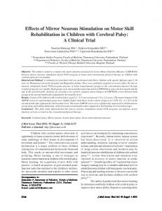
pyramidal tract neurons that project to the cord — including some that connect monosynaptically to spinal a-motoneurons which in turn directly drive muscles — show mirror properties. Mr Bananas’ spinal cord, including a-motoneurons, is being actively modulated by M1 as Mr Bananas watches Fred. Is the spinal cord also part of a distributed cognitive circuit that represents various motor acts? Vigneswaran et al. [1] advance an alternative (though not mutually exclusive) theory based on their finding that over half of the mirror pyramidal tract neurons found in both PMv and M1 increase their discharge during action execution (when Mr Bananas performs the movement himself; Figure 1A), but decrease their discharge during action observation (when Mr Bananas watches Fred; Figure 1B). Because less excitation is delivered from M1 to the spinal cord during action observation than during action execution, Mr Bananas does not move as he watches Fred: monkey see, monkey not do. This begs an interesting question, however. Why does the cerebral cortex send any activation at all to the spinal cord during action observation? Why not leave the spinal cord in the dark about the observed action unless Mr Bananas wants to make the movement himself? A similar discovery concerning transmission of apparently cognitive information below the cerebral cortex has unfolded over the past decade. In the 1970s and 1980s, some neurons in M1 [10], and many more in the dorsal premotor cortex (PMd) [11], were found to begin to discharge when a monkey receives a cue instructing him to move in a particular direction, and then continue to discharge even if the monkey has to wait several seconds before making the movement. This would be similar to the time between when a sprinter preparing to race hears, ‘‘On your mark.Get set.’’ and then ‘‘Go!’’ Such ‘directional set’ activity initially was viewed as representing the cognitive intention to execute movement in a particular direction. But directional set activity has now been found in pyramidal tract neurons, in brainstem reticulospinal neurons that project to the cord [12], and even in interneurons in the spinal cord [13]. Being distributed widely in the motor system, such directional set activity now may be viewed as priming the entire motor
Current Biology Vol 23 No 4 R152
A
PTN 1 PTN 2
B
PTN 1 PTN 2
Current Biology
Figure 1. Mirror pyramidal tract neurons. (A) Action execution. Two neurons in the motor cortex that send their axons to the spinal cord via the pyramidal tract (pyramidal tract neurons, PTNs) each fire a burst of action potentials (PTN 1 and PTN 2) as a monkey executes a precision pinch, grasping a raisin between the tips of the thumb and index finger. (B) Action observation. Each pyramidal tract neuron also responds as the monkey watches a human grasp a raisin with a precision pinch. But while PTN 1 fires another burst, the firing of PTN 2 is comparatively suppressed. The total excitation delivered to the spinal cord by the pyramidal tract neurons therefore is less during action observation, when the monkey does not execute the movement. (Monkey drawn by A. Goodman.)
system to execute the upcoming movement as quickly and efficiently as possible when the ‘‘Go’’ cue arrives. Our understanding of mirror neurons may evolve in a similar direction as future studies explore additional possibilities. During action observation, visual inputs lead to activation of mirror neurons but not to movement execution. Perhaps mirror neurons can be activated as well by internal
inputs. Humans can imagine making a movement — ‘motor imagery’ — without actually performing the movement. Studies using functional magnetic resonance imaging show activation in the same part of M1 during motor imagery as occurs during actual execution of the same movement [14]. Perhaps just as visual inputs activate mirror neurons during action observation, internal inputs activate mirror neurons during motor imagery. Furthermore, by imagining the same movement being performed over and over — ‘mental rehearsal’ — humans may actually improve their performance in tasks ranging from the seemingly non-cognitive, such as weightlifting, to the highly skilled, such as surgery and piano playing. Improved execution indicates that the mental rehearsal induced some degree of motor learning, presumably reflecting changes in synaptic strengths resulting from activity-dependent plasticity. If mirror pyramidal tract neurons eventually are found to be activated during mental imagery and mental rehearsal, then some of this synaptic plasticity may be occurring in the spinal cord [15]. Finally, both humans and monkeys can learn to perform a complex movement by watching others perform the movement. Mirror pyramidal tract neurons may cause activity-dependent synaptic plasticity in the spinal cord that helps Mr Bananas learn to perform a new movement after watching Fred: monkey see, monkey do.
3.
4.
5.
6.
7.
8.
9.
10.
11.
12.
13.
14.
15.
a neurophysiological study. Exp. Brain Res. 91, 176–180. Gallese, V., Fadiga, L., Fogassi, L., and Rizzolatti, G. (1996). Action recognition in the premotor cortex. Brain 119, 593–609. Casile, A. (2012). Mirror neurons (and beyond) in the macaque brain: An overview of twenty years of research. Neurosci. Lett. http:// dx.doi.org/10.1016/j.neulet.2012.11.003, Epub ahead of print. Rizzolatti, G., Fadiga, L., Gallese, V., and Fogassi, L. (1996). Premotor cortex and the recognition of motor actions. Cogn. Brain Res. 3, 131–141. Umilta, M.A., Kohler, E., Gallese, V., Fogassi, L., Fadiga, L., Keysers, C., and Rizzolatti, G. (2001). I know what you are doing: A neurophysiological study. Neuron 31, 155–165. Kohler, E., Keysers, C., Umilta, M.A., Fogassi, L., Gallese, V., and Rizzolatti, G. (2002). Hearing sounds, understanding actions: action representation in mirror neurons. Science 297, 846–848. Ferrari, P.F., Gallese, V., Rizzolatti, G., and Fogassi, L. (2003). Mirror neurons responding to the observation of ingestive and communicative mouth actions in the monkey ventral premotor cortex. Eur. J. Neurosci. 17, 1703–1714. Kraskov, A., Dancause, N., Quallo, M.M., Shepherd, S., and Lemon, R.N. (2009). Corticospinal neurons in macaque ventral premotor cortex with mirror properties: a potential mechanism for action suppression? Neuron 64, 922–930. Tanji, J., and Evarts, E.V. (1976). Anticipatory activity of motor cortex neurons in relation to direction of an intended movement. J. Neurophysiol. 39, 1062–1068. Weinrich, M., Wise, S.P., and Mauritz, K.H. (1984). A neurophysiological study of the premotor cortex in the rhesus monkey. Brain 107, 385–414. Buford, J.A., and Davidson, A.G. (2004). Movement-related and preparatory activity in the reticulospinal system of the monkey. Exp. Brain Res. 159, 284–300. Prut, Y., and Fetz, E.E. (1999). Primate spinal interneurons show pre-movement instructed delay activity. Nature 401, 590–594. Munzert, J., Lorey, B., and Zentgraf, K. (2009). Cognitive motor processes: the role of motor imagery in the study of motor representations. Brain Res. Rev 60, 306–326. Wolpaw, J.R. (2010). What can the spinal cord teach us about learning and memory? Neuroscientist 16, 532–549.
References 1. Vigneswaran, G., Philipp, R., Lemon, R.N., and Kraskov, A. (2013). M1 corticospinal mirror neurons and their role in movement suppression during action observation. Curr. Biol. 23, 236–243. 2. di Pellegrino, G., Fadiga, L., Fogassi, L., Gallese, V., and Rizzolatti, G. (1992). Understanding motor events:
Department of Neurology, University of Rochester, 601 Elmwood Ave, Box 673, Rochester, NY 14642, USA. E-mail:
[email protected] http://dx.doi.org/10.1016/j.cub.2013.01.004
Plant Development: Brassinosteroids Go Out of Bounds Patterning in plants requires defining boundary domains that separate and organize the development of the neighboring organs. Two papers now show how the interplay between brassinosteroid phytohormones and frontier genes contributes to boundary formation in plants. Nicolas Arnaud and Patrick Laufs The formation of boundaries is a recurrent process during both animal
and plant development [1,2]. Boundaries act as a frontier between two different cell types, thus allowing cell specialization and the apparition of