prosthetic groups. .... The specific dehydrogenases for 5-exo-hydroxycamphor. (II) and 5-endo- ..... the 5-endo-hydroxy compound during growth suggests that it.
THE
JOURNAL OF BIOLQ~ICAL Vol. 240, No. 10, October
Printed
CHEMISTRY 1965
in U.S.A.
III.
AN
ELECTRON
Mixed
Function
TRANSPORT
COMPLEX
H. E. From
the Division
of Biochemistry,
K.
CONRAD,
LIEB,
for publication,
FMNH, E2.2Fe2+ Sum:
+ + DPNH
H+ + FMN=% E2.2Fe3+
O2 +
I +
+
DPN+
FMN
2H+
+
+
AND
+ FMNHz EP.2Fe2+
E2.2Fe3+
+ H+ + 02 + I + DPN+
+
(1) +
V +
2H+ Hz0
+ V + Hz0
(2) (3)
(4)
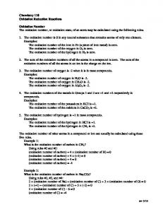
Enzymes El and Ez are readily separated by fractionation of the soluble cell extracts. When these are recombined in the presence of FMN, they couple very poorly in the catalysis of lactonization with a large proportion of the DPNH electrons being lost via auto-oxidation of the reduced flavin (1,5). In preliminary reports (5-7) we described the isolation of an enzyme complex which catalyzes lactonization. This complex, illustrated in Fig. 2, contains Ei, Ez, FMN, and iron, and is char* This work was supported in part by grants from the National Science Foundation (G-24037) and from the United States Public Health Service (AM-00562). The second paper in this series is Reference 1. 1 Name assigned by R. Y. Stanier and N. J. Palleroni, University of California, Berkeley, on basis of gelatin liquification (-), egg yolk test (-), growth: 40” (-), 4” (-), mannitol (-), creatine (+), hippuric acid (+), nicotinic acid (+), trigonellin (+). 2 The tentative formulation of Reaction 2 with 2 iron atoms gives electron transport stoichiometry. Alternate formulations with a single iron atom in EZ are possible (5). A definitive formulation and study of the precise form of the enzyme-iron-oxygen complex must await analytical verification of iron content of Ez and determination of the oxidation state of the iron.
4029
CAMPHOR I. C.
KETOLACTONIZATION*
GUNSALUS
and Chemical 61803 March
Engineering,
University
of Illinois,
26, 1965)
acterized by the nonauto-oxidizability of the bound FMN following its reduction with DPNH. When a cyclic ketone lactonization substrate is added to a mixture of DPNH and complex, there is a marked enhancement of the rate of DPNH oxidation resulting from the initiation of electron flow through the complex to oxygen by the lactonization mechanism. The binding of FMN by Ei plus & in a fashion such that FMN is only very slowly auto-oxidizable permits a detailed study of the nature of the coupling between EL and &, the mode of uncoupling by inhibitors, and the flow of electrons through the complex to endogenous and exogenous electron carriers. To facilitate these studies it has been necessary to separate the complex from other enzymes acting on camphor metabolites, a process which results in extensive loss of activity but which yields a dehydrogenase-free coupled system with sufficient activity for characterization of the system. This paper presents the details of assay, isolation, and characterization of this electron transport complex which catalyzes lactonization of cyclic ketone substrates in the presence of DPNH. The specificity range of ketolactonase for camphor derivatives is also considered. MATERIALS
AND
METHODS
Ma,teriaZs-Reagents were obtained as follows: from Sigma, DPN, DPNH, FMN, Tris (Sigma 121), and glucose-6-P; from Distillation Products, methylene blue, Tiron3 2, B-dichlorophenolindophenol, and 2,2’-bipyridine; from Mann Research Laboratories, enzyme grade ammonium sulfate; from Brown Company, DEAE-cellulose; and from Buehler, Ltd., No. 40-6435, levigated alumina. The cyclic ketone lactonization substrates were isolated from broths of Pseudomonas putida, strain CiB, grown on (+)-camphor (1). Glucose-6-P dehydrogenase was prepared from Leuconostoc mesenteroides, strain 39, by the method of DeMoss (8). Methods-Protein was estimated by the biuret method of Gornall, Bardawill, and David (9) or by the optical method of Warburg and Christian (10) with the ratios of absorbances at 280 and 260 rnp. Heme, flavin, and iron were determined as described previously (1). Enzyme Units-The unit of enzyme activity used throughout this work is defined as the amount which converts 1 pmole of substrate per min as measured by rate of substrate loss or product accumulation under the reaction conditions selected as standard. Specific activity is expressed as enzyme units per mg of protein. 3 The abbreviations zenedisulfonate; DCI,
used are: Tiron, 4,5-dihydroxy-m-ben2,6-dichlorophenolindophenol.
Downloaded from www.jbc.org by guest, on July 13, 2012
In the early steps of D ( +)-camphor oxidation by Pseudomonas putida’ both alicyclic rings are cleaved by lactonization reactions (2) as shown in Fig. 1. The inducible mixed function oxidase system (3) which converts D( +)-camphor (I) and its 5-keto derivative (III) into their respective lactones has been separated into two cofactors, reduced diphosphopyridine dinucleotide and flavin mononucleotide, and two enzymes, El and Ez, referred to as a DPNH oxidase and ketolactonase (4). The enzymes have been purified and characterized (I). The oxidase catalyzes an FMN-coupled reduction of O2 to HzOz by DPNH. It has an approximate molecular weight of 50,000 and contains no bound prosthetic groups. The ketolactonase has an approximate molecular weight of 80,000 and contains nonheme iron which is reduced by FMNHz and thus rendered bipyridine-reactive (4). Reduced & reacts with molecular oxygen yielding an activated form of oxygen which participates in the oxygenation reaction. The reaction sequence in the camphor lactonization has been formulated2 (4) as: +
IN
Department of Chemistry Urbana, lllinois (Received
DPNH
Oxidation
Mixed Function Oxidation.
o*?!Jo- jQo COOH lx FIG. 1. The lactonixation by pseudomonads.
reaction
X
in metabolism
of camphor
r------DPNH-+--E,
‘t-----r-----r--
M’B
$ ----------I FMN -
tiB II 4
FIG.
aation.
E,*Fe
j-
0,
BIPYRIDINE TPdoN
2. Elect,ron transport in mixed function MB, methylene blue.
oxidative
lactoni-
Oxidase (EJ, ketolactonase (I&), and catalase are assayed as described in the previous paper (1). The action of ketolactonase (EJ requires an electron donor, a cyclic ketone substrate, and 02. The natural electron donor is reduced FMN; in the Ez assay described previously this is generated by the El-catalyzed reduction of FMN by DPNH. Because of poor coupling between Ez and FMNHz it is not possible to achieve levels of El which are nonlimiting in t,he Ez assay; however, when the levels of FMN and E1 are empirically set at 0.01 mM and 0.17 unit per assay, respectively, the rate of lactonization is directly proportional to the amount of Ez added from 0.0007 to 0.005 unit per assay. If the Ez sample to be assayed contains some El, enough of the IatOer is added to bring the total concentration to 0.17 unit per assay volume. Assay of Coupled Oxidase Ketolactonase Electron Transport Complex-The lactonizing activity of the El-E2 complex is manifested by a stimulation of the rate of DPNH oxidation or 02 consumption when substrate, e.g. camphor, is added to a buffered mixture of the enzyme complex and DPNH. For routine assay the oxidation of DPNH is followed by the rate of decrease in absorbance at 340 rnp in a cuvette with 0.5-cm light path containing the enzyme in 50 mM Tris buffer, pH 7.2, and I pmole each of DPNH and cyclic ketone. Buffer and DPNH are brought to 1.10 ml, and the absorbance is set at 0.50 on a Zeiss PM& II spectrophotometer. The enzyme is added in 0.10 ml, the cuvette contents mixed, and the absorbance read at 15-set intervals for 1 min. Then the cyclic ketone substrate is added in 0.10 ml, and
10
the increased rate of DPNH oxidation is followed for an additional minute. Under these conditions the number of units of enzyme added to the cuvette equals 0.417 times the difference between the decrease in absorbance per min in the presence of cyclic ketone and in its absence. The rates are linear from 0.015 to 0.15 unit per assay. The substrates used are camphor (I), diketocamphane (III), or the 5-exo- (II) or 5-edno-hydroxycamphor (IV), depending upon the purpose of the assay. To assay coupling activity by oxygen uptake stimulated by cyclic ketone, the assay mixture described above is placed in a 15-ml Warburg cup with the substitution of 1 pmole of DPN plus 20 pmoles of glucose-6-P and 1.6 units of glucose-6-P dehydrogenase for DPNH. The reaction is started by tipping in the glucose-6-P from the side arm, and the rate of oxygen consumption is followed for 30 min at 30”. Separate flasks are run to compare rates with and without substrate. To determine the stoichiometry of product formation, 2,5-diketocamphane (III) is used as substrate in the Warburg runs, and when the oxygen uptake rate with Compound III becomes equal to the rate without III, 1.0 ml of 5% H3P03 is added, precipitated protein is spun off, and Aeas of the Compound III sample is read against the sample with no substrate. Bate of Compound IX formation is calculated with E = 15,000 M-’ cm-r (4). The specific dehydrogenases for 5-exo-hydroxycamphor (II) and 5-endo-hydroxycamphor (IV) (11) are measured spectrophotometrically by rate of increase in absorbance at 340 mp in a cuvette with 0.5~cm light path containing, in micromoles per 1.3 ml: Tris buffer, 50, pH 9.0; DPN, 1.0; and Compound II or IV, 1.0. Buffer, DPN, and enzyme are brought to 1.2 ml, and the absorbance at 340 rnp is set at 0.1 on a Zeiss PM& II spectrophotometer. The hydroxycamphor is added in 0.1 ml and the absorbance is read at 15-set intervals; the number of units of enzyme added to the cuvette equals 0.417 times the absorbance increase per min. Isolation of Lactonizing Complex-Cell extracts of Pseudomonas putida,’ strain CrB, grown on mineral salt-camphor medium are prepared by grinding with levigated alumina as described previously (I). The fractionation used for ketolactonase is also employed; the complex accumulates in a different fraction. Assays of coupling activity are made by following DPNH oxidation with (+)-camphor as substrate. All purification steps are carried out at 5” with buffers at pH 7.2. A cell extract containing approximately 5 g of protein is washed into a column of DEAE-cellulose (100 to 200 mesh, packed in 0.001 M phosphate under gravity to a height of 50 cm in a 7%mm diameter column, then compressed to 21 cm with Nz pressure) with 0.05 M phosphate. The complex is eluted with an increasing linear gradient formed from 4 liters each of 0.1 M KC1 and 0.45 M KCl, both in 0.005 M phosphate buffer, and 60-ml fractions are collected. Fractions emerging in 0.25 to 0.34 M KC1 are combined and the protein is precipitated by addition of solid ammonium sulfate to 75% saturation. The precipitate is collected by centrifugation at 20,000 x g and dissolved in 0.05 M Tris buffer to give a final protein concentration of 10 mg per ml. This solution is fractionated by addition of solid ammonium sulfate. The fraction precipitating between 0.35 and 0.5 saturation contains the coupled lactonizing activity. Data for a typical purification are shown in Table I. RESULTS
Separation of Lactonization Complex from Cell Extracts-The removal of the alcohol dehydrogenases and recovery of the un-
Downloaded from www.jbc.org by guest, on July 13, 2012
DCI
Vol. 240, No.
III
October 1965
H. E. Conrad, K. Lieb, and I. C. Gunsalus TABLE Separation
of lactonizing
complex
from
4031
I hydroxycamphor
dehydrogenases
Stage 1, cell extract from alumina-ground cells; Stage 2, DEAE-cellulose eluate emerging and Stage 3, ammonium sulfate fraction precipitating between 0.35 and 0.50 saturation. Unit9
from the column from 0.25 to 0.34 M KCl;
Recovery
Specific activitya
Measurement
Protein....................................... Coupling activity.. Oxidase (El). Ketolactonase (Ez). 5-exo-Hydroxycamphor dehydrogenase. 5-endo-Hydroxycamphor dehydrogenase.
stage 1
stage 2
stage 3
5000” 386 2720 357 453 543
470.0” 10.8 135.0 178.0 4.5 2.7
109.0” 2.0 120.0 13.0