Cell Systems, Volume 6. Supplemental Information. Modeling Cell-to-Cell Communication. Networks Using Response-Time Distributions. Kevin Thurley, Lani F.
Cell Systems, Volume 6
Supplemental Information
Modeling Cell-to-Cell Communication Networks Using Response-Time Distributions Kevin Thurley, Lani F. Wu, and Steven J. Altschuler
Supplemental Figures and Tables
Figure S1: Supplementary analysis of simple multi-step models. Related to Figure 2. (A) Analysis of the reversible chain model, analogous to Figure 2B-D. (B) Fitting error (Root-mean squared sum of residuals) of best-fit gamma distributions to the indicated models (see Figure 2A-E), analogous to Figure 2G. (C) Fitting error and shape parameter 𝛼 for the parallel chain model (Figure 2D) with varying number of molecules (parameter 𝑚).
1
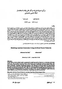
Figure S2: Comparison of intracellular and intercellular feedback. Related to Figure 3. (A) Model scheme: The parallel chain model (Figure 2D; 𝑛 = 10 steps, 𝑚 = 100 molecules) is implemented either with inter-cellular feedback between cells (as in Figure 3B-D, left panels), or with intra-cellular feedback. The inter-cellular feedback is implemented by allowing molecules in the final state (here 𝑥10 ) to alter all rate parameters in all cells. For intra-cellular feedback, the reaction rate depends on the number of molecules in an intermediate state 𝑥𝑖 and there is no cell-cell communication. (B-C) Arrival time distributions and values of synchronization time and delay for the indicated models: no feedback (FB); intracellular feedback (negative or positive) from state 𝑥𝑖 with 𝑖 = 1,5,9 ; intercellular feedback (negative or positive).
2
Figure S3: Supplementary analysis of intercellular interaction motifs. Related to Figure 3. (A) Additional analysis of network motifs (see Figure 3B-D). (B-D) Additional network motifs (see Figure 3B-D).
3
Figure S4: Supplementary analysis of response-time modeling applications. Related to Figure 5. (A) Simulations are carried out with the best-fit gamma distributions (“response-time model) or exponential distributions (“single-step model”) to the arrival-time distributions arising in the gate and transition motifs (see Figure 4), all with the same delay value of 𝑡delay = 3. Black bars indicate stimulus duration. (B) Maximal fraction of activated cells after stimulation (“Amplitude”) at varying stimulus duration (at 𝑡delay = 3) and delay (at duration 𝑡d = 5)(see Star Methods). (C) Simulations of IFN-γ secretion onset (Figure 5E-G) with varying feedback strength 𝐾𝑣 . Bold: Feedback strength used Figure 5G. (D-E) Validation of the generalized Gillespie algorithm used in Figure 5E-G. (D) Simulations of the 10-step process with indicated number of cells, and comparison to the exact solution (gamma distribution, see Equation 1). (E) Root-mean-square deviation (mean and standard deviation from 4 simulations) in histograms such as shown in (D).
4
Figure S5: Supplementary data for IL-2 competition simulations. Related to Box 2. (A) Simulation of the model shown in Box Figure, panel B, with indicated extracellular IL-2 concentration. (B) Best-fit gamma distribution parameters α and β to curves as shown in Box Figure, panel C, which were obtained from simulations as shown in (A), for a range of IL-2 concentrations. Dashed lines are interpolating curves used for the implementation of the response-time model (see Star Methods). (C) IL-2 competition (or “tug-of-war”) simulation from (Feinerman et al., 2010), reprinted for comparison with simulations shown in Box Figure, panel D (right).
5
Table S1: Parameter values used in Figure 5. Description Value
Reference
Half-saturation constant of IL-2 interaction
0.05
Shape parameter of CD25 up-regulation
34.9 (Figure 5E, bottom)
(Dorner 2009)
et
al.,
Rate parameter of CD25 up-regulation
0.21 (Figure 5E, bottom)
(Dorner 2009)
et
al.,
Shape parameter IL-2 secretion onset
4.33 (Figure 1A)
(Han et al., 2012)
Rate parameter IL-2 secretion onset
1.36 (Figure 1A)
(Han et al., 2012)
Shape parameter initial IFN-γ secretion 14.7 (Figure 1A) onset
(Han et al., 2012)
Rate parameter initial IFN-γ secretion onset
0.45 (Figure 1A)
(Han et al., 2012)
Fraction of IL-2+ cells
0.1
(Han et al., 2012)
Fraction of early IFN-γ+ cells
0.35
(Han et al., 2012)
Fraction of late IFN-γ+ cells
0.55
(Han et al., 2012)
Average duration of IL-2 secretion
4 hr
(Han et al., 2012)
Average duration of IFN-γ secretion
4 hr
(Han et al., 2012)
When figure panels are referred to, the parameter values are computed in those figures based on data in the cited reference.
6