JTh2A.49.pdf
CLEO 2017 © OSA 2017
Modification of UV Surface Plasmon Resonances in Aluminum Hole-Arrays with Graphene Yunshan Wang, Sourangsu Banerji, Jieying Mao, Sara Arezoomandan, Berardi Sensale-Rodriguez, and Steve Blair Department of Electrical and Computer Engineering, The University of Utah Author e-mail address:
[email protected],
[email protected]
Abstract: In this work we study the UV transmission through monolayer graphene films transferred on top of aluminum hole-arrays. Interaction of graphene pi-plasmons with surface plasmon resonances leads to strong wavelength shifts. OCIS codes: (240.6680) Surface plasmons; (300.6540) Spectroscopy, ultraviolet.
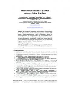
1. Introduction Active optical devices enabled by plasmon resonances have gained increasing interest during recent years. In this context, ultra-violet (UV) plasmonics has earned much attention due to its applications in areas such as sensing and light-sources. Tunable UV devices can enable enhanced functionalities including multiplexed sensing, wavelengthtunable light emission, and so on. At diverse spectral ranges, ranging from the THz to the visible, graphene has been proposed or demonstrated as a material capable of actively tuning the transmission properties of electromagnetic structures [1-3]. Interestingly, in the UV range, graphene shows an abnormal optical absorption due to pi-plasmon resonance [4] (see Fig. 1a). At these wavelengths either electric bias or chemical doping can tune the absorption peak position [5]. From this perspective, studying the interaction of graphene with UV plasmonic structures can pave a road for designing new UV tunable devices. Here we report our studies on the interaction of graphene piplasmons with the surface plasmon resonance on Al hole-arrays.
Fig. 1. (a) Band structure of graphene (left) together with typical conductivity spectra (right). (b) Sketch of the analyzed samples consisting of CVD graphene transferred on Al hole-arrays. 2. Methods For the purpose of this study, Al hole-array samples with varied periodicity were analyzed with and without graphene (see Fig. 1b). In all samples, 100 nm Al thin film was sputtered (Denton Discovery 18) onto 1” diameter UV fused silica coverslips (Esco Optics, Inc) at a deposition rate 3.7 Å/s. Periodic nanoapertures were milled into the Al films by Focused Ion Beam (FEI Helios) under iodine gas injection. Designed sizes of the nanoapertures are 100 nm in diameter and the milling depth is 50 nm into the fused silica substrate. Hole periodicities of 240, 280, 320 nm are employed. Monolayer graphene grown via CVD on a copper foil (BGT Materials) was used in our study. A layer of PMMA was spun onto graphene for the purpose of transfer. After etching the copper foil, graphene was suspended in DI water with PMMA on top. Transfer of graphene onto the hole-array was carried out in DI water, followed by 10 mins baking at 150°C on a hot plate. PMMA was then removed by means of acetone rinsing. After fabrication, our samples were optically characterized. For transmission measurements, a fiber coupled light source (Laser-Driven Light Source Model EQ-99-FC) was used to produce light from 230 nm to 800 nm. A biconvex lens focuses the light onto the 30x30 µm2 hole-array pattern. After passing through the pattern, transmitted light is received by a lens-coupled optical fiber connected to either a Maya2000 Pro (200 to 400 nm) or AvaSpec-USB2-DT spectrometer (350 to 800 nm) so to obtain transmittance. The intensity spectrum measured without a sample was used as the reference signal. Transmittance though the Al hole-array was measured before and after graphene transfer. Moreover, a reference hole-array pattern was created close to the edge of the coverslip. Spectrum through
JTh2A.49.pdf
CLEO 2017 © OSA 2017
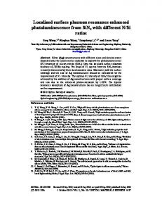
the reference pattern was compared before and after graphene transferring indicating no change in the optical setup conditions. 3. Results and discussion 3.1. Experimental results Depicted in Fig. 2a is a typical transmission through the hole-array with and without graphene (280 nm periodicity). Two important features are noticed across all the measured samples: (i) in all cases, the longer-wavelength resonant dip, which has its origin on SPRs in the Al/substrate interface (bottom interface), is not altered when adding graphene. This is a result of the geometric placement of the graphene layer being on top of the Al film. On the other hand, (ii) the short-wavelength resonant dips, which are associated with SPRs at the top interface, red-shift when graphene is added. This is a result of the interaction of graphene pi-plasmons with Al SPRs. Shown in Fig. 2b is the extracted top interface SPR wavelength-shift as a function of SPR wavelength showing the latter described effect. Moreover, when analyzing Fig. 2b it is observed that a maxima occurs in the 288 nm to 310 nm wavelength range (i.e. 4.3 to 4.0 eV). This is attributed to an enhanced graphene optical conductivity owed to pi-plasmons. However, as depicted in Fig. 1a, the UV peak in σ(E) for pristine graphene occurs at around 4.7 eV. The reason why our observed wavelength-shift maxima occurs at a longer wavelength is a result of σ(E) being strongly influenced by the carrier density and our samples having a large unintentional doping as a result of the transfer process. In this regard, transmission measurements of graphene on quartz samples show a minima at 290 nm (4.27 eV), which is consistent with observations in the literature for highly doped graphene [5].
Fig. 2. (a) Typical transmittance spectra through the hole-array after/before graphene transfer (280 nm periodicity). (b-c) Extracted SPR wavelength-shift as a function of SPR wavelength; (b) experiment, (c) simulation. 3.2. FDTD simulations In order to provide further insight into the effect of adding graphene into the Al hole-array, numerical calculations were carried out through the finite-difference time-domain (FDTD) method using Lumerical FDTD Solutions. The Al permittivity was measured through ellipsometry and was imported into Lumerical directly as the film optical data. The metal film was placed on top of a silica substrate, using Palik silica data. An incident plane wave along the vertical direction with transverse magnetic (TM) polarization was used to illuminate the hole-array as a Total-FieldScattered-Field (TFSF) source. For simulation of a periodical hole-array, a unit cell containing a single nanohole was used, along with periodic boundary conditions on the sides. Perfectly Matched Layer (PML) boundaries were set in the vertical direction. A frequency-domain field and power (DFT) monitor sheet was set on top of film and placed outside of the source boundary to avoid incident light collection. The far-field transmission spectrum was obtained through the power passing through the DFT monitor as a function of wavelength. For modeling graphene, measured optical constants were extracted from Ref. [4] and imported into Lumerical. In this regard, so to reduce the computational cost, graphene was modeled as a 3D material with a thickness 2 nm. Our simulations showed similar trends to those observed in the measured data. Depicted in Fig. 2c is the simulated top interface SPR wavelengthshift as a function of SPR wavelength. A maxima is observed at around 260-270 nm, which is consistent with the used graphene model (Ref. [4]), which exhibits a peak at 264 nm. As observed in our experiments SPR wavelengthshift and graphene optical conductivity are strongly correlated and exhibit their maxima at similar wavelengths. 5. References [1] R. Yu et al, “Active modulation of visible light with graphene-loaded ultrathin metal plasmonic antennas.” Scientific Reports 6, 32144 (2016). [2] R. Yan et al, “Exceptional terahertz wave modulation in graphene enhanced by frequency selective surfaces.” ACS Photonics, 3, 315 (2016). [3] M. Liu et al, “A graphene-based broadband optical modulator.” Nature, 474, 64 (2011). [4] V. G. Kravets et al, “Spectroscopic ellipsometry of graphene and an exciton-shifted van Hove peak in absorption”. PRB, 81, 155413 (2010). [5] K. F. Mak et al, “Tuning many-body interactions in graphene: The effects of doping on excitons and carrier lifetimes.” PRL, 112, 207401 (2014).