Research Paper
Modulation of Cyclophosphamide-Induced Cellular Toxicity by Diphenylmethyl Selenocyanate In Vivo, an Enzymatic Study Pramita Chakraborty, Ugir Hossain Sk, Nabendu Murmu, Jayanta Kumar Das, Smarajit Pal, and Sudin Bhattacharya1 Departments of Cancer Chemoprevention [P. Chakraborty, Sk U. Hossain, N. Murmu, J. K. Das, S. Bhattacharya] and Clinical Biochemistry Unit [S. Pal], Chittaranjan National Cancer Institute, Kolkata, India
AIM: Cyclophosphamide (CP) is one of the most widely used alkylating antineoplastic agents that damage normal cells while killing cancerous cells in vivo. The use of CP in treating cancer patients is limited due to its severe toxicities induced mainly by oxidative stress. Diphenylmethyl selenocyanate is a synthetic organoselenium compound shown to act as a potent antioxidant in chemically induced murine toxicity and carcinogenesis models in vivo. In the present study, this compound has been evaluated for its protective potential against CP-induced toxicity in Swiss albino mice. METHODS: CP was administered intraperitoneally (50 mg/kg) and diphenylmethyl selenocyanate was given orally (3 mg/kg) in a pretreatment and concomitant treatment schedule, and the effects were assessed by estimating lipid peroxidation level, phase II detoxifying enzyme system, blood hemoglobin level, serum transaminase activity, and nitrite content. RESULTS: Diphenylmethyl selenocyanate significantly (P < 0.05) increased glutathione-S-transferase, glutathione peroxidase, and catalase levels whereas decreased the lipid peroxidation levels in both liver and lung tissues of the animals given CP. Superoxide dismutase was increased significantly in liver (P < 0.05) but not in the lung. The selenium compound also significantly (P < 0.05) increased the blood hemoglobin level whereas decreased the transaminase activity in serum and the nitrite content in peritoneal macrophages. CONCLUSION: Our result suggests that diphenylmethyl selenocyanate has the potential to prevent CP-induced cellular toxicity.
Keywords: cyclophosphamide diphenylmethyl selenocyanate oxidative stress antioxidant
Journal of Cancer Molecules 4(6): 183-189, 2009.
Introduction Cyclophosphamide (CP2) is an alkylating agent widely used in cancer chemotherapy [1,2]. Its cytotoxic effects are the result of chemically reactive metabolites that alkylate DNA and protein, producing cross-links [3]. The injury of normal tissues is the major limitation of using CP, which gives rise to numerous side effects [4,5]. It has been reported that oxidative stress mediated disruption of redox balance after CP exposure generates biochemical and physiological disruptances [6-8]. Received 10/1/08; Revised 12/2/08; Accepted 12/2/08. 1 Correspondence: Dr. Sudin Bhattacharya, Department of Cancer Chemoprevention, Chittaranajan National Cancer Institute, 37, S. P. Mukherjee Road, Kolkata 700 026, West Bengal, India. Phone: 91-3324765101 ext 316. E-mail:
[email protected] 2 Abbreviations: CP, cyclophosphamide; CDNB, 1-chloro-2,4dinitrobenzene; GSH, reduced glutathione; DTNB, 5,5’-dithio-bis(2nitrobenzoic acid); β-NADPH, β-nicotinamide adenine dinucleotide phosphate (reduced); TBA, thiobarbituric acid; ALT, alanine transaminase; AST, aspartate transaminase; LPO, lipid peroxidation; TBARS, TBA reactive substances; GST, glutathione-S-transferase; GPx, glutathione peroxidase; CAT, catalase; SOD, superoxide dismutase; NO, nitric oxide (NO•); Hb, hemoglobin.
2009 MedUnion Press − http://www.mupnet.com
Several studies suggest that antioxidant supplementation can influence the response to chemotherapy as well as the development of adverse side effects that result from treatment with antineoplastic agents [9]. Selenium is an essential trace mineral for animals and humans to fight against many diseases including cancer. Epidemiological and animal studies indicated that many human diseases could be prevented by increasing dietary selenium mainly due to the antioxidative or cancer chemopreventive properties of selenium compounds [10,11]. Both inorganic and organic selenium compounds have the potential to reduce hydrogen peroxide, lipid and phospholipid hydroperoxide levels, thereby mitigating the propagation of free radicals; reactive oxygen and nitric oxide mediated cellular damages [12,13]. However, the use of the main known dietary sources of selenium, such as selenomethionine, selenocysteine, and inorganic selenium such as sodium selenite is limited due to their toxicity [14-16]. These observations suggested the need for creating synthetic organoselenium compounds with improved efficacy and safety. Diphenylmethyl selenocyanate, a synthetic organoselenium compound, was reported earlier as a potential antioxidant and cancer chemopreventive agent against chemically induced toxicity and carcinogenesis model in vivo [17-19]. The acute oral LD50 value of
183
Chakraborty et al. J. Cancer Mol. 4(6): 183-189, 2009
diphenylmethyl selenocyanate was found to be >500 mg/kg as compared to 28 mg/kg for sodium selenite, showing that it is relatively much less toxic than sodium selenite. The present study was undertaken to evaluate the chemoprotective efficacy of diphenylmethyl selenocyanate against CP-induced toxicity in Swiss albino mice.
Materials and Methods Animals Adult (5-6 weeks) Swiss albino male mice (23 ± 2 g), bred in the animal colony of Chittaranjan National Cancer Institute, were maintained at controlled temperature under an alternating light and dark condition. Standard food pellets (Lipton India Ltd., Calcutta, West Bengal, India) and drinking water was provided ad libitum. The experiments were carried out following strictly the Institute’s Guideline for the Care and Use of Laboratory Animals. Reagents CP was obtained from Cadila Pharmaceuticals (Bhat, Ahmedabad, India). Diphenylmethyl bromide, potassium selenocyanate, 1-chloro-2,4-dinitrobenzene (CDNB), EDTA, reduced glutathione (GSH), pyrogallol, 5,5’-dithio-bis(2nitrobenzoic acid) (DTNB), SDS, bovine serum albumin, thioglycolate broth, β-nicotinamide adenine dinucleotide phosphate (reduced) (β-NADPH), glutathione reductase, RPMI 1640, N-(1-napthyl) ethylenediamine dihydrochloride, and sodium azide (NaN3) were obtained from Sigma-Aldrich Chemicals (Bangalore, India). Hydrogen peroxide (30%), thiobarbituric acid (TBA), propylene glycol, hexane, orthophosphoric acid, and sodium nitrite were obtained from Merck Specialities Private Ltd. (Worli, Mumbai, India). Sulphanilamide, Tris-HCl and acetone were obtained from Sisco Research Laboratories (Mumbai, Maharashtra, India). Diethyl ether, K2HPO4, and KH2PO4 were obtained from Spectrochem Private Ltd. (Mumbai, Maharashtra, India). Fetal calf serum was obtained from Hyclone Laboratories (Logan, UT, USA). Serum alanine transaminas (ALT) and aspartate transaminase (AST) assay kits were obtained from Span Diagnostics Ltd. (Udhna, Surat, India). Synthesis of the compound Diphenylmethyl selenocyanate was prepared following a literature procedure [20]. Briefly, diphenylmethyl bromide was treated with potassium selenocyanate (KSeCN) in acetone at 60-70°C for 5 h. Acetone was removed under reduced pressure and the resulting solid was extracted with diethyl ether. Usual work up then afforded the desired compound, which was crystallized from hexane to get a colorless crystalline solid with a melting point at 68-69°C.
Drug preparation Synthetic organoselenium compound diphenylmethyl selenocyanate was used as a suspension in 5.5% propylene glycol in water. Experimental design Animals were divided into four groups containing six animals (n = 6) in each group. Vehicle treated group (Group I): Each animal received distilled water intraperitoneally and 5.5% propylene glycol in water orally for 10 days. CP treated group (Group II): CP was administered intraperitoneally at a dose of 50 mg/kg in water for 10 days. Pretreatment group (Group III): Diphenylmethyl selenocyanate was administered
184
orally at a dose of 3 mg/kg 7 days prior to CP treatment and then continued along with CP for 10 days. Concomitant treatment group (Group IV): Diphenylmethyl selenocyanate was administered orally at a dose of 3 mg/kg for 10 days and CP was given as in Group II. The mice were sacrificed on day 11, and the parameters described below were studied.
Biochemical estimation Quantitative estimation of lipid peroxidation (LPO): LPO level was estimated in liver and lung microsomal fraction. The level of lipid peroxides formed was measured using TBA and expressed as nmol of TBA reactive substances (TBARS) formed per mg of protein using the extinction coefficient of 1.56 × 10⁵ M-1cm-1 [21]. Estimation of GSH level: GSH level was estimated in liver and lung cytosols spectrophotometrically by determination of DTNB reduced by –SH groups by measuring the absorbance at 412 nm. The level of GSH was expressed as nmol/mg of protein [22]. Estimation of glutathione-S-transferase (GST) activity: GST activity was determined from the increase in absorbance at 340 nm with CDNB as the substrate and specific activity of the enzyme was expressed as CDNB-GSH conjugate per min per mg of protein [23]. Estimation of glutathione peroxidase (GPx) activity: GPx activity was measured by NADPH oxidation using a coupled reaction system consisting of GSH, glutathione reductase and H2O2 [24]. Briefly, 100 µl of enzyme sample was incubated for 10 min with 800 µl reaction mixture (0.25 M potassium phosphate buffer containing 2.5 mM EDTA and 2.5 mM NaN3, 10 mM reduced glutathione, 2.5 mM NADPH, and 2.4 units of glutathione reductase). The reactions started by adding 100 µl H2O2 and followed the decrease in NADPH absorbance at 340 nm for 3 min. The enzyme activity was expressed as µmol NADPH utilized/min/mg of protein. Using extinction coefficient of NADPH at 340 nm as 6200 M-1cm-1. Estimation of CAT activity: CAT activity was determined spectrophotometrically at 250 nm and expressed as unit/mg of protein where the unit was the amount of enzyme that liberated half the peroxide oxygen from H2O2 in seconds at 25°C [25]. Estimation of SOD activity: SOD activity was determined by quantification of pyrogallol autooxidation inhibition and the amount of enzyme necessary for inhibiting the reaction by 50%. Autooxidation of pyrogallol in 50 mM Tris-HCl buffer (pH 7.5) is measured by increased in absorbance at 420 nm [26,27]. Assay of nitrite production in peritoneal macrophages: Nitric oxide (NO•) produced by macrophages quickly reacts with oxygen to produce nitrate (NO3-) and nitrite (NO2-) ions. Accumulation of nitrite in the medium was measured spectrophotometrically based on Griess reaction [28]. In brief, 50 µl of Griess reagent mixture of 0.1% N-(1-napthyl) ethylenediamine dihydrochloride, 1% sulphanilamide and 2.5% orthophosphoric acid was reacted with 50 µl of sample (cellfree supernatant) at room temperature for 10 min and the NO2- concentration was determined by absorbance at 550 nm in comparision with the sodium nitrite standards. Data were represented as µmol of nitrite produced in 50 µl cell-free supernatant. Determination of serum ALT and AST activity: Serum was collected from blood sample of mice by centrifugation. The serum ALT and AST levels were measured spectrophotometrically by standard enzymatic method using commercial kits (Span Diagnostics Ltd.). Hemoglobin (Hb) level: Hb content of blood samples was measured with the Sahli’s method [29]. Estimation of protein concentration: Protein concentration was estimated spectrophotometrically [30] with bovine serum albumin as standards. Print ISSN 1816-0735; Online ISSN 1817-4256
Chemoprotective Effect of Diphenylmethyl Selenocyanate
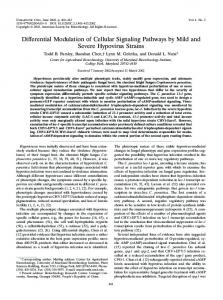
Figure 1: Effect of diphenylmethyl selenocyanate on the LPO
Figure 2: Effect of diphenylmethyl selenocyanate on the GSH
level of liver (A) and lung (B) in CP-treated mice. Data represent the mean ± SD of six independent experiments. aP < 0.05 with respect to Group I. bP < 0.05 with respect to Group II.
level of liver (A) and lung (B) in CP-treated mice. Data represent the mean ± SD of six independent experiments. aP < 0.05 with respect to Group I. bP < 0.05 with respect to Group II.
Statistical analysis The differences in mean values of different groups were tested and the values are expressed as mean ± SD. The data were analyzed using the Student’s t-test and P < 0.05 was considered to be significant.
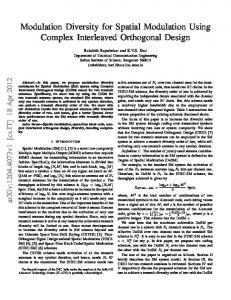
GST activity The activity of GST in the liver and lung of animals treated with CP showed a decrease of 39.88% and 33.44%, respectively, as compared to vehicle treated group (Figure 3). The GST activity elevated sharply by 33.63% and 38.41% in liver and lung by pretreatment with the selenium compound. However, concomitant treatment with this compound elevated GST activity in liver and lung by 25.69% and 27.73%, respectively.
Results Microsomal LPO level The level of LPO in the liver and lung microsomes of the animals, treated with CP at a dose of 50 mg/kg for 10 days, increased significantly (P < 0.05) by 41.05% and 37.36% respectively (Group II) as compared to vehicle treated group (Figure 1). Seven-day pretreatment with the selenium compound, reduced the elevated level of LPO in liver and lung by 39.73% and 40.32% (Group III) and concomitant treatment with the compound inhibited LPO in liver by 33.11% and in the lung by 35.21% respectively (Group IV). GSH level The GSH level of animals treated with CP showed a decrease of 53.80% in liver and 55.62% in lung (Group II) as compared to vehicle treated group (Figure 2). Pretreatment with the selenium compound increased GSH level significantly (P < 0.05) in liver by 41.88% and in lung by 54.98% (Group III). Concomitant treatment with the same compound elevated GSH level in liver and lung significantly by 36.78% and 37.85% (Group IV), respectively. 2009 MedUnion Press − http://www.mupnet.com
GPx activity GPx activity of CP-treated group (Group II) decreased significantly by 51.20% and 46.66% in liver and lung as compared to vehicle treated group (Group I, Figure 4). Seven-day pretreatment study revealed that the selenium compound increased the enzyme activity in liver and lung significantly by 58.11% and 61.47% (Group III) in comparison to CP-treated group. Concomitant treatment with the compound increases the enzyme activity by 52.42% in liver and 46.45% in lung (Group IV). CAT activity CAT activity in liver and lung of CP-treated animals showed a significant (P < 0.05) decrease of 53.61% and 56.65% with respect to vehicle treated group (Figure 5). Pretreatment significantly (P < 0.05) increased the enzyme activity in liver and lung by 52.22% and 67.56% (Group III) and concomitant treatment increased the enzyme activity by 43.75% in
185
Chakraborty et al. J. Cancer Mol. 4(6): 183-189, 2009
Figure 3: Effect of diphenylmethyl selenocyanate on the GST
Figure 4: Effect of diphenylmethyl selenocyanate on the GPx
activity of liver (A) and lung (B) in CP-treated mice. Data represent the mean ± SD of six independent experiments. aP < 0.05 with respect to Group I. bP < 0.05 with respect to Group II.
activity of liver (A) and lung (B) in CP-treated mice. Data represent the mean ± SD of six independent experiments. aP < 0.05 with respect to Group I. bP < 0.05 with respect to Group II.
liver and 55.24% in lung respectively (Group IV) in comparison to CP-treated group.
compared to CP-treated animals. There was also a significant (P < 0.05) elevation in serum AST activity by 39.33% in CP-treated animals as compared to vehicle treated group. Pretreatment and concomitant treatment with the selenium compound reduced the enzyme activity significantly by 45.92% and 34.79%, respectively, as compared to CP-treated group. Hb level in the CP-treated group was decreased significantly (P < 0.05) by 50.35% as compared to vehicle-treated group. The level of Hb was increased significantly (P < 0.05) by 46.96% in case of pretreatment and 36.07% in case of concomitant treatment with selenium compound as compared to CP-treated group.
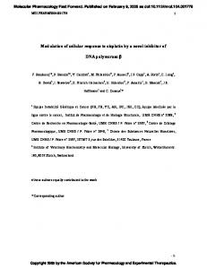
SOD activity Administration of CP for 10 days significantly (P < 0.05) reduced the SOD activity in liver by 47.59% (Group II) as compared to vehicle treated group (Figure 6A). The activity of this enzyme was significantly (P < 0.05) elevated by 46.31% in case of 7-day pretreatment with the selenium compound (Group III) and by 42.66% in case of concomitant treatment with the compound (Group IV). No significant alteration of SOD activity was observed in the lung cytosol. Nitrite production in peritoneal macrophages The nitrite level of the animals treated with CP increased significantly (P < 0.05) by 44.03% after 10 days compared to vehicle treated group (Figure 6B). The level of nitrite production was decreased significantly (P < 0.05) by 48.33% and 44.43% respectively in case of pretreatment (Group III) and concomitant (Group IV) treatment with the selenium compound, as compared to the level of CP-treated animals. Modulation of serum ALT and AST activity and blood Hb level by diphenylmethyl selenocyanate in CP-treated mice A significant (P < 0.05) elevation of 60% in ALT activity was observed after 10 days of CP treatment as compared to vehicle treatment (Table 1). Pretreatment and concomitant treatment with the selenium compound reduced the enzyme activity significantly by 50.45% and 40.98%, respectively, as
186
Discussion Several studies indicate that CP has a prooxidant character, and generation of oxidative stress after CP administration leads to decrease in the activities of antioxidant enzymes and increase in lipid peroxidation in liver, lung and serum of mice and rats [31-36]. The present investigation was carried out to evaluate the chemoprotective potential of diphenylmethyl selenocyanate against oxidative stressmediated cellular toxicity induced by CP in Swiss albino mice. LPO is used as a biomarker to show the index of oxidative stress, and causes cell membrane damage resulting in gradual loss of cell membrane fluidity, decreased membrane potential and increased permeability to ions [37]. Induction
Print ISSN 1816-0735; Online ISSN 1817-4256
Chemoprotective Effect of Diphenylmethyl Selenocyanate
Figure 6: Effect of diphenylmethyl selenocyanate on the SOD Figure 5: Effect of diphenylmethyl selenocyanate on the CAT activity of liver (A) and lung (B) in CP-treated mice. Data represent the mean ± SD of six independent experiments. aP < 0.05 with respect to Group I. bP < 0.05 with respect to Group II.
of LPO has been reported in different tissues of experimental animals after CP administration [38-41]. CP and its metabolite acrolein cause inactivation of microsomal enzymes and result in increased reactive oxygen species (ROS) generation and LPO [42,43]. We noted significant increases of TBARS in liver and lung microsomes after 10 days of CP (50 mg/kg) treatment. Pretreatment and concomitant treatment with diphenylmethyl selenocyanate resulted in significant decrease in TBARS levels, and this may be due to the free radical scavenging potential of the compound. The protective activity was found to be more pronounced in the pretreated group. However sodium selenite, given at an equivalent dose (same amount of selenium given) did not show any preventive effect on hepatic lipid peroxidation (data not shown). GSH is the principal intracellular nonenzymatic antioxidant. It can detoxify toxic endogenous and exogenous substances, including free radicals and xenobiotics [44]. CP metabolism produces highly reactive electrophiles and the decreased value of GSH in CP-treated group most probably due to the electrophilic burden on the cells and also due to the formation of acrolein, which deplete GSH content [45]. Treatment with diphenylmethyl selenocyanate reduced the electrophilic burden and thereby increased GSH levels in liver as well as in lung. GST catalyzes conjugation of GSH with highly reactive electrophiles, and thereby plays a major role in the detoxification of alkylating agents [46]. GST acti-
2009 MedUnion Press − http://www.mupnet.com
activity of liver (A) in CP-treated mice. Effect of diphenylmethyl selenocyanate on the nitrite release from peritoneal macrophages of CP treated mice (B). Data represent the mean ± SD of six independent experiments. aP < 0.05 with respect to Group I. bP < 0.05 with respect to Group II.
vity has been shown to be decreased significantly by CP administration, treatment with diphenylmethyl selenocyanate restored the activity near to vehicle-treated group both in liver and lung, making the cells more effective with respect to detoxification of toxic metabolites. Moreover, the elevated level of GSH could effectively provide thiol group for the possible GST-mediated detoxification reactions. The selenium-containing enzyme GPx protects cells against ROS. In this study, the activity of GPx decreased significantly after CP treatment. This may be due to the increase in LPO induced by CP. GPx which scavenges hydroperoxides and lipid peroxides was mostly utilized by the enhanced lipid peroxides resulting in the depletion of its level. Treatment with diphenylmethyl selenocyanate prevented the depletion of GPx level in liver and lung, and thus provided protection of biomembrane from oxidative attack. Here also sodium selenite did not show any effect on GPx in liver when given at an equivalent dose or same amount of selenium (data not shown). This showed that diphenylmethyl selenocyanate was much more effective than sodium selenite. SOD and catalase are present in all oxygen-metabolizing cells and their function is to provide a defense against the potentially damaging reactivities of superoxide and hydrogen peroxide. SOD detoxifies the superoxide radicals to H2O2, which has been eliminated by CAT. In the present study, activity of SOD was decreased significantly in the liver of
187
Chakraborty et al. J. Cancer Mol. 4(6): 183-189, 2009
Table 1: Modulation of serum ALT and AST activity and blood Hb level by diphenylmethyl selenocyanate in CP-treated mice Vehicle-treated group (Group I) CP-treated group (Group II) Pretreatment group (Group III) Concomitant treatment group (Group IV)
ALT (U/ml) 43.6 ± 2.9 109.0 ± 6.3a 54.0 ± 2.8b 64.3 ± 3.2b
AST (U/ml) 129.3 ± 8.3 212.6 ± 9.2a 115.0 ± 6.0b 138.6 ± 4.0b
a
Hb (g/dl) 14.1 ± 0.1 7.0 ± 0.4a 13.2 ± 1.0b 10.9 ± 0.7b
b
Data represent the mean ± SD of six independent experiments. P < 0.05 with respect to Group I. P < 0.05 with respect to Group II.
CP-treated animal but no significant changes was observed in the lung whereas catalase activity was found to be depleted in both liver and lung of the CP-treated animals. Administration of diphenylmethyl selenocyanate significantly elevated the SOD and CAT activities, suggesting that it had the ability to restore the activities of these two enzymes. Pretreating the animals with diphenylmethyl selenocyanate enhanced the detoxifying enzyme activity more effectively than in the animal receiving the selenium compound from the day of CP treatment. NO• is a short-lived and highly reactive free radical. NO• is synthesized from L-arginine by the enzyme nitric oxide synthase (NOS). NO• reacts spontaneously with available superoxide radical to form the more potent and versatile oxidant peroxynitrite. Peroxynitrite (ONOO-) can damage DNA [47], initiate membrane LPO [48], deplete antioxidant enzyme activity, and reduce GSH content [49,50]. Enhancement of NO• production by peritoneal macrophages in CPtreated group (Group II) was observed after 10 days as compared to vehicle-treated animal. Significant decrease of NO• level from peritoneal macrophages was observed when the animals were treated with diphenylmethyl selenocyanate. As observed earlier pretreatment with the selenium compound showed better protective effect. The results indicate that the selenium compound has the potential to reduce the NOS-mediated excessive production of NO•. ALT and AST are marker enzymes for assessment of liver function. Elevated levels of serum enzymes are indicative of cellular damages and loss of functional integrity of cell membrane in liver [51]. Damage to liver cells causes release of cellular enzymes into serum. Hepatic activation of CP leading to the formation of toxic metabolites caused damage to the liver tissues as shown by increased AST and ALT levels. Treatment with diphenylmethyl selenocyanate resulted in significant reduction in the level of these transaminase activities. Chemotherapy-induced anemia is one of the most common side effects experienced by cancer patients, occurring in approximately 70-90% of those undergoing treatment for the disease [52]. This is because chemotherapeutic drugs kill rapidly dividing cells in the body, including cancer cells and normal cells which include red blood cells, at the same time suppress bone marrow ability to produce new ones, resulting in decrease of blood Hb level. It was observed that feeding the animals with diphenylmethyl selenocyanate in both pre and concomitant treatment schedules enhanced the Hb level which was depleted by CP treatment significantly. The greater efficacy shown by the pretreatment group might be the result of the compound providing some added protection to the target cells before exposure to the chemotherapeutic agent. From this observation, it is possible to conclude that diphenylmethyl selenocyanate exerts its chemoprotective effect against CP-induced cellular toxicity in part by scavenging the free radicals generated by CP reactive metabolites. Because CP does not act through oxidative stress mechanism, it is expected that the selenium compound would be beneficial to the host but without reducing the efficiency of the treatment. Further studies are in progress to find out whether diphenylmethyl selenocy-
188
anate has the ability to enhance or inhibit the antitumor efficacy of CP in tumor-bearing mouse model.
Acknowledgments The authors wish to thank the Director of Chittaranjan National Cancer Institute for supporting this study.
References 1. 2. 3.
4. 5. 6.
7.
8.
9.
10. 11.
12.
13.
14. 15. 16.
17.
18.
19.
20.
Baumann F, Preiss R. Cyclophosphamide and related anticancer drugs. J Chromatogr B: Biomed Sci Appl 764: 173-192, 2001. Fleming RE. An overview of cyclophosphamide and ifosfamide pharmacology. Pharmacotherapy 17: 1465-1545, 1997. Hales BE. Comparison of the mutagenicity and teratogenicity of cyclophosphamide and its active metabolites, 4-hydroxycyclophosphamide, phosphoramide mustard and acrolein. Cancer Res 42: 3016-3021, 1982. Bukowski R. The need for cytoprotection. Eur J Cancer 32A: S2S4, 1999. Fraiser LH, Kanekel S, Kehrer JP. Cyclophosphamide toxicity; characterizing and avoiding the problem. Drug 42: 781-795, 1991. Das UB, Mallick M, Debnath JM, Ghosh D. Protective effect of ascorbic acid on cyclophosphamide-induced testicular gametogenic and androgenic disorder in male rats. Asian J Androl 4: 201-207, 2002. Ghosh D, Das UB, Ghosh S, Mallick M, Debnath J. Testicular gametogenic and steroidogenic activities in cyclophosphamide treated rat: a correlative study with testicular oxidative stress. Drug Chem Toxicol 25: 281-292, 2002. Haque R, Bin-Hafeez, Ahmad I, Parvez S, Pandey S, Raisuddin S. Protective effect of Emblica officinalis Gaertn in cyclophosphamide treated mice. Hum Exp Biol 20: 643-650, 2001. Weijl NI, Cleton FJ, Osanto S. Free radicals and antioxidants in chemotherapy-induced toxicity. Cancer Treat Rev 23: 209-240, 1997. Barber DA, Harris SR. Oxygen free radicals and antioxidants: a review. Am pharm 34: 26-35, 1994. Jose LQ, Jesus RH, Maurizio B, Jose M, Ramirez-Tortosa CM. Antioxidant nutrients and adriamycin toxicity. Toxicol 180: 79-95, 2002. Matsumoto K, Inagaki T, Hirunuma R, Enomoto S, Endo S. Contents and uptake rates of Mn, Fe, Co, Zn, and Se in Sedeficient rat liver cell fractions. Anal Sci 17: 587-591, 2001. Sugie S, Tanaka T, El-Bayoumy K. Chemoprevention of carcinogenesis by organoselenium compounds. J Health Sci 46: 422425, 2000. Ip C. Lessons from basic research in selenium and cancer prevention. J Nutr 128: 1845-1854, 1998. Narajji C, Karvekar MD, Das AK. Biological importance of organoselenium compounds. Ind J Pharm Sci 69: 344-351, 2007. Muller A, Cadenas E, Graf P, Sies H. A novel biologically active seleno-organic compound-I. Biochem Pharmacol 33: 3235-3239, 1984. Das RK, Bhattacharya S. Inhibition of DMBA-croton oil twostage mouse skin carcinogenesis by diphenylmethyl selenocyanate through modulation of cutaneous oxidative stress and inhibition of nitric oxide production. Asian Pacific J Cancer Prev 5: 151-158, 2004. Das RK, Das S, Bhattacharya S. Protective effect of diphenylmethyl selenocyanate against carbon tetrachloride induced hepatotoxicity in vivo. J Environ Path Toxicol Oncol 23: 287-296, 2004. Das RK, Sk UH, Bhattacharya S. Diphenylmethyl selenocyanate inhibits DMBA-croton oil induced two-stage mouse skin carcinogenesis by inducing apoptosis and inhibiting cutaneous cell proliferation. Cancer Lett 230: 90-101, 2005. Pederson CT. Preparation of some 4-substituted selenosemicarbazides. Acta Chemica Scandinavica 17: 1459-1461, 1963.
Print ISSN 1816-0735; Online ISSN 1817-4256
Chemoprotective Effect of Diphenylmethyl Selenocyanate
21.
22.
23.
24.
25.
26.
27.
28.
29. 30.
31.
32.
33.
34.
35.
36.
Okhawa H, Ohishi N, Yagi K. Assay for lipid peroxides in animal tissue by thioberbituric acid reaction. Annal Biochem 95: 351358, 1979. Sedlack J, Lindsay RN. Estimation of total protein bound and non-protein sulphydryl groups in tissue with ellman reagent. Annal Biochem 25: 192-205, 1968. Habig WH, Pabst MJ, Jacoby WB. Glutathione-S-transferase, the first enzymatic step in marcapturic acid formation. J Biol Chem 249: 7130-7139, 1974. Paglia DE, Valentine WN. Studies on the quantitative and qualitative characterization of erythrocyte glutathione peroxidase. J Lab Clin Med 70: 158-169, 1967. Luck H. A spectrophotometric method for estimation of catalase. In: Bergmeyer HV, ed. Methods of enzymatic analysis. New York: Academy Press 1963: 886-888. Marklund S, Marklund G. Involvement of the superoxide anion radical in autooxidation of pyrogallol and a convenient assay for superoxide dismutase. Eur J Biochem 47: 469-474, 1974. McCord JM, Fridovich I. Superoxide dismutase: an enzymatic function for erythrocuprein (hemoprotein). J Biol Chem 244: 6049-6055, 1969. Green LC, Luzuriaga RKD, Wagner DA, Rand W, Istfan N, Young VR, Tannenbaum SR. Nitrate biosynthesis in man. Proc Natl Acad Sci USA 78: 7764-7768, 1981. Wintrobe MM. Clinical hematology, 7th ed. Philadelphia: Lea & Febiger 1975: 114-115. Lowry OH, Rosenbrough NJ, Farr AL, Randall RJ. Protein measurement with the folin-phenol reagent. J Biol Chem 193: 265-276, 1951. Patel JM, Block ER. Cyclophosphamide-induced depression of the antioxidant defense mechanisms of the lungs. Exp Lung Res 8: 153-165, 1985. Venkatesan N, Chandrakasan G. Modulation of cyclophosphamide induced early lung injury by curcumin, an antiinflammatory antioxidant. Mol Cell Biochem 142: 79-87, 1995. Kaya H, Oral B, Ozguner F, Tahan V, Babar Y, Delibas N. The effect of melatonin application on lipid peroxidation during cyclophosphamide therapy in female rats. Zentralbl Gynakol 121: 499-502, 1999. Lear L, Nation RL, Stupans I. Effects of cyclophosphamide and adriamycin on rat hepatic microsomal glucuronidation and lipid peroxidation. Biochem Pharmacol 44: 747-753, 1992. Mathew S, Kuttan G. Antioxidant activity of Tinospora cordifolia and its usefulness in the amelioration of cyclophosphamide induced toxicity. J Exp Clin Cancer Res 16: 407-411, 1997. Premkumar K, Pachiappan A, Abraham SK, Santhiya ST, Gopinath PM, Ramesh A. Effect of Spirulina fusiformis on cyclophosphamide and mitomycin-C induced genotoxicity and oxidative stress in mice. Fitoterapia 72: 906-911, 2001.
2009 MedUnion Press − http://www.mupnet.com
37. 38.
39.
40.
41.
42. 43. 44.
45.
46.
47. 48.
49.
50.
51.
52.
Halliwell B, Gutteridge JM. Free radicals in biology and medicine, 2nd ed. Oxford: Clarendon Press, 1989. Haque R, Bin-Hafeez B, Parvez S, Pandey S, Sayeed I, Ali M, Raisuddin S. Aqueous extract of walnut (Juglans regia L.) protects mice against cyclophosphamide-induced biochemical toxicity. Hum Exp Toxicol 22: 473-480, 2003. Patel JM. Stimulation of cyclophosphamide-induced pulmonary microsomal lipid peroxidation by oxygen. Toxicol 45: 79-91, 1987. Selvakumar E, Prahalathan C, Mythili Y, Varalakshmi P. Mitigation of oxidative stress in cyclophosphamide-challenged hepatic tissue by DL-alpha-lipoic-acid. Mol Cell Biochem 272: 179185, 2005. Venkatesan N, Chandrakasan G. Modulation of cyclophosphamide induced early lung injury by curcumin, an antiinflammatory antioxidant. Mol Cell Biochem 142: 79-87, 1995. Adams JD, Klaidman LK. Acrolein induced oxygen radical formation. Free Radic Biol Med 15: 187-193, 1993. Uchida K. Current status of acrolein as a lipid product. Trends Cardiovasc Med 9: 109-113, 1999. Ali-Osman F. Quenching of DNA cross-link precursors of chloroethylnitrosoureas and attenuation of DNA interstrand cross-linking by glutathione. Cancer Res 49: 5258-5261, 1989. McDiarmid MA, Iype PT, Koldner K, Jacobnson KD, Strickland PT. Evidence for acrolein-modified DNA in peripheral blood leucocytes of cancer patients treated with cyclophosphamide. Mutat Res 248: 93-99, 1991. Touliatos JS, Neitzel L, Whitworth C, Rybak LP, Malafa M. Effect of cisplatin on the expression of glutathione-S-transferase in the cochlea of the rat. Eur Arch Otorhinolaryngol 25: 6-9, 2000. Weiming XU, Liu LZ, Loizidou M, Ahmed M, Charles IG. The role of nitric oxide in cancer. Cell Res 12: 311-320, 2002. Radi R, Bcckman JS, Bush KM, Freeman BA. Peroxynitrite induced membrane lipid peroxidation: the cytotoxic potential of superoxide and nitric oxide. Arch Biochem Biophys 288: 481507, 1991. Vander VA, Smith D, O’ Neill CA, Kaur H, Darley-Usmar V, Cross CE, Halliwell B. Interaction of peroxynitrite with human plasma and its constituents: oxidative damage and antioxidant depletion. Biochem J 303: 295-301, 1994. Tamir S, Tannenbaum SR. The role of nitric oxide (NO•••) in the carcinogenic process. Biochem Biophys Acta 1288: F31-F36, 1996. Kumar G, Banu SG, Kannan V, Pandian RM. Antihepatotoxic effect of β-carotene on paracetamol induced hepatic damage in rats. Ind J Exp Biol 43: 351-355, 2005. Groopman JE, Itri LM. Chemotherapy-induced anemia in adults: incidence and treatment. J Natl Cancer Inst 91: 1616-1634, 1999.
189