Gerard M. Cooke and Bernard RobaireS. From the Centre for the Study .... gue-Dawley rats (300 g) were obtained from Charles River Canada. Inc., St. Constant ...
Vol. 260,No. 12,Issue of June 25,pp. 7489-7495,1985 Printed in U.S.A.
THEJOURNAL OF BIOLOGICAL CHEMISTRY 0 1985 by The American Society of Biological Chemists, Inc.
Modulation of Epididymal A4-Steroid 5wReductase Activityin Vitro by the Phospholipid Environment* (Received for publication, December 19,1984)
Gerard M. Cooke and BernardRobaireS From the Centre for the Study of Reproduction, Departments of Pharmacology and Therapeutics and of Obstetrics and Gynecology, McGill University and the Royal Victoria Hospital, 3655 Drummond Street, Montreal, Quebec, Canada H3G 1 Y6
one (DHT). In the epididymis, this activity is essential for Epididymal 5a-reductaseconvertstestosteroneto 5a-dihydrotestosterone. Theenzyme is localized to the the maturation of spermatozoa (l), and theimportance of this nuclear and microsomal membranes, and using two enzyme to male fertility has stimulatedseveral investigations approaches, we investigated the relationship between into its properties (2-11). 5a-Reductase activity is localized 5a-reductase activity and the membrane environment. in thenuclear and microsomal membranes of rat epididymis, In the first, nuclear and microsomal membrane frac- and removal of the enzyme from the membrane environment tions were treated withphospholipases to modify spe- causes apartial loss of specific activity (6). This finding cifically the structure of the phospholipid component suggested that membrane components are necessary for opof the membranes, and the effectsof these treatments timal activity. Evidence to support this was obtained in a on the kinetic parameters of 5a-reductase were ex- recent study where we found that pentachlorophenol inhibited amined. The second approach was to observe the effects of phospholipids of known structure on solubi- 5a-reductase activity: and quinacrine appeared to enhance activity (12). Pentachlorophenol disturbs membrane function lized 5a-reductase activity. Treatment of the membrane fractions with phospho- (13,14), possibly by mediating changes in membrane structure of both the nuclear and (15), and quinacrine is an inhibitor of phospholipase A2 (16). lipase C increased the Km(app) The investigations presented here were done to elucidate microsomal 5a-reductases for testosterone. Phospholipase Az treatment also increased the KmtpPp) of the the relationship between 5a-reductase activity and the phosof the pholipid environment. To determine how phospholipids influmicrosomal enzyme, but in contrast, theKm,app) nuclear 5a-reductase for testosterone was unaffected. ence epididymal 5a-reductase activity, we have used two approaches. In the first, nuclear and microsomal membrane This demonstrateda fundamental difference in the role of the membrane environment in the expression of 5a- structure was altered by using phospholipases, and theeffect reductase activity in these subcellularcompartments. of these modifications on 5a-reductase activity was examined. The ability of phospholipids to enhance the activity In the second, 5a-reductase wasremoved from the memof solubilized 5a-reductase was highly specific and branes, and the effects of adding phospholipids of known structure related. Only phosphatidylcholines contain- structure to theactivity of the solubilized enzyme were detering either unsaturated acyl chains or saturated acyl mined. chains of 12 carbon atoms were found to activate 5aPhospholipase A2 cleaves one of the acyl chains from phosreductase. The most potent activator was dilauroyl pholipids (17) and wasused to alter the structure of the phosphatidylcholine, which reduced the values nonpolar region of the membrane bilayer. Phospholipase C of both nuclear and microsomal 5a-reductases for tes- cleaves the phosphate-ester polar group from phospholipids tosterone, without affecting the concentration of active This is the first time that an (17) and was used to modify the polar region of the membrane. 5a-reductase (Vmsr(app)). activator of 5a-reductase hasbeen found. These find- Analysis of the kinetic parameters of Sa-reductase following ings suggest that epididymal Sa-reductase activitymay these treatments, hasshown that thenuclear and microsomal be regulated by changes in the phospholipid environ- enzymes are dependent on their phospholipid environments in fundamentallydifferent ways. ment. In order to identify which phospholipids are important to 5a-reductase activity nuclear and microsomal 5a-reductases were solubilized, treated with exogenous phospholipids of known structure, and the specific activity determined. We The enzyme A*-steroid 5a-reductase(3-oxo-5a-steroid NADP’ A4-oxidoreductase,EC 1.3.1.22) (5a-reductase’) cat- found that a remarkable structure-related specificity exists in alyzes the conversion of testosterone to 5a-dihydrotestoster- the ability of phospholipids to activate the nuclear and microsomal 5a-reductases. The results presented below show for the first time that 5a-reductase activity can be modulated by * This work was supported by grants from the Medical Research Council of Canada and Fonds de la Recherche en Santh du Quebec. changes in thephospholipid content of the microenvironment. Some of this work was presented at the 7th International Congress of Endocrinology, Quebec City, July 1984. The costs of publication of this article were defrayed in part by the payment of page charges. This article must therefore be hereby marked ‘‘aduertkement” in accordance with 18 U.S.C. Section 1734 solelyto indicate this fact. $ T o whom correspondence and reprint requests should be addressed. The abbreviations used are: 5arreductase,3-oxo-5a-steroid: NADP+ A‘oxidoreductase; DHT, 5a-dihydrotestosterone; DLPC, dilauroyl phosphatidylcholine.
EXPERIMENTALPROCEDURES
Materials-Organic solvents (J.T. Baker Chemical Co.) were purchased from Canlab, Montreal, Quebec, Canada. Steroids were bought from Steraloids Inc., Wilton, NH and were recrystallized before use. [1,2,6,7-SH]Testosterone(93.9 Ci/mmol) was purchased from New England Nuclear and purified by thin layer chromatography prior to
7489
* G. M. Cooke and B. Robaire, unpublished data.
7490
Phospholipid Dependence of Sa-Reductase Activity
use. OCS (organic counting scintillant) was purchased from Amer- Activity-Modification of the membrane environment with sham Cow. Phospholipase A2 from Naja-Naja venom, phospholipase phospholipase A2 or C (5 units/ml, 1 h) caused significant C from Clostridiumperfringem, and phospholipids were bought from Sigma. All other chemicals used were from Fisher Scientific, Mon- decreases in thespecific activities of nuclear and microsomal 5a-reductases (Table I). The loss of specific activity (40%) treal, and were of reagent grade. Animals and Tissue Preparation-Seventy-two adult male Spra- was similar for both enzymes and both phospholipase treatgue-Dawley rats (300 g) were obtained from Charles River Canada ments. Inc., St. Constant, Quebec, maintained on a 14-h light, 10-h dark Time course studies revealed that membrane-bound nuclear schedule, and provided with food and water ad libitum. 5a-reductase activity was linear for at least 120 min (rate = Rats were killed by decapitation and the epididymides removed, weighed, and homogenized immediately in a buffer containing 116 77.5 pmol DHT/h/mg protein). When phospholipase AZ (5 mmol/liter NaCl, 4.5 mmol/liter KCl, 1.3 mmol/liter MgC12, 3.0 units/ml) was includedin these incubations, 5a-reductase mmol/liter CaCI2, 10 mmol/liter phosphate buffer, 5% v/v glycerol, activity was decreased in a linear fashion for the first 60 min pH 6.9 (Krebs-Ringer phosphate buffer, KRP), and 0.1 mmol/liter (rate = 51.4 pmol DHT/h/mg protein), but between 60 and NADPH. Subcellular fractions were prepared by differential centrif- 120 min,a plateau was evident (rate = 4.0 pmol DHT/h/mg ugation as previously described (18). Solubilization of nuclear and protein). Similar effects were seen for phospholipase C (rate microsomal 5a-reductase activity was achieved in the absence of detergent, using a mixture of 0.1 mol/liter sodium citrate, 0.1 mol/ 0-60 min = 43.4, rate 60-120 min = 3.9 pmol DHT/h/mg liter KCl, and 20% v/v glycerolin KRP, pH6.9, containing 0.5 mmol/ protein). The membrane-bound microsomal 50-reductase acliter NADPH(6). Membrane fractions were treated with this mixture tivity was also linear for 120 min (rate = 167.9 pmol DHT/ for 1 h at 4 "C with constant stirring. The solubilized material was h/mg protein), andthe phospholipases caused the same obtained as the supernatantfollowing centrifugation (176,000 X g ., changes as were observed for the nuclear enzyme (phosphofor 1 h) at 4 "C in an L8-70 ultracentrifuge (Beckman Instruments, lipase AZ: rate 0-60 min = 115.2; rate 60-120 min = 15.9; Palo Alto, CA). Enzyme Assays-5a-Reductase activity was assessed radiometri- phospholipase C: rate 0-60 min = 123.2, rate 60-120 min = cally in KRP buffer, pH 6.9 (containing NADPH, 0.5 mmol/liter) at 27.8 pmol DHT/h/mg protein). Therefore, the phospholipases 22 "C for 1 h. Testosterone concentrations ranged from 20 to 500 decreased 5a-reductase activity in a linear fashion for 60 min, nmol/liter. Phospholipase A2 or C was added to membrane prepara- and thisexposure time was used in subsequent studies. tions at 0, 0.25, 1, 2, 5, or 10 units/ml. Phospholipids dissolved in The phospholipase-mediated decrease in specific activity chloroform were placed in incubation tubes, and the solvent was was not due to a change in reaction pH caused by phospholipid removed under a stream of nitrogen. After the addition of a11 other reagents except the enzyme the tubes were vortexed (5 s), and breakdown products, as thiswas monitored throughout a 2-h reactions were initiated by the addition of enzyme. Incubations were exposure period and was identical to untreated controls (pH range 6.7-6.9). Furthermore, the phospholipasesdid not cause terminated with ethylacetate (10ml) containingcarriersteroids (testosterone, DHT, 4-androstenedione, 5a-androstane-3a,17j3-diol, the nicotinamide cofactor concentration to be altered, as and 5a-androstane-3,9,17/3-diol,30 pg each). Purification and quan- residual NADPH was measured following termination of astification of substrate andthe product DHT was by thin layer says (12) and was foundto be the same as controls (92-108%). chromatography and scintillation counting exactly as described elseThe biphasic effect of the phospholipase treatment and the where (18). The spectrophotometric measurement of residual NADPH at the absence of any alteration in reaction pH and cofactor level end of the incubation period was done as previously described (121, indicated that theinactivation of 50-reductase was not caused and the assessment of any changes in reaction pH, caused by the by direct inhibition by either the phospholipase protein or by metabolism of phospholipids by phospholipases, was done using a pH contaminants of the phospholipase preparations. Rather, it meter model 291 (Fisher Scientific). appeared that the phospholipase-mediated modification of Data Analysis-Values of KmC,,,,,, and V,.,,, were obtained using nonlinear least-squares analysis (19, 20). Statistical comparisons membrane structure was responsible forthe loss of 5a-reduc(Student's t test) were done using arcsin-transformed proportional- tase activity. ized data (21). Effect of Phospholipase A2 on Kinetic Parameters of Mem-
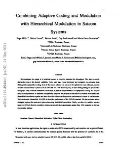
brane-bound Sa-Reductases-Phospholipase Az modifies the nonpolar region of the membrane (17), and to determine the Effects of Solubilization on Sa-ReductaseSpecific Activity- role of this region in 5a-reductase activity, nuclear and miRemoval of 5a-reductase from the membrane environment crosomal membrane fractions were exposed to phospholipase using a detergent-free medium caused a decrease in the spe- Az at concentrations of 0, 0.25, 1, 2, 5, or 10 units/ml for 1 h cific activities of both nuclear and microsomal 5a-reductases at 22 "C. The testosterone concentrations were also varied, (Table I). The loss of specific activity, approximately 40%, allowing kinetic analyses to be done. It can be seen from Fig. la that phospholipase A2 treatment did not alter the Km(app) was identical for both enzymes. Effectsof Phospholipaseson Membrane-boundSa-Reductase of the nuclear 5a-reductase. The four kinetic studies provided a mean KmCapp) value of 143 nmol/liter, and phospholipase A2 at all concentrations tested did not significantly alter this TABLE I Effect of solubilizationor phospholipase treatment on 5a-reductase value (the mean Km(,, values ranged from 128to 154 nmoll liter). However, significant increases in Km(,,,)were seen for activity Epididymal nuclear and microsomal membranes were either solu- the microsomal 5a-reductase (Fig. lb). The mean value for bilized (6) or treated with phospholipase Az or C (5 units/ml) for 1 h the untreated microsomal enzyme was160 nmol/liter, and at 22 "C, and the 5a-reductase activity was determined using a tea- there was a dose-dependent increase of the Km(app) value to tosterone concentration of 500 nmol/liter. 5a-Reductase activity is 288 nmol/liter with increasing amounts of phospholipase Az. presented as themean & S.E. of at least four separate studies. The Vmax(app) values of both 5a-reductases werereduced Ba-Reductase activity maximally by all the phospholipase AS treatments (Fig. 1, c and d ) , but the decreases were consistently greater for the Nuclear Microsomal nuclear enzyme (43-53%) than for the microsomal enzyme p m l / h / m g protein (%) (20-30%). Therefore, alteration of the nonpolar region of the 71.9 f 2.8 (100) 28.0 f 2.4 (100) Control nuclear membrane environment did not affect the 5a-reduc42.6 f 7.8 (59) 16.4 f 2.7 (59) Solubilization 44.6 & 1.7 (62) 16.5 2 2.8 (59) Phospholipase AP tase-testosterone interaction, but this interaction was altered 43.5 2 2.5 (61) 15.5 f 1.2 (55) Phospholipase C by such modification of the microsomal membrane. RESULTS
7491
Phospholipid Dependence of 5a-Reductase Activity 2 so
-
a
C
b
3 0 0 ,
-e
3
e
0 V
-
-2
3
E" Y
0
U
125
3
a
a
9
E
E
Y
3001
L
L
C
-*
b
150
Y
n "
0 0 0.25 1
5 2
10
0 0.25 1
Phospholipase A2 (U/ml)
2
5 10
Phospholipase C (U/ml)
0
0 0.25 1
2
5 10
Phoaphollpase C tU/ml)
I 100
100
= e
e
-
0
-
0
0
-
z
3
a a
-e
a a
50
n
3
50
a
0
X
:
:
>
"
E"
> 0
0 0.25 1 2 Phosphollpase Ai
5 10 (Wml)
50
X
> n
100
=:
L
L
0 0.25 1
2
5
1
Phorpholpase A 2 (Wml)
0 0 0 . 2 5 1 52
10
Phorphoilpare C (U/ml)
Phosphollpase C (U/ml)
FIG. 1. Effects of phospholipase An on the kinetic parameters of membrane-boundepididymal nuclear and microsomal were treated with phosSa-reductase activity. Membrane fractions pholipase A, (0, 0.25, 1, 2, 5, or 10 units (U)/ml) and 5a-reductase activity assessed overa range of testosterone concentrations(25-500 nmol/liter) for 1 h at 22 'C. Values of K,,,(,) and V-(,) were
FIG. 2. Effect of phospholipase C on the kinetic parameters of membrane-bound nuclear and microsomal Sa-reductase activity. Experimentaldetails,dataanalysis,abbreviations, and actual values of kinetic parameters were exactly as described in the legend to Fig. 1. Phospholipase C concentrations were 0, 0.25,1,2,5, obtained by nonlinear least-square analysis. Four studies were done. or 10 units/ml. Hatched bars indicatenuclearwhile stippled bars Significant differences betweenthe kinetic parameters of 5a-reduc- indicate microsomal Sa-reductase activity. tase in untreatedandphospholipaseA,-treatedmembraneswere determined by t tests on arcsin-transformed proportionalizedK,,,(-) environment is important for the enzyme-substrate interacand V-(-)values, i.e. untreated values in each study = 100%. Actual tion and for catalytic capacity. values of for the nuclear enzyme were89, 180, 121, and 180 Effects of Phospholipids on Solubilized Sa-Reductase Actiunmol/liter. The corresponding Vmu(.pp) values were 43, 106, 72, and 130 pmol/h/mg protein. Similarly, actualK,,,(-) values for the micro- ity-The use of phospholipases of broad substrate specificity somal enzyme were 99, 198, 141, and 202 nmol/liter, and the corre- showed that phospholipids areimportant to 5a-reductase sponding VmU(-) values were 126, 312,223,and 314 pmol/h/mg activity. In order to identify which ones were important, we protein. Confidence limits (95%)are shown. Hatched bars indicate investigated the effects of phospholipids of known structure nuclear while stippled burs indicate microsomal 5a-reductase activity. on 5a-reductase activity. 5a-Reductase was solubilized in the *, p < 0.05;NS, not significant. absence of detergent, and phospholipids with different polar
groups were added at a final concentration of 500 pg/ml. It Effect of Phospholipase C on the Kinetic Parameters of can be seen in Fig. 3 and Table I that solubilization caused Membrane-bound 5a-Reductase Activity-Phospholipase C decreases in the specific activities of both the nuclear and removes the polar group from phospholipids, generating di- microsomal 5a-reductases. The inclusion ofegg lecithin, a acylglycerols (17). This treatmentincreased the Km(app) values mixture of phosphatidylcholines, in assays of solubilized 5aand decreased the V-(aw) values for both &-reductases. As reductase enhanced activity. The 2.2-fold increase in nuclear can be seen from Fig. 2a, the mean Km(app) value of the nuclear 5a-reductase activity was sufficient to completely restore the 50-reductase was significantly increased by phospholipase C membrane-bound specific activity, but the enhancement of concentrations of 2, 5, and 10 units/ml. The mean Km(app) the microsomal 5a-reductase (1.3-fold) was not sufficient in value of the untreatednuclear 5a-reductase of 143 nmol/liter this respect. Phospholipids containing serine, ethanolamine, was elevated to 253 nmol/liter by 10 units/ml of phospholipase glycerol, and phosphatidic acid in the polar groups were C. However, in the case of the microsomal 5a-reductase, all without effect, and phosphatidylinositol caused small inthe phospholipase C concentrations increased the Km(app) sig- creases in specific activity (1.3- and 1.15-fold for the nuclear nificantly, and theeffects were greater than those evident for and microsomal 5a-reductases, respectively). Retinol inhibited the 5a-reductases (45 and 42%, respectively), and cardithe nuclear 5a-reductase (Fig. 2b).The untreatedmean K,,,,) value for the microsomal Sa-reductase of 160 nmol/liter was olipin had no effect. These findings suggested that only phosincreased to 392 nmol/liter by phospholipase C at 10 units/ pholipids containing choline were capable of activating solubilized 5a-reductase activity in vitro. ml. The structure of the acyl chains of the phosphatidylcholines values were greater Conversely, the decreases in V(., for the nuclear than for the microsomal 5a-reductase (Fig. 2, that would optimize the activation of 5a-reductase was unc and d). Therefore, removal of the polar groups from mem- known and, therefore, a series of phosphatidylcholines was brane phospholipids affected the kinetic parameters of both obtained, in which the two acylchains were identical. In these 5a-reductases, and thus, the polar region of the membrane compounds, the acyl chains were either (a) saturated and
7492
Phospholipid Dependence of 5a-Reductase Activity
respectively, for dicapryloyl phosphatidylcholine and 8 and 4%, respectively, for dicaproyl phosphatidylcholine). However, the addition of 2 carbon atoms to the acyl chain length a had aremarkable effect. Dilauroyl phosphatidylcholine, which possesses acyl chain lengths of 12 carbon atoms, caused a 420 C fold increase in nuclear 5a-reductase activity anda 50% increase in the microsomal 5a-reductase activity. Furtherc r more, the enhancement of the nuclear 5a-reductase by dilaun o royl phosphatidylcholine caused the specific activity to be 0 MEM SOL LEC PS PGPE PI PA RET CARD elevated to a level considerably greater than was evident in 5 the original membrane preparation (220% increase). This phospholipid also elevated the specific activity of the microsomal Sa-reductase, almost to itsmembrane-bound level. The requirement for the dilauroyl structure was extremely specific because increasing the length of the acyl chains to 14 carbon atoms ormore rendered the phosphatidylcholine incapable of activation. The importance of the degree of unsaturation of the acyl chains to 5a-reductase activity was also investigated. Distea" MEM SOL LEC PS PE PG PI PA RET CARD royl phosphatidylcholine contains 18 carbon atoms in the FIG. 3. Effects of lipids on the specific activities of solubi- saturated acyl chains and had no effect on the specific activity lized nuclear andmicrosomal Sa-reductases. Membrane protein of the 5a-reductases (Fig. 4). The presence of either one or was solubilized usipg a mixture of sodium citrate (0.1 mol/liter), KC1 (0.1 mol/liter), and glycerol (20%v/v) in KRP buffer for 1 h at 4 "C two double bonds in these acyl chains (dioleoyl and dilinoleoyl as described in Ref. 6. Lipids were added to 5a-reductase assays at phosphatidylcholine) caused enhancement of both nuclear final concentrations of 500 pg/ml. Testosterone concentrationwas 50 (212 and 198%) and microsomal (123 and 127%) Sa-reducnmol/liter. MEM, membrane-bound specific activity; SOL, solubi- tases. The activation of the nuclear enzyme restored the lized activity; LEC, egg lecithin; P S , phosphatidylserine; PE, phos- specific activity to the membrane-bound level, but this was phatidylethanolamine; PG, phosphatidylglycerol; PI, phosphatidylinot achieved in the case of microsomal Sa-reductase. Hownositol; PA, phosphatidic acid; RET, retinol; CARD, cardiolipin. ever, the unsaturated phospholipid configuration was more Hatched bars indicate nuclear while stippled bars indicate microsomal favorable than the equivalent saturated distearoyl phospha5a-reductase activity. tidylcholine. Activation of Solubilized 5a-Reductase by Dilauroyl Phosphatidylcholine-The activation of nuclear and microsomal 5a-reductases by dilauroyl phosphatidylcholine observed in the above investigations may have been due to a direct effect on the enzymes or to a process that required modification of the enzyme-phospholipid complex. In evaluations of the activation by dilauroyl phosphatidylcholine with time, the phospholipid (500 rg/ml) was added to incubations that had been in progress for 27 min. In Fig.5, it can be observed that 8 111 420 16 121808: l 18:2 a dilauroyl phosphatidylcholine had an apparently immediate and linear effect on the Sa-reductase activities. The activation, as assessed by the slopes, was %fold for the nuclear and
-e
5
40
e
\
\
-
FIG. 4. Effects of phosphatidylcholines with defined acyl chain structure on the specific activities of solubilized nuclear and microsomal Sa-reductases. Phosphatidylcholines (500 pg/ml) were included in assays of 5a-reductase. Experimental details were as described for Fig. 3. The values for membrane-bound specific activity (MEM) and solubilized specific activity (SOL) are identical 18, and 16, 20 refer to thelength to Fig. 3. The numbers 8, 10, 12, 14, of the two saturated acyl chains of dicapryloyl, dicaproyl, dilauroyl, dimyristoyl, dipalmitoyl, distearoyl, and diarachidoyl phosphatidylcholine. 18:l and 182 refer to the acyl chain lengths and degree of unsaturation of dioleoyl and dilinoleoyl phosphatidylcholines. Hatched bars indicate nuclear while stippled bars indicate microsomal 5a-reductase activity.
increased in length in intervals of two carbon atoms or (b) unsaturated and contained 18 carbon atoms. It can be seen from Fig. 4 that phosphatidylcholines containing saturated acyl side chains of 8 (dicapryloyl-) or 10 (dicaproyl-) carbon atoms were mildlyinhibitory, decreasing the specific activities of both nuclear and microsomal 5a-reductases (19 and 35%,
Incubation Time (mln)
lncubatlon Time (mln)
FIG. 5. Time course studies of the activation of solubilized
nuclear and microsomal Sa-reductases by dilauroyl phosphatidylcholine. Assays of solubilized nuclear ( a ) and microsomal ( b )
5a-reductase were either untreated (u or DLPC ), (500 pglml final concentration, W) was added after 27 min of incubatlon time. The testosterone concentration was 50 nmol/liter.
7493
Phospholipid Dependenceof Sa-Reductase Activity 300
5
P
a
300
1
b
I
c. L 200
200
0
-i
w
:
n
0 100
100
0
0
5 50 100500
0
5 50 100500
0
c 8 I
g 0 2
A
12 '
I
b
P
100
75
0
a
10
w
50
n
8
n8
:
6
25
4
FIG. 7. Effect of DLPC on the kinetic parameters of solubilized nuclear and microsomal 5a-reductases. DLPC (0,5,50,
2 -I
n
-0
60
120
180
PROTEIN Cpg) FIG. 6. Activation by DLPC of solubilized nuclear and microsomal &-reductases at different protein concentrations. Varying amounts of protein solubilized from the nuclear ( a ) and microsomal (b) membranes were incubated with testosterone (55 nmol/liter) for 1 h a t 22 'C, in the presence ( 0 - - -0) and absence (U of ) DLPC (500 pg/ml).
2-fold for the microsomal 5a-reductase. These data suggested that a direct effect was operative and thatno further modification of the phospholipid or the enzyme-phospholipid complex was required. The activation by dilauroyl phosphatidylcholine was also linear with proteinconcentration. In the absence of this phospholipid, solubilized nuclear and microsomal 5a-reductase activities were linear up to protein concentrations of 180 pg/ml incubation volume (Fig. 6). However, in the presence of dilauroyl phosphatidylcholine (500 pg/ml) linearity was observed up to 30 pg protein/ml, beyond this protein concentration linearity was lost (data notshown). Kinetic analyses of the activation of solubilized nuclear 5areductase, revealed that dilauroyl phosphatidylcholine confor centrations of 5 , 50, and 100 pg/ml reduced the Kmtapp) testosterone by20, 60, and 83%, respectively, and yet no (Fig. 7, a and c). change was observed in the value of Vmar(app) At a higher concentration (500 pg/ml), the was further was increased by reduced (to 1%of control), and the Vmax(epp) 30%. The microsomal 5a-reductase was affected similarly (Fig. 7, b andd),butthe decrease in caused by the same dilauroyl phosphatidylcholine concentrations were comparatively smaller (3, 35, and 69%). Again, the Vmlu(epp) was not altered by the phospholipid except at 500 pg/ml. It was
100,and 500 pg/ml, final concentration) was included in assays of solubilized nuclear (a and c ) and microsomal ( b and d ) 5a-reductase. Testosterone concentrations were 20,30,40,55, 70,85, 100, 250, and 500 nmol/liter (1). The results of a typical study are shown above. Km(.pp) and Vmm(.pp) values were obtained by nonlinear least-squares analysis. Hatched bars indicate nuclear while stippled bars indicate microsomal 5a-reductase activity.
clear that dilauroyl phosphatidylcholine, at concentrations of 100 pg/ml and less, did not activate 5a-reductase molecules rendered inactive by the solubilization procedure butenhanced the enzyme-substrate interaction of existing active 5a-reductase molecules. Hill plots (22) of the data for dilauroyl phosphatidylcholine concentrations of 0, 50, and 100 pg/ ml revealed parallel lines with Hill coefficients of 0.84 for the nuclear 5a-reductase and 0.88 for the microsomal 5a-reductase. This demonstrated that testosterone binds to noninteracting sites on the 5a-reductase and that dilauroyl phosphatidylcholine does not cause positive or negative cooperativity. Effects of Phospholipases on Solubilized5a-Reductase Actiuity-It was possible that therelative unresponsiveness of the solubilized microsomal 5a-reductase to activation by phospholipids was due to thepresence of membrane phospholipid that had accompanied the solubilization of protein. We, therefore, treated solubilized nuclear and microsomal protein fractions with phospholipase Az or C (1 unit/ml) and examined the effect of these treatments on the 5a-reductase activity. The specific activity for both enzymes was reduced (60%with phospholipase Az, 45% with phospholipase C), suggesting that phospholipids present in the solubilized preparations were maintaining 5a-reductase activity. However, since the phospholipases caused identical decreases for both enzymes, it was unlikely that theendogenous phospholipids could account for the differences in sensitivity to activation by exogenous phospholipids.
Phospholipid Dependence of SLY-ReductaseActivity
7494
nuclear enzyme, the affinity of the microsomal enzyme for testosterone was reduced. These findings are consistent with the possibility that the active site of the microsomal enzyme is in closer association with the nonpolar environment of the membrane and requires the formation of an enzyme-substrate-phospholipid complex for optimal activity. The nuclear 5a-reductase would not seem to require the formation of such a complex for catalytic activity, and the active site may be oriented in the polar region of the membrane structure. Removal of phospholipid polar groups alters the active concentration and the substrate interaction of both 5a-reductases. I I I In other studies, it has been shown that themembrane envin 0 30 60 90 120 Y ronment is necessary for several steroidogenic enzymes. The Incubation Time (min) >r adrenal lip-hydroxylase (23), testicular5-ene 3&hydroxysteroid dehydrogenase, 17-hydroxylase, C-17,20 lyase, and 178hydroxysteroid dehydrogenase (24,25) and 5-ane38-hydroxysteroid dehydrogenase (26) are all inactivated by phospholipase treatment. However, in these studies the role of the phospholipid in the regulation of the catalytic process was not investigated. The kinetic analyses in this study revealed a fundamental difference in the role of the membrane in the activity of nuclear and microsomal 5a-reductases of the epididymis. The relationship between phospholipid structure and the ability to enhance 5a-reductase activity was extremely spe0 cific. The finding that phosphatidylcholines were more effi0 30 60 90 120 cient in activating 5a-reductase than were other phosphoIncubation Time (min) lipids has been observed for the liver 5a-reductase (27-29), FIG.8. Time course studieson the effect of DLPC on mem- but the adrenal and ovarian (luteal) cholesterol side-chain brane-bound Sa-reductase activity. Assays of nuclear (a)and or) cleavage enzymes (cytochrome P-450,,) were activated by microsomal ( b ) 5a-reductase were either untreated (M DLPC treated (500 pg/ml, final concentration, 0-- -0). DLPC was cardiolipin and phosphatidylinositides (30-33); phosphatidyladdedafter 27 min of incubation time (arrow).The testosterone cholines were poor activators by comparison. Interestingly, concentration was 50 nmol/liter. neither cardiolipin nor phosphatidylinositol was an efficient activator of epididymal Sa-reductase. A common feature of Activation of Membrane-bound Sa-Reductase by Dihuroyl these studies is the necessity for phospholipids to be present Phosphatidylcholine-The enhancement of solubilized 5a-re- at concentrations higher than the critical micelle concentraductase activity by dilauroyl phosphatidylcholine and inacti- tion. In thecase of 5a-reductase, the specificity for phosphavation of both membrane-bound and solubilized 5a-reductase tidylcholine may be due to the ability of this phospholipid to by phospholipase treatment promptedan investigation of the form lamellae (34), and this bilayer may be a more natural effect of adding dilauroyl phosphatidylcholine to membrane- environment for the enzyme. The presence of unsaturated acyl chains in the phosphatibound 5a-reductase. This phospholipid was added to incubations that had been in progress for 27 min. It can be seen dylcholine structure (dioleoyl and dilinoleoyl) revealed that from Fig.8 that both the nuclear and microsomal 5a-reductase activation of 5a-reductase was evident, whereas the correactivities were enhanced immediately and linearly upon inclu- sponding saturated phosphatidylcholine (distearoyl) was sion of the phospholipid. The elevation in specific activity, as without effect. This was also found for the activation of assessed by slopes, was 60% for both enzymes. These results mitochondrial cytochrome P-450,, (35, 36). A possible explasuggest that the incorporation of dilauroyl phosphatidylcho- nation for this preference involved the ability of the enzyme line into the membrane environment has a direct effect on to incorporate into the phospholipid micelle. For cytochrome P-450,, the incorporation into the micelleswas faster if the activity of 5a-reductase. unsaturated acyl chains were present (37). DISCUSSION The acyl chain length of 12 carbon atoms was critical for The studies presented here have been concerned with the activation of 5a-reductase by saturated phosphatidylcholines. relationship between epididymal Sa-reductase activity and In anotherstudy (38)this specificity for the dilauroyl configthe membrane environment. The loss of specific activity that uration was evident for estrone glucuronyltransferase, but is a consequence of solubilizing the enzyme from the mem- other chain lengths were also activatory, although to lesser brane demonstrated that both nuclear and microsomal 5a- extents. This enzyme was also less sensitive to activation by reductases required an ordered membrane structure for opti- dilinoleoyl than by distearoyl phosphatidylcholine. It is posmal activity. Furthermore, the use of phospholipases that sible that theacyl chain length of phospholipids is important modify membrane structure in specific ways has shown that for the concentration and presentationof the substrate to the the phospholipids surrounding 5a-reductase may be regulat- enzyme. However, the absolute requirement for 5a-reductase ing 5a-reductase activity, and the influence exerted is differ- of the 12-carbon acyl chain wouldsuggest that dilauroyl ent for the nuclear and microsomal 5a-reductases. Modifica- phosphatidylcholine is not merely elevating the relative subtion of the nonpolar region of the membrane with phospho- strate concentration, as the other phospholipids would be lipase Az decreased the active population of 5a-reductases in expected to do this. The fact that dilauroyl phosphatidylchowithout altering the Vm.l(a,,,,) both membrane preparations. However, while such modifica- line decreases the value of Km(epp) tions did not affect the enzyme-substrate interaction of the suggests that it is not reactivating 50-reductase molecules 1004
a
I
C
Y
I
Phospholipid Dependence of 5cu-Reductase Activity
7495
that were rendered inoperative by the solubilizing processbut is enhancing activity through an active site-oriented process. The activation of both membrane-bound and solubilized 5areductase by dilauroyl phosphatidylcholine suggests that this phospholipid provides a more suitable membrane structure for the 5a-reductase mechanism. This may be because the acyl chains are the optimal length to bring the ionic charges on the phospholipid and the enzyme into proximity. Alternatively, this phospholipid may create a more suitable solvent or electrochemicalgradient for the 5a-reductase proton transfer, thereby effectively reducing the activation energy of the reaction. The ability of dilauroyl phosphatidylcholine to activate membrane-bound Sa-reductase supports the possibility that the activity of this enzyme is regulated by the phospholipid environment. In conclusion,these studies have demonstrated for the first time that epididymal 5a-reductase is not only dependent on an ordered membrane structure for activity but that specific phospholipids can regulate the activity. We suggest that changes in membrane phospholipid content regulate 5a-reductase activity and thatsuch changes would be instrumental in the expression of DHT-dependent processes.
13. Arrhenius, E., Renberg, L., and Johansson, L. (1977) Chem. Biol. Interact. 18.23-34 14. Arrhenius, E., Renberg, L., Johansson, L., and Zetterqvist, M. A. (1977) Chem. Biol. Interact. 18,35-46 15. Van Dam, K., and Meyer, A. J. (1971) Annu. Rev. Biochem. 4 0 , 115-160 16. Vargaftig, B. B., and Dao Hai, N. (1972) J. Phurm. Pharmaol. 24,159-161 17. Mahler, H. R., and Cordes, E. H. (1966) Biological Chemistry, pp. 508-524, Harper and Row, New York 18. Robaire, B. (1979) Can. J . Physiol. Pharmacol. 57,998-1003 19. Wilkinson, G. N. (1961) Biochem. J. 80,324-332 20. Cleland, W. W. (1967) Adu. Enzymol. 29, 1-32 21. Sokal, R. R., and Rohlf, F. J. (1973) Introduction to Biostatistics. The Assumption of ANOVA, pp. 215-217, W. H. Freeman & Co., San Francisco, CA 22. Koshland, D. E. (1970) in The Enzymes (Boyer, P. D., ed) Vol. 1, pp. 341-396, Academic Press, New York 23. Wang, H.-P., Pfeiffer, D. R., Kimura, T., and Tchen, T. T. (1974) Biochem. Biophys. Res. Commun. 67,93-99 24. Machino, A., Nakano, H., and Tamaoki, B-I. (1969) Endocrinol. Jpn. 1 6 , 3 7 4 6 25. Samuels, L. T., Bussmann, L., Matsumoto, K., and Hueeby, R. A. (1975) J. Steroid Biochem. 6,291-296 26. Larner, J. M.,and Weibe, J. (1983) J. Steroid Biochem. 18,541-
REFERENCES Danzo, B. J., and Davies, J. (1975) in Handbook of Physiology, Section 7: Endocrinology (Hamilton, D. W., and Greep, R. O., eds) Vol. 5, pp. 319-338, Williams & Wilkins, Baltimore, MD Kinoshita, Y. (1981) Endocrinol. Jpn. 28,499-513 Suzuki, K., and Tamaoki, B-I. (1974) J. Steroid Biochem. 19, 249-256 Djoseland, 0.(1976) Steroids 2 7 , 47-64 Brown, D. V., Amman, R.P., and Wagley,L. M. (1983) Biol. Reprod. 28,1257-1268 Scheer, H., and Robaire, B. (1983) Biochem. J. 2 1 1 65-74 , Scheer, H., and Robaire, B. (1983) Biol. Reprod. 2 9 , 1-10 Robaire, B., Ewing, L. L., Zirkin, B.R., and Irby, D.C. (1977) Endocrinology 1 0 1 , 1379-1390 Scheer, H., and Robaire, B. (1980) Endocrinology 107,948-953 Foldesy, R. G., and Leatham, J. H. (1981) J. Endocrinol. 91,4351 Robaire, B., Scheer, H., and Hachey, C. (1981) Bioreguhtors of Reproduction (Jagiello, G., and Vogel, H. J., eds) pp. 487-498, Academic Press, New York Cooke,G. M., and Robaire, B. (1984) J. Steroid Biochem. 20, 1279-1284
27. Golf, S. W., Graef, V., and Staudinger, H. (1974) Hoppe-Seyler’s Z. Physwl. Chem. 355, 1499-1507 28. Golf, S. W., and Graef, V. (1978) J. Steroid Biochem. 9, 10871092 29. Graef, V., Golf, S. W., and Rempeters, G. (1978) J. Steroid Biochem. 9,369-371 30. Tanaka, T., and Strauss, J. F. (1982) Endocrinology 110, 15921598 31. Farese, R. V., and Sabir, A.M. (1980) Endocrinology 106, 18691879 32. Lambeth, J. D. (1981) J. Biol. Chem. 266,4757-4762 33. Pember, S. O., Powell, G . L., and Lambeth, J. D. (1983) J. Bwl. Chem. 258,3198-3206 34. Gurr, M. I., and James, A. T. (1971) Lipids os Components of Macromolecules. Lipid Biochemistry, pp. 175-179, Cornel1University Press, Ithaca, NY 35. Yamakura, F., Kido, H., and Kimura, T. (1981) Biochim. Biophys. Acta 649,343-354 36. Lambeth, J. D.,Kamin, H., and Seybert, D.W. (1980) J. Biol. Chem. 255,8282-8288 37. Tuckey, R. C., and Kamin, H. (1982) J. Biol. Chem. 2 5 7 , 28872893 38. Tukey, R.H., Billings, R.E., Autor, A. P., and Tephly, T. R. (1979) Biochem. J. 179.59-65
1. Orgebin-Crist, M.C.,
2. 3. 4. 5.
6. 7. 8.
9. 10. 11. 12.
550