Molecular Basis for Effects of Carcinogenic Heavy Metals on Inducible Gene Expression Joshua W. Hamilton, Ronald C. Kaltreider, Olga V. Bajenova, Michael A. lhnat, Jennifer McCaffrey, Bruce W. Turpie, Erin E. Rowell, Jannet Oh, Michael J. Nemeth, Carrie A. Pesce, and Jean R Lariviere Department of Pharmacology and Toxicology, Dartmouth Medical School, Hanover, New Hampshire, and Norris Cotton Cancer Center, Dartmouth-Hitchcock Medical Center, Lebanon, New Hampshire Certain forms of the heavy metals arsenic and chromium are considered human carcinogens, although they are believed to act through very different mechanisms. Chromium(VI) is believed to act as a classic genotoxic and mutagenic agent, and DNANchromatin appears to be the principal target for its effects. In contrast, arsenic(lll) is considered nongenotoxic, but is able to target specific cellular proteins, principally through sulfhydryl interactions. We had previously shown that various genotoxic chemical carcinogens, including chromium(VI), preferentially altered expression of several inducible genes but had little or no effect on constitutive gene expression. We were therefore interested in whether these carcinogenic heavy metals might target specific but distinct sites within cells, leading to alterations in gene expression that might contribute to the carcinogenic process. Arsenic(lll) and chromium(V1) each significantly altered both basal and hormone-inducible expression of a model inducible gene, phosphoenolpyruvate carboxykinase (PEPCK), at nonovertly toxic doses in the chick embryo in vivo and rat hepatoma H411E cells in culture. We have recently developed two parallel cell culture approaches for examining the molecular basis for these effects. First, we are examining the effects of heavy metals on expression and activation of specific transcription factors known to be involved in regulation of susceptible inducible genes, and have recently observed significant but different effects of arsenic(lll) and chromium(Vl) on nuclear transcription factor binding. Second, we have developed cell lines with stably integrated PEPCK promoter-luciferase reporter gene constructs to examine effects of heavy metals on promoter function, and have also recently seen profound effects induced by both chromium(V1) and arsenic(lil) in this system. These model systems should enable us to be able to identify the critical cis (DNA) and trans (protein) cellular targets of heavy metal exposure leading to alterations in expression of specific susceptible genes. It is anticipated that such information will provide valuable insight into the mechanistic basis for these effects as well as provide sensitive molecular biomarkers for evaluating human exposure. Environ Health Perspect 1 06(Suppl 4):1 005-1015 (1998). http://ehpnet1.niehs.nih.gov/docs/1998/Suppl4/ 1005-1015hamilton/abstract.html Key words: chromium, arsenic, mitomycin C, phosphoenolpyruvate carboxykinase, transcription factors, genetic constructs, transfection
This paper is based on a presentation at the Symposium on the Superfund Basic Research Program: A Decade of Improving Health through Multi-Disciplinary Research held 23-26 February 1997 in Chapel Hill, North Carolina. Manuscript received at EHP 1 1 December1997; accepted 27 March 1998. The authors dedicate this manuscript to the memory of the late Dr. Karen E. Wetterhahn, who was an invaluable scientist, mentor, colleague, and friend. Dr. Wetterhahn and her laboratory provided reagents, laboratory resources, and valuable advice on this project. Most important, Dr. Wetterhahn was the original director and the major impetus behind the Dartmouth Superfund Basic Research Program Project which made this research possible. The authors also wish to thank A. Barchowsky and his lab for their advice and assistance in the gel shift analysis. This work was supported by grants to JWH from NIH-NIEHS (ES07373), NIH-NCI (CA49002), and the Norris Cotton Cancer Center. JWH was also partially supported by the N6rris Cotton Cancer Center; RCK and OVB were supported by fellowships from NIH-NIEHS (ES07373) and AJW was partially supported by the Dartmouth College Chemistry Department. Support for the Dartmouth College Molecular Biology Core Facility was provided by NIH-NIEHS (ES07373) and the Norris Cotton Cancer Center Core grant (CA23108). Address correspondence to J.W. Hamilton, Department of Pharmacology and Toxicology, 7650 Remsen, Dartmouth Medical School, Hanover NH 03755-3835. Telephone: (603) 650-1316. Fax: (603) 650-1129. E-mail:
[email protected]
Abbreviations used: EMSA, gel electrophoretic mobility shift assay; MMC, mitomycin C; MMS, methyl methanesulfonate; PEPCK, phosphoenolpyruvate carboxykinase; TNFa, tumor necrosis factor a; TNFOi, tumor necrosis factor P; U.S. EPA, U.S. Environmental Protection Agency; UV, ultraviolet.
Introduction The heavy metals chromium(VI) and arsenic(III) are considered human carcinogens and share several properties in regard to their carcinogenicity. They are both associated with certain tumors in humans based principally on epidemiologic evidence that is quite strong and that preceded animal carcinogenicity data by many years (1-3). Both are considered lung carcinogens whose effects are synergistic with cigarette smoking or exposure to other lung carcinogens such as benzo[a]pyrene or other carcinogenic metals. Human exposure to these metals occurs principally in occupational settings and through environmental contaminations such as from toxic waste sites, including numerous Superfund sites, or from naturally contaminated drinking water. Chromium and arsenic are listed as 2 of the 8 heavy metals that are among the top 22 compounds of particular concern to the U.S. Environmental Protection Agency (U.S. EPA) Office of Solid Waste and Emergency Response concerning Superfund sites. Thus, there is now increasing concern about human health effects from exposure to heavy metals found in high concentrations at these sites, such as the high levels of chromium and arsenic at waste sites in Woburn, Massachusetts, and northern New Jersey. However, potential human exposures and health risk from these sites are less well defined. Thus, understanding the overall human health impact of environmental exposure to these heavy metals is of paramount concern. Chromium(VI) is a known human lung carcinogen and may also contribute to nasopharyngeal cancers (1,2,4,5). A strong association between chromium exposure and an increase in lung cancer was first observed in the chrome ore industry in Germany in the 1920s. Since then, there have been numerous studies linking inhalation exposure to chromium, particularly chromium(VI) compounds, with increases in lung and nasal cancers. However, early recognition of this association and significant decreases in exposure through industrial hygiene practices have reduced these incidences. Arsenic is considered to be a probable human lung, skin, and bladder carcinogen (3,6). Lung exposure has been principally through occupational settings in the mining, processing, and smelting of arsenic-containing ores, and in the manufacture of arsenic-containing pesticides, whereas skin exposure has been through
Environmental Health Perspectives * Vol 106, Supplement 4 * August 1998
1 005
HAMILTON ET AL.
many of these same occupational settings as well as topical application of arsenical medications. There is also an association between environmental arsenic exposure, e.g., through arsenic in well water, and an increased risk of certain skin and possibly bladder cancers (3,7). Arsenic exposure has also been implicated in certain other tumor incidences including scrotal cancer (copper smelters) and hepatic angiosarcomas (vineyard workers) (3,7). As with the other carcinogenic metals, there is a strong synergistic association between arsenic exposure and cigarette smoking for risk of lung cancer. However, in the case of arsenic, this is complicated by the fact that there were high levels of arsenic in tobacco prior to the 1970s as a result of use of arsenical pesticides in tobacco agriculture (3). Arsenic has only recently been shown to be carcinogenic in animal models (3). The form and valence of chromium is a major factor in assessing its biologic effects, and chromium(VI) is the principal form responsible for the mutagenic and carcinogenic properties, as first proposed in the uptake-reduction model by Wetterhahn and co-workers (1,4,8,9). Chromium(VI) has been shown to be genotoxic, mutagenic, and carcinogenic in a large number of short- and long-term assay systems using various end points and systems (1,2,4,8,9). Like many organic carcinogens such as benzo[a]pyrene and aflatoxin B1, chromium(VI) appears to act as a classic initiator in these test systems, and this is believed to be the principal mechanism by which chromium(VI) increases carcinogenic risk. Our laboratory has hypothesized that genotoxic carcinogens, including chromium(VI), exert preferential effects on the expression of a specific dass of genes, i.e., inducible genes, as a result of nonrandom DNA damage targeted to members of this gene class (8-12). Previous studies in our laboratory have shown that a number of different genotoxic chemical carcinogens that induce different types of DNA damage, including the genotoxic metals chromium and nickel, significantly alter both basal and inducible expression of several model inducible genes but have no effect on expression of constitutive genes. Assuming that the effects on gene expression are a result of carcinogen-induced DNA damage, what is their molecular basis? We have postulated that inducible genes are strongly affected by chemicallyinduced DNA damage as a result of both targeting of DNA damage and the intrinsic structural and biochemical properties of those genes, and have proposed a model in 1 006
which these effects occur as a consequence of chromatin structure and/or nuclear architecture (10,13). This nuclear model provides a framework for understanding how two different genes in the same nucleus may reside in very different physical and chemical environments, and may therefore have different sensitivities to chemical attack. Thus, even low levels of DNA damage in vivo might be sufficient to have significant effects on expression of certain sensitive genes. We previously demonstrated that treatment of 14-day chick embryos with a single administration of chromium(VI), at a dose that produced no overt toxicity but that caused significant levels of DNA damage (14), had profound effects on expression of several inducible genes while having no effect on expression of several constitutively expressed genes (10). Both the basal and inducible expression of the inducible 5'-aminolevulinate synthase, cytochrome P450 CYP2HI, and phosphoenolpyruvate carboxykinase (PEPCK) genes were markedly affected by the chromium treatment, whereas the albumin, transferrin, and ,-actin genes were refractory to this treatment. The effects on expression of the inducible genes were seen at both the steady-state mRNA and transcriptional levels, and the time courses for these effects closely matched the time course for chromium-induced DNA damage and repair (10,12,14). Interestingly, certain effects most closely correlated with chromium-DNA monoadduct formation, whereas other effects were more closely associated with chromium-DNA cross-link formation (10,12,14). In contrast to chromium, the mechanistic basis for the carcinogenic effects of arsenic is still poorly understood. Arsenic has generally been shown to be negative in bacterial and mammalian mutagenesis assays, and there is little evidence of DNA damage induced by arsenic, although it has been demonstrated to be a clastogen and weak inducer of sister chromatid exchanges in some systems (3,6). Arsenic causes morphologic transformation, anchorage independence, aneuploidy, and gene amplification in mammalian cell culture assays (6). These effects may be related to its ability to alter DNA synthesis and DNA repair, as well as fidelity of chromosome segregation. Arsenite [As(III)] is approximately 4-fold more toxic than arsenate [As(V)], although these two forms can be inter-converted by redox pathways in vivo (3). Arsenic(III) is readily methylated in vivo, and the methylated forms have a low
interaction with macromolecules and are
readily excreted. However, the rate of methylation differs among tissues and species. The primary mechanism of arsenic(V) toxicity is uncoupling of oxidative phosphorylation, by substitution for inorganic phosphate. Arsenic(III) also can cause oxidative bursts in endothelial and other cells, and is heat shock-mimetic, inducing the classic heat shock and stress responses of cells (15). Thus, its biologic effects are more closely associated with cytoplasmic rather than nuclear effects, and DNA does not appear to be the primary target for arsenic toxicity or carcinogenesis. In this regard arsenic appears to act more as a classic promoter such as the phorbol ester 12-O-tetradecanoylphorbol-13acetate or hydrogen peroxide, although its specific effects may be more complex. Arsenic displays distinct interactions with specific proteins in vivo, which may be the basis for much of its specific toxic effects as well as its carcinogenicity. Most of the intracellular effects of arsenic are attributed to arsenic(III) rather than arsenic(V). Arsenic(III) binds avidly to thiols in cells, both to small molecule thiols such as glutathione as well as thiolcontaining proteins (3,16,17). Arsenic has a particular avidity for hydrolases and can compete with phosphate to inhibit enzymes such as phosphoraldehyde and glucose dehydrogenase (3). Many of the cellular effects of arsenic can be ascribed to its ability to elicit a heat-shock response (15,16). Arsenic induces phosphorylation of hsp27, increases expression of hsp27, hsp70, and hsp90 through the heat-shock factor, induces heme oxygenase, mdrl, and quinone reductase gene expression, and induces metallothionein expression by both a transcriptional and posttranscriptional mechanism, although arsenic is not a ligand for metallothionein protein binding (3,18,19). Arsenic exhibits a strong preferential binding to the vicinal dithiol of the glucocorticoid receptor, inhibiting binding of glucocorticoid hormone but not altering hsp9O binding (16,17). This effect is extremely specific, as there is not a similar effect on the closely related receptors for androgen, estrogen, mineralocorticoids or progesterone. Because at least one of the model inducible genes we have used in our studies, i.e., PEPCK is regulated by glucocorticoids, we predicted that this induction pathway would also be markedly affected by arsenic treatment. Other effects on PEPCKexpression are also predicted to be likely, as a result of arsenic effects on other
Environmental Health Perspectives * Vol 106, Supplement 4 * August 1998
EFFECTS OF HEAVY METALS ON GENE EXPRESSION
specific response pathways and regulatory proteins within the cell. Thus, there is reason to believe that both chromium and arsenic can have very selective effects on activation of regulatory pathways and expression of certain genes in vivo, albeit through different mechanisms. We are particularly interested in testing the hypotheses that specific DNA regulatory regions within the promoters of targeted genes are responsible for mediating the cis effects, and that specific transcriptional factors are responsible for mediating the trans effects of these heavy metals on inducible gene expression. Such effects may be very important in understanding the overall effects of these and other heavy metals on the carcinogenic process. The purpose of these studies is to explore these hypotheses in detail.
benzamidine), 7 pg/MI L-1-p-tosylamino-2phenylethyl chloromethyl ketone, and 0.1 mM dithiothreitol (DTT). Nuclear protein soluble extract was isolated by a procedure of Dignam et al. (23), as modified by Barchowsky (24). Measurement of mRNA Levels and
used as a template for the PCR ampliflcation of the PEPCK promoter. The sequences for the upper (forward) primers were 5'-CTAGCTAGCCAATCACCCCT (NheI, -592 of the rat PEPCKpromoter region) and 5'-CTAGCTAGCCATGT CCCTGCCCCC (NheI, -1162 of the rat PEPCKpromoter region); and the sequence EMSAAnalysis of the lower (reverse) primer was 5'Steady-state mRNA levels were measured GGAAGATCTCTACCTTTCTT (Bgl II, by a quantitative solution hybridization +87 of the rat PEPCKpromoter region). All assay using gene-specific 5'-[32P]-end- plasmids used were grown in Escherichia coli labeled synthetic oligonucleotide probes, JM109 strain. The PEPCKpromoter fragexactly as previously described (20). This ments were digested with NheI and BgIII assay has been shown to provide a sensitive, enzymes and ligated into pGL3. Plasmids linear, and quantitative measure of specific containing inserts were sequenced with an steady-state mRNA levels (fmol of mRNA ABI PRISM DyeDeoxy Terminator Cycle per milligram of total RNA), and can accu- Sequencing kit (Applied Biosystems, Foster rately determine differences in mRNA City, CA). All sequences were checked expression of less than 20% (10,20,25,26). against GenBank PEPCKsequences using The c-fos, c-jun, gadd45, gaddl53, and the Blast sequence analysis program to Materials and Methods PEPCKsolution hybridization probes we ensure sequence specificity. Plasmid DNA used are synthetic cDNA oligonucleotides was purified using Qiagen columns Animal and Cell Treatment corresponding to nucleotides 222-248 (Qiagen, Valencia, CA) and transfected into and Sample Preparation (27), 622-648 (28), 697-719 (29), 517- H4IIE rat hepatoma cells using the lipoAll chemicals were purchased from Sigma 540 (30), and 268-291 (31), respectively, fectamine technique (GIBCO-BRL). To Chemical Co. (St. Louis, MO). Fertile of the published sequences. Double- obtain stable transfectants, cells were White Leghorn chicken eggs (Truslow stranded DNA consensus sequences for cotransfected with two types of plasmids: Farms, Inc., Chestertown, MD) were incu- AP-1 and nuclear factor-kappa B (NF-KB) pGL3 reporter vectors with different probated as previously described (20). Test for use as EMSA probes were obtained moters plus pSV2neo that confers resistance compounds were administered in solution from Promega (Madison, WI). The Spl to neomycin. Cells were selected in G418 onto the inner shell membrane as pre- double-stranded probe was synthesized and resistant colonies were subcloned and viously described (21). Chromium(VI) was based on the Spl/EGRI site within the checked for PEPCK-luciferase insertion. administered as sodium dichromate mdrl gene promoter. Protein concentra- For the luciferase assay, cells were lysed in (Na2Cr207.2H20) at a dose of 50 tions were determined by a standard the luciferase reporter buffer in the wells pmol/kg in 100 pl H20. Arsenic(III) was absorbance assay (bicinchoninic acid according manufacturer's instructions administered as sodium arsenite (NaAsO2) [BCA] assay; Pierce, Rockford, IL). The (Luciferase assay system, Promega). Luciat a dose of 100 pmol/kg in 100 pl H20. EMSA analysis was performed essentially as ferase activity was measured using a 96-well Dexamethasone was administered as dexa- previously described (32) with the follow- plate luminometer (Dynatech Laboratories, methasone-21-phosphate in 10 1l H20 at ing modifications (33): a high ionic load- Chantilly, VA). Protein concentrations were a dose of 5 jmol/kg embryo weight. For ing buffer was used containing 10 mM determined by a standard absorbance DC steady-state mRNA studies, after embryo Tris, 20 mM KCl, 0.2 mM DTT, 0.2 mM (detergent compatible) protein assay treatment the livers were removed and EDTA, 4% glycerol, 0.2 pg/pl bovine (Bio-Rad, Hercules, CA). immediately frozen at -75°C. H4IIE rat serum albumin, 0.02 pg/pl poly d(IC) hepatoma and MDA-MB-435 (MDA) cells (GIBCO-BRL, Gaithersburg, MD), and Statistica Analysis were grown and treated essentially as previ- 0.04 pg/pl salmon sperm DNA (pH 8.0). Data were analyzed for statistical significance ously described (22). UV-C (ultraviolet C) A high ionic-strength running buffer was by ANOVA, Student's t-test, and nondoses (254 nm) were administered to cells also used containing 50 mM Tris, 380 parametric analysis, where appropriate, using the Instat and Prism software programs in 150-mm cell culture dishes with a UV mM glycine, 2.0 mM EDTA (pH 8.0). (GraphPad Software, San Diego, CA). Stratalinker (Stratagene, Lajolla, CA) or using a calibrated germicidal lamp at the Generation and Analysis of PEPCKResults desired dose. Total cellular RNA was sub- Luciferase Genetic Constructs sequently isolated using a guanidine iso- All DNA-modifying enzymes and lipofecta- Initial experiments were conducted to thiocyanate-cesium chloride gradient mine were purchased from GIBCO-BRL. examine whether there were preferential technique exactly as previously described The pGL3 vectors and the luciferase assay effects of chromium and arsenic on expres(20). For the electrophoretic mobility shift system were purchased from Promega. The sion of a model-inducible gene, PEPCK, in assays (EMSA), cells were washed with ice- PEPCK-luciferase reporter vectors were 14-day chick embryo livers. The chick cold phosphate-buffered saline (pH 7.4), constructed by cloning a region of the embryo represents a simple, inexpensive, containing 1.0 mM EDTA, protease PEPCKpromoter into the multiple cloning and easily manipulated in vivo model and inhibitor mix (4 pg/ml leupeptin; 4 site of the pGL3 basic vector. Rat genomic has been an excellent system for examining pg/ml soybean trypsin inhibitor; 1 mM DNA from H4IIE cells was isolated and effects of carcinogens on gene expression Environmental Health Perspectives * Vol 106, Supplement 4 * August 1998
1007
HAMILTON ET AL.
based on our previous studies (10-13, 34-36). Chick embryo development has been extensively characterized, providing a large background of information about its embryology, physiology, and biochemistry. This model has been used to investigate the genotoxicity of over 50 chemicals using a wide variety of genetic end points, including chromosomal damage, sister chromatid exchange, inhibition of DNA synthesis, unscheduled DNA synthesis, carcinogenDNA binding, and DNA alkaline elution (10,14,21,37-39). The chick embryo also possesses an active, highly inducible hepatic mixed-function oxidase enzyme system capable of metabolizing indirectacting mutagen-carcinogens to active forms (20,21,26,37,38,40). The chick embryo can also be used to specifically examine the developmental effects of agents on an embryonic system and mimics the human fetus in many important aspects with regard to its toxicology (21,37,38,40,41). Thus, this system represents an excellent nonmammalian whole animal toxicology model. The highest doses of chromium(VI) and arsenic(III) that caused no overt toxicity or lethality in dose-response experiments were determined to be 50 imol/kg and 100 pmol/kg, respectively (10,12,42). At these doses there is little or no effect on overall DNA, RNA, or protein synthesis, and the embryos can be hatched normally with no obvious toxic effects (10,12,42). Using these doses, 14-day embryos were
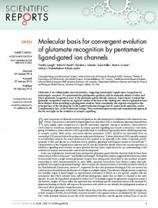
treated with a single administration of chromium or arsenic, and mRNA expression of various inducible and constitutive genes was measured over time (Figure lA-C). Chromium (Figure IA) or arsenic (Figure iB) treatment significantly increased the basal expression of PEPCK, but had no effect on expression of ,-actin (Figure 1C). Chromium and arsenic also significantly altered the response of the PEPCKgene to glucocorticoid induction (Figure 1A,B). Chromium increased the induction response of PEPCKto glucocorticoids initially (1-2 hr) but at later times almost completely suppressed the response of PEPCKto its normal induction signal (Figure IA). Arsenic had similar effects, although the magnitude of the response was greater and occurred over a more protracted time than the response to chromium (Figure 1B). These effects were then examined in the H4IIE rat hepatoma cell line to investigate the molecular basis for this phenomenon. This cell line expresses PEPCKin a basal and hormone-inducible manner similar to that of the liver in vivo, and has been used extensively to examine PEPCKregulation [reviewed by Granner et al. (43) and Lucas and Granner (44)]. Initial experiments established the toxicity dose-response to chromium and arsenic using a colonyforming assay as a measure of cell survival (Figure 2). These experiments established the maximal noncytotoxic doses as well as the minimal doses which produced
A
0-
0.01
10.0
100.0
1000.0
Figure 2. Cytotoxicity of chromium(VI) and arsenic(ll) in rat hepatoma H411E and human breast carcinoma MDA-MB-435 cell lines. Cells were treated with chromium or arsenic for 4 hr and cytotoxicity was assessed by a colony-forming assay as described in "Materials and Methods." Data are expressed as a percent of control colonies formed. Each data point represents the mean±SD of values from three separate plates. *, MDA cells treated with arsenic; *, MDA cells treated with chromium; A, H411E cells treated with arsenic; *, H411E cells treated with chromium.
C
0 0
CD
1.0
150-
0
U)
0.1
Time after chromium(VI) or arsenic(iII), hr
B
.CO
700-
0
.U) UL M_
Q-
W
150
900.
C
X
complete cell death. These were used in subsequent experiments to examine responses to low (i.e., noncytotoxic) versus high (i.e., cytotoxic) doses of heavy metals. Using these low doses, the effects of chromium and arsenic on PEPCK expression were examined in the H4IIE cells, and compared to the effects of mitomycin C (MMC), an organic DNA cross-linking agent with similar genotoxic properties as chromium(VI). Figure 3 shows that
L-
= 500
X
Zc.
C
tP) _
E °- 300 C., 100 t
125-
X
o5 100 -5
,z c CD