Sep 25, 2018 - Molecular cloning techniques recently applied to studies of mucins, however, have .... for cloning of the purified DNA following instructions supplied by the manufacturer. ...... Laboratory Manual, 2nd Ed., Chap. 9, pp. 9.17-9.19 ...
Vol. 268, No. 21, Issue of September 25, pp.. 20563-20569,1993 Printed in U.S.A.
THEJOURNALOF BIOLOGICAL CHEMISTRY
0 1993 by The American Society for Biochemistry and Molecular Biology, Inc.
Molecular Cloning, Sequence, and Specificity of Expression of the Gene Encoding theLow Molecular Weight Human Salivary Mucin (MUC7)* (Received for publication, March 31, 1993, and in revised form, June 9, 1993)
Libuse A. BobekS, Hsiaoyun Tsai, Aaron R. Biesbrock, and Michael J. Levine From the Department of Oral Bi0log.y and Dental Research Institute, School of Dental Medicine, State University of New York at Buffalo, Buffalo,New York 14214
Previous biochemical studies have determined that Human submandibular-sublingual saliva contains low and human saliva contains high and low molecular weight high molecular weight mucin glycoproteins, MGZ1(120-150 mucin glycoproteins(MG1 and MG2, respectively)that kDa) and MG1 (>lo00 kDa), respectively, that are structurare structurallydistinct. In this study, we describe the ally and functionally distinct (Prakobphol et al., 1982; Levine isolation and characterization of overlapping cDNA et al., 1987; Loomis et al., 1987; Cohen and Levine, 1989; cloneswhichcodeforthe MG2 proteincore. DNA Ramasubbu et al., 1991; Scannapieco and Levine, 1993). In sequencingrevealed a translated region 113 of 1 nucleotides encoding a protein of 377 amino acid residues addition, recent immunocytochemical studies suggested the with amolecularmassof 39 kDa. The first 20 N- existence of cell populations specific for each of the human terminal residueswere very hydrophobic and probablysalivary mucins; thus MG1 and MG2 may bedistinct secretory products (Cohen et al., 1990,1991). MG2 is composed of about comprise theMG2 leader peptide. The region encoding the secreted protein can be divided into three distinct30% proteinand 68% carbohydrate. It exists asa single domains; unique5’- and 3”translatedregions contain- polypeptide chain, an exception among the secretory mucins. ing 4 and 1 potential N-glycosylation sites, respec- In addition, two isoforms of MG2, designated as MG2a and tively, and a central region ofsix almost perfect tan- MGBb, have been identified which differ primarily in their dem repeats of 23 amino acid residues with a high content of the terminal sugars, sialic acid, and fucose (Ranumber of Thr and Ser. No sequence homology with masubbu et al., 1991). MG1 is comprised of 15% protein and any other human or animal mucins, and no significant homology to any other proteins was found. MG2 mRNA 78% carbohydrate and appears to exist as an oligomer of is about 2.5 kilobases long, and its expression appears monomeric units.’ Both MG1 and MG2 contain 0-linked and The0-linked carbohydrate to be species-, tissue-, and cell-specific. We propose to N-linkedcarbohydrateunits. name this gene MUC7 in accordance with the mucin units of MG1 and MG2 range in size from 4-16 residues to 2-7 residues, respectively (Levine et al., 1987). genes cloned to date named MUC1-MUCG. MG2 functions, inpart, topromote the clearance of various bacteria by masking their surface adhesins and thereby inhibiting colonization (Levine et al., 1987). MG1 functions in Inrecentyears considerable advances have been made mucosal and enamel surface coating and in adherence of toward our understanding of the structure and function of mucin glycoproteins. These were recently summarized in ex- microbial flora. The viscoelastic properties of both mucins tensive reviews by Strous andDekker (1992) and Devine and play a role in lubrication (Aguirre et al., 1989). The determination of the primary sequence of all mucins McKenzie (1992). Mucin-type glycoproteins are used by the has been hampered by their highdegree of glycosylation. organism mainly for protective purposes. There are secretory and membrane-bound mucins, both characterized by a high Molecular cloning techniques recently applied to studies of level of 0-linked oligosaccharides and by the presence of mucins, however, have greatly increased our insight into the repeating sequences (e.g. tandem repeats) inthe protein back- structure of their polypeptide backbones. The aim of this bone. Secretory mucins constitute theviscous gel that covers study was to obtainand characterize the cDNA clone(s) encoding the MG2 protein core (apo-MG2) and deduce its most mucosal surfaces of respiratory, gastrointestinal, and reproductive tracts. In general, most native secretory mucins primary structure from the nucleotide sequences. Preliminary have an oligomeric structure, composed of mucin monomers data dealing with screening of a human submandibular gland that are, most probably, linked together by disulfide bonds cDNA expression library with anti apo-MG2, isolation, and (Strous and Dekker, 1992). partial sequencing of one of the selected clones were previously reported (Reddy et al., 1992). We propose to call this * This study was supported, in part, by United States Public Health gene MUC7, since six other human mucins, MUC1-MUC6, Service grants DE07585 and DE08240. The costs of publication of this article were defrayed in part by the payment of page charges. have already been described in the literature (see “DiscusThis article must therefore be hereby marked “advertisement” in sion”). ~
~
~~~~
accordance with 18 U.S.C. Section 1734 solely to indicate this fact. The nucleotide sequencefs) reportedin thispaperha.s been submitted totheGenBankTM/EMBLDataBankwith accession numberfs) L13283. $ T o whom correspondence should be addressed Dept. of Oral Biology and Dental Research Institute, School of Dental Medicine, 112 Foster Hall, State University of New York at Buffalo, Buffalo, NY 14214. Tel.: 716-829-2114; Fax: 716-829-3942.
The abbreviations used are: MG2, mucin glycoprotein 2 (small molecular weight human salivary mucin); aa, amino acid(s); pfu, plaque-forming unit; PCR, polymerase chain reaction; XF and XR, lambda forward and reverse primers, respectively; bp, base pair; kb, kilobase; kpb, kilobase pair. M. S. Reddy, P. C. Jones, M. J. Levine, and R. C. Cohen, unpublished results.
20563
20564
cDNAEncoding Clones Salivary Human Mucin
treated with a Klenow fragment of E. coli DNApolymerase I (2 units) EXPERIMENTALPROCEDURES and four deoxynucleotide triphosphates mix at 37 “C for 10 min to Materials-The following werepurchased from Promega (Madison, repair the ends. This was followed by recirculation of the deleted WI): agarose, acrylamide, and bisacrylamide, Escherichia coli strains molecules by ligation with T4 DNA ligasefor 4 hat room temperature Y1090 and JM109, pGem-11Zf vectors, all restriction endonucleases, and transformation of competent E. coli JM109. Deleted plasmid Klenow fragment of E. coli DNA polymerase I, T4 DNA ligase, Tap1 clones were then characterized by isolation of plasmid DNAs and DNA polymerase. XF and XR primers and MG2-specific primers (see sizing of the linearized plasmids determined on an agarose gel. A set sequences below) were synthesized by the Center for Advanced Mo- of exo 111 deleted plasmid clones spanning the complete region of lecular Biology and Immunology (CAMBI) at theUniversity at Buf- target DNA was selected for DNA sequencing (see Fig. 1). falo. GeniusTMNonradioactive DNA Labeling and Detection Kit and Extension of the MG2-6-1 cDNA B’-end by PCR, Subcloning, and the Random Primed DNA Labeling Kit were from Boehringer Mann- Sequencing of Amplified Products-A MG2-specific primer (primer 7 heim. TA Cloning Kit was from Invitrogen (San Diego, CA). Exonu- derived from the antisense strand; seeFig. 1 for its location and clease 111, S1 nuclease, and Sequenase Kit were from U. S. Biochem- sequence) and a XF primer (sense strand, see sequence above) were ical COT. Allradioisotopes were from Du Pont-New EnglandNuclear. used in the PCR amplification of a phage aliquot of a human subImmobilon-N hybridization membrane was obtained from Millipore mandibular gland cDNA library serving as a DNA template. The Corp. (Bedford, MA), and nitro ME hybridization membrane was reaction was performed as above for amplification of MG2 inserts, from MSI (Westboro, MA). A temperature cycler was obtained from except that the mixture contained only 5 pl of the library stock (1 X COY (Ann Arbor, MI). 10’’pfu/ml) and theMgC1, concentration was raised to 2.5 mM. After Human Tissues-Human submandibular and sublingual gland tis- amplification, 10 pl of the PCR product were analyzed on 1%agarose sues were obtained from an individual ‘‘Sal” following prophylactic gel. The remainder of the reaction mixture was then run on a 3% low radical neck dissection (Bobek et al., 1991). Parotid gland tissues melting agarose gel and DNA isolated from the gel in two fractions were obtained from a patient with a history of chronic inflammatory (0.5 kbp and a smear smaller than 0.5 kbp) and purified. A “TA disease of the lateral neck. Tonsils and adenoids were obtained from cloning kit” containing a PCR I1 vector was used for cloning of the a 12-year-oldgirl suffering from chronic tonsillitis. Ovaries and uterus purified DNA following instructions supplied by the manufacturer. were from a 37-year-old woman undergoing hysterectomy. Placenta Several transformants with inserts were isolated and sequenced. was from a 29-year-old woman undergoing elective termination of DNA Sequencing and Sequence Analysis-Nucleotide sequences pregnancy, and stomach was obtained from a 64-year-old man. In- were determined by the dideoxynucleotide chain termination method formed consent was obtained from all patients prior to surgery. of Sanger et al. (1977) using the Sequenase Kit and L X - ~ ~ S - ~ A T P . Immediately following excision, all specimens were stored in liquid Double-stranded plasmids were used as DNA templates; both strands nitrogen until used. of the inserts were sequenced. Universal M13 forward and M13 Screening of Human Submandibular Gland cDNA Library with reverse sequencing primers were from the Sequenase Kit. MG2 MG2-6-1 cDNA Probe-The cDNA library in the Xgtll SfilNotvector primers aredescribed in Fig. 1.The University of Wisconsin Genetics (Bobek et al., 1991) was plated with a density of -5,000 pfu/90-mm Computer Group software was used to analyze DNA sequence inforplate. Plaques transferred to nitro MEmembrane were screened with mation (Devereux et al., 1984). a 2.0-kbp insert of a clone designated MG2-6-1. Labeling of the probe, Northern Blot Analysis-Human tissues were obtained from plaque hybridization and washing conditions, and blot developing sources indicated above. COS cells (monkey kidney) were obtained were performed as described in the GeniusTMnonradioactive DNA from Dr. M.-L. Hammarskjold, University at Buffalo. Rat submanlabeling and detection kit. Positive clones were plaque-purified by dibular gland RNA wasa gift from Dr. G. Bedi, University at Buffalo. additional rounds of screening with the same probe. Total RNA from most tissues was isolated by extraction with guaniPCR Amplification of cDNA Inserts-PCR was performed in a dinium isothiocyanate followed by centrifugation in a cesium chloride COY Tempcycler using XF (5’-GGTGGCGACGACTCCTGGAGCC gradient as described by Kingston (1991).RNA and DNA fromhuman CG-3’) and XR (5’-TTGACACCAGACCAACTGGTAATG-3’) submandibular gland (individual Sal) were isolated simultaneously as primers. A typical reaction mixture contained a10-pl aliquot (from 1 described by Davis et al. (1986) and below for Southern blot analysis. ml total) of eluted purified plaque (not boiled), 10 pl of 10 X T q I RNA was denatured by a formaldehyde/formamide procedure, sepaDNA polymerase buffer (500 mM KCI; 100 mM Tris-HC1, pH 9.0, 15 rated by 1.4% agarose, formaldehyde gel electrophoresis and subsemMMgC12, 0.1% gelatin, 1.0% Triton x-loo),8 p1 of 2.5 mM mix of quently transferred to an Immobilon-N hybridization membrane as all four deoxynucleotide triphosphates (0.2 mM final concentration), described by the manufacturer. The blot was prehybridized, hybrid100 pmol (1p ~ of) each of the XF and XR primers, 0.5 p1 (2.5 units) ized, and washed using high stringency hybridization conditions of T q I DNA polymerase and distilled water to a final volume of 100 (Reddy et al., 1992). The hybridization probe (3 X lo6 cpm) was a pl. The reaction mixture was overlaid with 100 p1 of mineral oil. 2.0-kb cDNA insert of clone 1-3 (selected with the MG2-6-1 cDNA Thirty cycles of amplification were performed at adenaturation probe) and labeled with [ L X - ~ ~ P I using ~TTP the Random Primed DNA temperature step of 94 “C for 1 min, annealing temperature of 52 “C Labeling Kit following the manufacturer’s instructions. for 2 min, and extension temperature of72 “C for 2 min. The Southern Blot Analysisof Human. Genomic DNA-DNA labeled as amplification cycle was followed by an extension reaction at 72 “C Sal was isolated from submandibular gland simultaneously with total for 10 min. The amplified products were analyzed on 1%agarose gels. RNA, essentially as described by Davis et al. (1986). Briefly, tissue Generation of Nested Deletions of Clone MG2-6-1-The insert of was ground to a fine powder with a mortar and pestle under liquid clone MG2-6-1 was blunt-ended by treatment with the Klenow frag- N,. After the addition of guanidinium isothiocyanate, the lysate was ment of E. coli DNA polymerase I, gel purified and ligated into a not sheared in order to keep the genomic DNA intact. After high BamHI-digested and blunt-ended pGemllZf+ vector, and theligation speed centrifugation of the lysate over a cesium chloride gradient, mixture was transformed into E. coli JM109 by standard protocols. DNA was purified from the cesium chloride layer and RNA from the Plasmids with a correct insert size were selected after preparation pellet. DNA labeled as “Dr”was isolated from white blood cells using and digestion of recombinant plasmids with restriction enzymes. the modified protocol of Blin and Stafford (1976) as described by For construction of a nested set of unidirectional deletions from Sambrook et al. (1989). Genomic DNAs were digested with indicated each end of the insert, a modified protocol of Henikoff (1987) as enzymes and fragments separated by size on 1%agarose gels. After described by Heinrich (1991) was followed. Briefly, for construction EtBr staining, denaturation, and neutralization of the gel, DNA was of the 5’-end deletions, about 5 pg of the recombinant plasmid was transferred to anImmobilon-N hybridization membrane as described double digested with NsiI (leaving a 3”overhanging end adjacent to by the manufacturer. The insert of MG2-6-1(2.0 kbp),double labeled and T P[ O - ~ ~ P J ~ Cusing T P ,the Random Primed DNA the sequencing primer site) in order to protect against exo I11 diges- with [ O - ~ ~ P I ~ A tion and XbaI (leaving a 5”overhanging end adjacent to the insert Labeling Kit, was used as a hybridization probe. The blot was preDNA) to allow the exo I11 digestion. Similarly for construction of the hybridized, hybridized (5 x lo6 cpm), and washed using the high 3’-end deletions, the same amount of recombinant plasmid was stringency hybridization conditions described by the manufacturer of digested with Sac1 to protect against exo 111 digestion and XhoI to the membrane. allow the exo I11 digestion. About 150 units of exo 111were added per pmol of susceptible ends, and the mixture was incubated at 37 “C. RESULTS Aliquot8 were removeda t 30-s intervals (for atotal of eight samples), Selection of cDNA Clones EncodingtheProtein Core of and the enzyme was inactivated by incubating at 70 “C for 10 min. MG2-As previously described (Reddy et al., 1992), screening This was followed by treatment with S1 nuclease (4 units to each sample) for 20 min at room temperature. Selected samples were of the human submandibular gland cDNA expression library
cDNA Clones Encoding Human Salivary Mucin with rabbit antibody against apo-MG2 yielded four plaquepurified clones, with inserts from 0.3-2.0 kbp, out of -100,000 pfu screened. In thisstudy, five additional clones were selected by screening of the same library using a 2.0-kbp insert of clone MG2-6-1 as a hybridization probe. The inserts were amplified by PCR (see “Experimental Procedures”),and their sizes were determined by mobility on agarose gels. None of the additional clones contained a longer insert than the previously selected MG2-6-1 clone. Southern blot analysis demonstrated that four of five additional clones cross-hybridized with the MG2-6-1 (results notshown). Analysis of Clone MG2-6-1: Generation of Nested Deletions and DNA Sequencing-As previously reported, apartial amino acid sequence deduced from the 5‘-end nucleotide sequence of this clone (subcloned in the BamHI site of the pGem 11Zf+) agreed with the N-terminal 26-aa sequence of a MG2-derived 95-kDa tryptic glycopeptide (Reddy etal., 1992). This finding confirmed that clone MG2-6-1 encodes a portion of apo-MG2. To sequence the entire insert, unidirectional exo 111 deletions were generated from both the 5’- and the 3’-ends, spanning the complete region of target DNA (see “Experimental Procedures” for details). Since it is known that most mucins contain repeated sequences, all transformations and subclonings were done in E. coli JM109, a recA host, to prevent recombinations and/or rearrangements of inserts. A set of deletions from each end of MG2-6-1 insert, as indicated in Fig. 1, was selected for DNA sequencing. Additionally, three fragments of clone MG2-6-1 were separately subcloned (also indicated in Fig. 1) and sequenced, so that nucleotide
A. Hc
S Hc
I
3
Hd E I I II 31 1 3 9 9 53567
2350
sequences of both strands of the entire insert could be determined. The insert contained 1968 nucleotides, spanning nucleotides 382-2350 in Fig. 2, with a polyadenylation signal (double underlined) and a poly(A) tail (starting at nucleotide 2350, indicated as lowercase a). The deduced amino acid sequence demonstrated an open reading frame starting from the firstnucleotide, suggesting that this was not a full-length clone. The open reading frame continued to a termination codon at nucleotides 1228-1230, encoding 281 amino acid residues and leaving an unusually long 3”untranslated region. Computer analysis of the sequence showed that there are no long open reading frames in any of the three forward reading frames downstream of the stop codon. The coding region can be divided into three distinctregions; unique 5’ and 3’ regions and a centralregion of six almost perfect tandem repeat arrays each of69 nucleotides encoding 23 amino acids. The first repeat spans nucleotides 589-657; the last, nucleotides 9341002 (see also Fig. 1).The deduced amino acids of the repeats have a consensus sequence of Pro8, Thr5,Alas, Ser4, andGlul indicating a high potential for 0-glycosylation sites. The regions 5’ and 3’ to therepeats contain potentialN-glycosylation sites of consensus sequence Asn, X , Ser/Thr, where X represents any amino acid except Pro. As indicated by an * in Fig. 2, there are four of these sites located 5’ to therepeats (Asn-97,128,135, and 146) and one located 3’ to the repeats (Asn-312).The N-terminal26-aasequence of an MG2-derived 95-kDa tryptic glycopeptide (Reddy et al., 1992) spans nucleotides 523-600 or amino acid 143-168 (solid underline in Fig. 2).
9
12
A
2
~
1923
r
20565
N
c! I
?
I
1459
10
7
B. “5-5 5
41 1 96 “C 5‘ 45 kDa-
3‘ 382 I 5’
-
“6-1 I
””””“”””””“”””
3‘
95 kDa
C1 ’‘ 2 ‘“3 ’‘4 ’ 5
C.
‘ 161
382
I
2002
.-”-
I I I
1520 1045 855 607
806
1600
S
E
8
U 382
567
1923
1459
Hc
I I N
I“--------1 2350
FIG. 1. A , schematic representation and restriction map of MG2 cDNA (to scale) derived from a composite of two MG2 cDNA clones (MG2-5-5 and MG2-6-1). B,BamHI; E, EcoRI; Hc, HincII; Hd, = HindIII; N , NotI; and S , SfiI. Horizontal arrows (not to scale) with numbers indicate the oligonucleotideprimers (their directionandnumber)used in this study. Primer 7, 5’-CCACTAAGGTAGGGTTGACC-3’ spanning nucleotides 396-415 of the antisense strand; primer 9,5’-AGATGTGATTCATGAGTGC-3’, spanning nucleotides 1266-1284 of the sense strand primer 10, 5‘-GCACTCATGAATCACATCT-3’ spanning nucleotides 1266-1284 of the antisense strand; primer 12, 5‘ACTCTTGCACCTGACACTTCTGAA-3‘ spanning nucleotides 1042-1068 of the sense strand. Numbers below the line indicate the actual base pair numbers. E , schematic representation of MG2 cDNA clones MG2-5-5 and MG2-6-1. The dashed lines represent the 5’- and 3’untranslated regions; thethick lines represent the translated regions.Numbers above these lines indicate the base pair numbers as in Fig. 2. The locations of the N termini of the 45- and 95-kDa tryptic glycopeptides and the six tandem repeats are indicated below the translated of MG2-6-1 selected for DNA sequencing. regions. C, The 3’- and 5’-unidirectionalsets of deletions and three separately subcloned fragments Additionally, (as indicated under “Results”),the nucleotide sequencesof several PCR generated clones that were shorter than the MG2-5-5 were also determined.
cDNAEncoding Clones
20566
Salivary Human
Mucin
The totalor partial DNA sequence of other selected clones was determined toconfirm the accuracy of the MG2 sequence. For example, an insertof clone MG2-3-1 (-300 bp; containing the tandem repeat region starting from the middle of repeat CTGCCGCTGmGTGTGCATCTGTGCACTGAGTGCTGAGTGCTTGCTTCTCGTTCAG?GAAGGTCGAGAAAG!XAT 174 26 >GluGlyArgGluArgAsp 1 and ending in the middle of the repeat 4) showed a 100% sequence identity with the same region in clone MG2-6-1. CAn;AACTACGTCACAGAAGGCRTCRTCACCAATCACCAATCACC~AAATCTCACmTATGAATTACCACATTATCCT 243 The insert of clone MG2-1-3 (-2.0 kbp) was sequenced parH i S G l u L e u A r g H i s A r g A r g H i s H i s H i ~ G l n S e ~ P r o L y s ~ e r H i s P h e G l u L e u P ~ o H i s ~ r P4~9o tially; 200 bp at the5‘- and 3‘-ends and250 bp upstream and GGACTGCTAGCTG4CCA04AGCCGTTCTGCAACATTAGAAAGTCCTATPAATGTCT~ACMCGCTGTA~CCT312 downstream of the stop codon, using MG2-specific primers 9, G l y L e u L e u A l a H i s G l n L y s P r o P h e I l e A r g L y s S e r ~ ~ L y s ~ s ~ u H i s L y s A72 rg~ 10, and 12. The 5’-end of this sequence started at the same pointas clone MG2-6-1andthisinsert also contained a 381 AAGCTTCCACCTTCACCTARTAAGCCCCCCAAATTCCC4AT ~ o H i s G l n P r o P r o L v S H i s P r o A s p95 poly(A) tail. The sequence differed from MG2-6-1 at nucleotide position 1100 (T in MG2-1-3 uersus C in MG2-6-1), a 1 AAAAATAGCAGTGTGGTCACCTACCTTCTGCAAAGT~TACAACCCPAATTCCA~TCTGACTTTCTGCAACCCATCA450 middle nucleotide of a Thr codon changing it to Ile codon, 1 1 8 LysAsnSerSerValValAsnProThrLeuValAlaTh~h~lnIleProSerValThrPheProSer and at the very last nucleotide before the poly(A) tail (G in G C T T C C A C C A A A A T T A C T A C C C T T C C A A A T G T G A C T m C T T A 519 MG2-1-3, T in MG2-6-1). These changes could be an artifact A l a S e r T h r L y s I l e T h r T h r L e u P r o A s n V a l T h r P h e L e u P r o G l ~ s ~ l a T h ~ h ~ I l e S e r 1S4e1r of either cDNA synthesis or PCR amplification with TuqI polymerase. AGAC4AAATGTTAACACAAGCTCTTCTGCAACTCTAGCTACATTAGCACCAG~AATTCCCCAGCTCCACAAGAC 588 164 ArqG1I1ASnVa.AlaProU Extension of the MG2-6-1 5‘-End Sequence-As indicated above, screening of the human submandibular gland cDNA ACCACAGCTGCCCCACCCACCTTCTGCAACTACACCAGCTCCACCATC~CCTCAGCTCCACCAGAG 657 library eitherwith the apo-MG2 antibodyor the insert of the ~ P r o P r o T h r P r o S e ~ A l a T h r T h T P T O o G l187 u 1.. . . . . . . . . . . . . . . . . . . . . . . . . . . . . . . . . . . . . . . . . . . . . . . . . . . . . . . . . . . . . . . . . . . MG2-6-1 cDNA clone did not yield a full-length clone. The ACCACAGCTGCCCCACCCACACCTTCTGCAACTGCAACTACACAAGCTCCACCATCTTCCTCA~TCCACCAGAG726 5‘-end of the MG2 cDNA was amplified by a PCR method, T h r T h r A l a A l a P r o P r o T h r P r o S e r A l a T h r T h r ~ l a P r o P ~ o S e r S e r S e r A l a P r o P r o G l u210 using the cDNA library as a template, with an assumption 2 .................................................................... that this sequence exists in the library. A unique antisense 795 ACCACAGCTGCCCCACCCACACCTCCTGCAACTACACCAGCTCCACCATCTTCTGCAACCTCAGCTCCACCAGAG ThrThrAlaAlaProProThrProEEQAlaThrThrProAlaProProSerSerSerAlaProP~oGlu 233 primer derivedfrom the 5’-end sequence of the MG2-6-1 3 .................................................................... (primer 7; see Fig. 1)and a XF universal primer (derived from ACCACAGCTGCCCCACCCACACCTTCTGCAACTGCAACTACACCA~TCCACTATCTTCTGCAACCTCAGCTCCAC~AGAG 864 256 Th~hrAlaAlaProProThrProSerAlaTh~ThrP~oAl~Pro~SerSerSerAlaPro P r ~ l u the sense strand) were used in the PCR amplification of an 4 . ................................................................... aliquot of the total cDNA library as described under “ExperACCACAGCTGTCCCACCCACACCTTCTGCAACTGCAACTACCCTAGACCCATCATCCGCCTCAGCTCCACCA~G933 imental Procedures.” Analysis of PCR products by agarose T h r T h r A l a V a l P r o P r o T h r P r o S e r A l a T h r T h r ~ P r o ~ S e r ~ S e r A l a P r o P r o G279 lu 5 . . .................................................................. gel electrophoresis revealed a distinctband of 0.5 kbp (includes primers and 50 bp of a X sequence) and a smear of 1002 ACCACAGCTGCCCCACCCACACCTTCTGCAACTACACCAGCTCCACCGTCTTCCCCAGCTCCAC~GAG T h r T h r A l N 4 l a P r o P r o T h r P r o S e r A l a T h ~ h r P r o A l a P ~ o P r o S e r S e r ~ A l a P r o ~ G 302 lu smaller PCR products. The PCR products were gel-purified 6. ................................................................... in two fractions and directly subcloned into a PCR I1 vector 1071 ACCACAGCTGCCCCAATTACCACACCTAATTCTGCAACTTCTGCAACCCCAACTACTCTTGCACCTGACAC~C~CT ThrThrAlaAlaProIleThrThrProAsnS~rSerProThrThrLe~laProAspThrSerGluThr325 using a “TA Cloning Kit.” Plasmid DNAs from severaltransformants were isolated and their inserts sequenced. The inTCAGCTGCACCCACACACCAGACTACTACTTCGGTCACTACTC~CTACTACTACTAAACAACCAACT 1140 serts of the shorter clones shared a 100% sequence homology SerAlaAlaProThrHisGlnTh~hiThrSerValThrThrGlnThrThrThrThrL~~sGlnProThr348 with the insertof the longest clone, MG2-5-5which contained 415 nucleotides. Their 3’-end sequences overlapped with the 1209 TCAGCTCCTGGCCAAAATAAAAWCTCGATTTCTTTCTGCAATATATATGAAGAATCTACTMCAGAATTATT SerAlaProGlyGlnAsnLysIleSerArgPheLeuLeu~rMetLysAsnLeuLeuAsnArgIleIle 371 5‘-end sequence of clone MG2-6-1, indicating that theycontained the MG2 5’-extended end. Thus we have avoided the 1278 GACGACATGGTGGAGCAATAGTATATTGTATGTATGTTGTAAAGTGTTCTGTCATTCTGCAATACAAGATGTGATTCAT rather complicated anchored PCR method (Loh et al., 1989) 377 AspAspMetValGluGlnEnd that was utilized in the synthesis of cDNA corresponding to GAGTGCAGRACTACCACCTTCTGCAATCT~AGCACCAATCCCAACCAATCCCAACATGAAA~ATATTACTCAGATTTAAAGC1347 the 5’-end of the human tumor-associated polymorphic epiA C T A T C A T T A A T C T T C T G C A A T C A C T A A T T A T T C A C C A C F A C A1 4T1 6 thelial mucin, MUCl (Gendler et al., 1990). The nucleotide 1485 CCCACAAGCCAGATGCAGGTCTGGGGTTCAAAATAACTCT~~ATCCTACAGAGATAGC~ACTGA~ and deduced amino acid sequence of clone MG2-5-5 is shown G C A A A G P A A G T C C T T A G A T R A A G A G A G A A T A T ~ T R T G G G G T1554 as part of Fig. 2. It spans nucleotides 1-415 with nucleotides 1623 TAGPAACTCACAAAACCACTACCTTCTGCAAGTACCCCCATC~TCCCACCTGAACCATCTAATCCTATAAAC 382-415 overlapping with the insert of clone MG2-6-1. When the amino acidsequence was deduced from this DNA seATRAAGGGGTAAAATTGGAACTCTCCAGA’IGAACAAAGACATCTAAATATCTGTAGATAGMCATTTCTGCAAA1692 quence, a portion agreed with the N-terminal23-aa sequence 1761 TCTATCTAAATATATTGATAGACCTGTCATTCTATTGATTAATGAC~CCCTTCTGCAATAGATAATTATCTT of the another MG2-derived tryptic (45 kDa) glycopeptide 1830 CCATmAAATAAAATTTTATTTCACAAATATGAGCCAAGAAAGA~AAAGTTCTGCAAGATTCTGCAATCAAGTGA~T (Reddy et al., 1992) which spans nucleotides 304-372 or amino 1899 TAGAAGTGAATGACAATATCTCGCAGCCAAGCACGAATGTA acids 70-92 (solid underline in Fig. 2; see also its position in 1968 TATAATTGTPTPAACTGCAA~T~CATTTA~GTGTTCTGCAAGTGTCTAAGTTCTGCAAAAWC~TCTAA~TACC Fig. 1). This finding further confirmed that the extended 2037 TGATTCTAGCCTCTGTGAAC~CAAGAATATCTTTCTGCAAGTGTATGTTCACATGG~TTATAATATTTCTGCAACACT sequenceencodesa part of apo-MG2. The MG2-5-5clone 2106 ATCAA?TCAATTCTGCAAAATTCACATAAATTCTGCAACCATGTGAAATGTATTCAACAATGGA~TATTCTGCAATTCTAAAACA~ contains the ATG (Met) initiation codon (nucleotides 97-99), 2175 an upstream in-frame stop codon (TAA, nucleotides 28-30), TAGTATACATTCTGCAATGAACGTATTTTAAACCATGCCARACTACTGCTTTCTGCAAAATGTCAAGT~GCA~~GTC and a strong Kozakconsensus translationstart sequence 2244 TCTGAAAATAAAAACCCTGACTTTAGTTGTAGTTGT~CAATAAAAG~A~TACTTCTGCAAGGTATACGGAGATGT~ (Kozak, 1986) immediately upstream of the initiation codon 2313 AATTPGGGATATGGAGGCAT~TATTATCTTCTGTCACTACTACTTAAAACTCTGATGATTATGTTAGATI (AAAGA). The actual 5’-endof the MG2 mRNA has not yet 2350 -CTWffiCTAAAAGCAA
GCAACTGGATTGAACACCCTAAGAAGPAAGATTCTGCAACACACTGT MetLvsThr
36
105 3
-
mGCTAACT~GATTKPAATGGCAAWa
FIG. 2. Combined nucleotide sequences and deduced aa seMG2-6-1 cDNAclones. The quences of the MG2-5-5 and sequence was arranged in such a way, so that the tandem repeats are alligned under each other. The amino acids underlined in the repeats 2-6 are the amino acids that differ from the repeat 1. The base pairs 1-12 with a strike through linerepresent anEcoRI adaptor. A putative leader peptide and a polyadenylation signal are double underlined;
the N-terminal aa sequences of the 45 kDa (amino acids 70-92) and 95 kDa (amino acids 143-168) tryptic glycopeptides are single underlined. The potential N-glycosylation sites areindicated by an asterisk (*); the six tandem repeats of 69 nucleotides or 23 amino acids are indicated by 1 through 6 and dots (. .. ..); the vertical arrow (at nucleotide 382) indicates the beginning of clones MG2-6-1 and MG21-3.
Salivary M u c i n
Encoding Clones cDNA Human been determined. No further potential N-glycosylation sites were found in this clone. Overall Structure of the MG2 cDNA-As shown in Fig. 2, the combinednucleotidesequence of clones MG2-6-1 and MG2-5-5 contains a 5"untranslated region of 84 nucleotides (first 12 bases represent cDNA adaptor) followed by a translated region encoding a protein of 377 amino acids. This protein moiety has a molecular mass of 39 kDa and a computer-calculated isoelectric point of 9.33. The translated region is followed by 1120 nucleotides of the 3"untranslated region. The Kyte-Doolittle hydropathy plot of the MG2 cDNA translated region is shown in Fig. 3. The first 20 amino acids of the deduced protein are very hydrophobic; in fact, they comprise the most hydrophobic stretch in the entire protein. This is immediately followed by a stretch of very hydrophilic residues, suggesting that the first 20 amino acids comprise the putative leader peptide of apo-MG2. The exact location of the boundary between the leader peptide and the secreted protein is still uncertain since the N-terminal sequence of the natural secreted protein is not known. Table I compares the amino acid composition of the putative secreted apo-MG2
derivedfrom the cDNAsequencewith MG2 isolatedfrom human saliva. The deduced protein consists of 62% Pro T h r + Ser + Ala which agrees very well with the 64-65% value determined biochemically for the salivaryMG2. Northern Blot Analysis-In a previous study, we have used insert of clone MG2-3-1 (0.3 kbp) as a hybridization probe and shown that the MG2 mRNA was expressed in human submandibular and sublingual glands but not in rat submandibular gland and monkey kidney cells (Reddy et al., 1992). In this study, mRNA from a human parotid gland, tonsils, stomach, uterus, placenta, and ovaries were also examined with an insert of clone MG2-1-3 (2.0 kbp) as a probe. EtBr staining of the gel revealed that the amountof RNA from all the tissues, except ovarieswas about equal (not shown). The probe specifically hybridized only with mRNAs isolated from the human submandibular and sublingual glands with a much stronger signal intensity in the sublingual gland (Fig. 4, lane 9). No hybridizationsignal wasdetected in the human parotid gland (lane8 ) ,rat submandibulargland (lane 7), or any other tissues examined. The major hybridization signal corresponds to an mRNA of-2.4 kb. A considerable smear below this band is shown in the lane with RNAfrom the sublingual gland, most probably indicating degradationof the RNA and/ or the mRNA polydispersity. If the MG2 mRNA is -2.4 kb long, than thecomposite nucleotide sequence of the two MG2 cDNA clones (2350 bp long without the poly(A) tail) represents a full-length or nearly full-length cDNA copy of the MG2 mRNA. Southern Blot Analysis of Genomic DNA-Genomic DNA
+
1 0
I
I
I
100
200
300
4 5 6
7 8
9 101112
I
translated region averaged over a window of 7 amino acids residues.
TABLE I Amino acid composition of apo-MG2" cDNA clone"
2 3
400
Residue Number FIG.3. Kyte-Doolittle hydropathy plot of the M G 2 cDNA
Residue
20567
Native MG2
kb 9.57.54.4+
residues/lOOO residues
Ala Arg (11) Asp + Asn G~Y Glu + Gln 'hCys His Ile Leu LYS Met Phe Pro Ser Thr
118 (42)b 31 56 (20) (3) 8 73 (26) (2) 6 21 34 (12) 21 22 (8) (17) 48 39 (14) (2) 6 17 17 (6) 210 (75) 123 (44) (61) 171
Trp 5
Tyr Val Pro + Thr
+ Ser + Ala
0 (0) 8 (3) 34 31 (11) 62%
133 22 61 .10 71 14
2.4+ 1.4-
46 23 1 230 105 186
0.24-
0
65%
Amino acid composition of apo-MG2 deduced from the nucleotide sequences of cDNA clones. Out of 377 total aa residues, 357 were included in the putative secreted protein (the first 20 residues comprise the putative leader peptide). A comparison is made with isoform MG2a isolated from saliva (Ramasubbu et al., 1991). Values in parentheses are actual numbers of residues as deduced from cDNA clone.
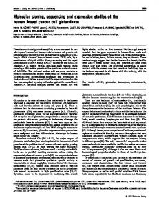
FIG.4. Northern blot analysis of total RNAs (10 pg/lane) using aninsert of M G 2 - 1 - 3 ( 2 . 0kbp) as a hybridization probe. Lanes 1-5 and 8-12 represent RNA extracted from human tissues; lane I , tonsils; lane 2, stomach; lane 3, ovary; lane 4 , uterus; lane 5, placenta; lane 8 , parotid gland; lane 9,sublingual gland; and lanes 1012, three different submandibular glands. Lane 6 and 7 represent RNA from monkey kidney cells (COS) and rat submandibular gland, respectively.
20568
Encoding Clones cDNA Human
Salivary Mucin
from two individuals, Sal and Dr, were digested with HincII and contain a high number of Ser and Thrresidues to which or BamHI. As indicated in Fig. 1, BamHI cuts the Sal MG2 the majorityof 0-linked glycans are attached. MG2 contains cDNA onlyonce (position 1459)while Him11 three times six tandem repeats of 23 amino acid residueswith a consensus (positions 399,531, and 1923). The 2.0-kbp insert of MG2-6- composition of Pron, Alas, Thrs,Ser4, Glul. Thistogether 1 was used as a hybridization probe. The results of the accounts for 54 hydroxylated aa residues. It was previously Southern blot hybridization (Fig. 5) suggest that MG2 is a estimated thatMG2 contains approximately 90-100 0-linked single copy gene. In addition, both the BamHI and HincII units (Levineet al., 1987), therefore almost half of these have hybridization patterns differ in the two individuals examined,to be located outside of the tandem repeatregion. The regions gene or 5' and 3' to the tandem repeats contain unique aa suggesting either a genetic polymorphism in the MG2 sequences a partial digest of the Sal DNA. For example with HincII, a as well as all the potential N-glycosylation sites and all Cys single band of -1400 bp was obtained with Drwhile a doublet residues. There are five N-glycosylation sites inMG2, four of of 1400 bp was obtained with Sal. However, further char- these are located 5' to the repeats and onelocated 3' to the polymor- repeats, as indicated by the * in Fig. 2. There are two Cys acterization of the structural basis for the MG2 phism, if any,is necessary. Recently, we haveperformed (residues 65 and70) in putative secretedpolypeptide of MG2 Southern blot analysisof human, monkey, rat, hamster, and that arelocated upstream of the tandem repeats. Since MG2 mouse genomic DNA using a 2.34-kbp insert of MG2 cDNA exists asa monomer, these maybe involved in an intramolecas a probe. We have found hybridization signals only in the ular but not an intermoleculardisulfide bond. human and monkeyDNA,suggesting the existence of an The nucleotide or deduced aa sequence of the MG2showed MG2 homolog in monkeys but not inphylogenetically distant no sequence homology with any other human or animal muspecies (results not shown). cins and no significant homology was found to any other gene sequences entered in the GenBank" data base. Mucins do not DISCUSSION show sequence homology either at the nucleotide or the proA comparison of sequencing data from all mucins studied tein level in their tandem repeats. However, regions outside thus far, including MG2 (MUC7), confirmed and extendeda of tandem repeats, especially the C-terminal domains, appear number of featurescharacteristic of mucins. First, mucin to share sequence homologies. For example, porcine submaxprotein backbones contain a high content of Ser, Thr, Pro, illary mucin (Eckhardt et al., 1991) and bovine submaxillary Ala, and Gly, usually accounting for more than 50% of the mucin-like protein(Bhargava et al., 1990)have a 82% setotal amino acid residues. In MG2, Pro, Ser, Thr, and Ala quence similarity at the protein level in a region spanning account for 62% of the total aa residues; however, there is residues 920-1154 in porcine apo-mucin anda corresponding very little Gly. Second, theproteinbackbonecontains a region inbovine apo-mucin including 30 Cys residues. In number of repeating aa sequences. In general, these are lo- addition,recentstudies showed thatthecysteine-rich Ccated in the central region of the deduced polypeptide chain terminal regions of ratintestinal mucin-like peptideand human MUC2 share extensive sequence similarity (Xu et al., Sal Dr Sal Dr 1992a, 1992b). This similarity is -73% over 730 aa residues B H B B H B H when a few short gaps were introduced (Gum et al., 1992). I Further, lesser sequence similarities over shorter regions were noted among the extreme C-terminal regions of the MUC2 and porcine submaxillarymucin, bovine submaxillary mucinlike protein and frog integumentary mucin B.l (Gum et al., 1992). Consequently, itwas speculated that conservationof a cysteine-dependent structural homology is necessary for the preservation of mucins essential function. As demonstrated by the Northern blot analysis,expression of MG2 (MUC7) isspecies specificsince nohybridization was detected in RNA from rat submandibular gland. In addition, its expression is also tissue and cell specific since hybridization was detected in RNA only from human salivary gland 3.0 tissues and only in those that contain mucous acinar cells ( e g . sublingual and submandibular glands) and notsalivary in glands containing only serous acinar cells (e.g. parotid gland). It is interesting to note that immunocytochemical studies with anti-MG2 antibody showed that MG2 was produced in the submandibular and labial glands but not in parotid or 1.o palatine glands (Cohenet al., 1991). Southern blot analysis of human genomic DNA suggested .5 a possible genetic polymorphism in the MG2 gene of the two individualsexamined. The majorgeneticpolymorphismin othermucin genes isduetodifferentnumber of tandem repeats in different individuals. This presumably arises by unequal crossing over in the homologous multiple tandem repeats. For example in the MUCl gene (polymorphic epitheFIG. 5. Southern blot analysis of genomic DNA using the lial mucin, PEM), the range in number of repeats is between insert of cDNA clone MG2-6-1 as a hybridization probe. Left 20 to 125 and 30 different alleles were found in 69 individuals panel, agarose gel stained with EtBr; right panel, Southern blot. Sal and Dr indicate DNA isolated from two different individuals; B and (Gendler et al., 1988). The MUC2 gene (human small intesH represent digestion of DNA with BarnHI and HincII, respectively. tinalmucin), however, exhibitsbothlengthand sequence
-
cDNA Clones Encoding Human Salivary Mucin polymorphisms and has different ethnic distribution (Toribara et aL, 1991). In summary, we have cloned and sequenced cDNAs encoding the protein core of the low molecular weight human salivary mucin, MG2. The translated region of 1131 nucleotides encodes a protein of 377 amino acids with a molecular mass of 39 kDa and a computer-calculated isoelectric point of 9.33. The first 20 residues comprise the putative leader peptide while the remaining 357 residues represent the putative secreted protein that contains six tandem repeats of 23 amino acid residues. More recently, we have succeeded in isolating a MG2 cDNA clone of 2340 bp which spans the sequences comprising both MG2-5-5 and MG2-6-1. The translated region of this clone is the same length as the translated region derived from the composite sequences of MG2-5-5 and MG26-1. To our knowledge this is the seventhhuman mucin cloned, and we named it MUC7 in accordance with the other cloned human mucins (MUC1-MUCG). Our recent mapping data show that the gene locus encoding the MUC7 is located on chromosome 4.3 The MUCl and MUC2 mentioned above contain tandem repeatsof 20 and 23 amino acids, respectively (Gendler et al., 1988 Gum et al., 1989). MUCl gene also codes for human pancreatic mucin (HPTM) (Lan et al., 1990) and human episialin (Ligtenberg et al., 1990). MUC3 is a novel intestinal mucin with a tandem repeat of 17 amino acids (Gum et al., 1990). MUC4 (Porchet et al., 1991) and MUC 5 (Crepin et al., 1990) are tracheobronchialmucins with tandem repeats of 16 and 8 amino acids, respectively. MUC6 is a gastric mucin which has a tandem repeatof 169 aa (Toribara et al., 19931, the longest repeat of all human mucins so far identified. Acknowledgment-We thank Dr. 2.-S. Xiao for help with the Northern blot. REFERENCES Aguirre, A., Mendoza, B., Reddy, M. S., Scannapieco, F. A,, and Levine, M. J. (1989) Dysphagia 4,95-100 Bhargava, A. K., Woitach, J. T., Davidson, E. A,, and Bhavanandan, V. P. (1990) Proc. Notl. Acad. Sci. U . S. A. 87, 6798-6802 Blin, N., and Stafford,D. W. (1976) Nucleic Acids Res. 3,2303-2308 Bobek, L. A., Aguirre, A., and Levine, M. J. (1991) Biochem. J. 2 7 8 , 627-635 Cohen, R. E.,,and Levine, M. J. (1989) in Human Saliua: Clinical Chemistry and Mtcrobtology (Tenovuo, J. 0.. ed) Vol. I, pp. 101-130, CRC Press, Boca Raton. FL Cohen, R. E . , Aguirre, A., Neiders, M. E., Levine, M. J., Jones, P. C., Reddy, M. S., and Harr, J. G. (1990) Arch. Oral Biol. 35,347-356 Cohen, R. E., Aguirre, A., Neiders, M. E., Levine, M. J., Jones, P. C., Reddy,
Y. A. Bobek, L. A. Bobek, and M. J. Levine, unpublished data.
20569
M . S., and Ham, J. G. (1992) Arch. Oral Biol. 36,127-136 Crepin, M., Porchet, N., Aubert, J. P., and Degand, P. (1990) Biorheology 2 7 , 471-484 Davis, L. G.,Dibner, M. D., and Battey,J. F. (1986) Basic Methods in Molecular Biology, Section 11-1, pp. 130-135, Elsevier, Amsterdam Devereux, P., Haeherli, P., and Smithies, 0.(1984) Nucleic Acids Res. 12,387391 Devine, P. L., and McKenzie, 1. F. C. (1992) BioEssays 14,619-625 Eckhardt, A. E., Timpte, C. S., Ahernethy, J. L., Zhao, Y., and Hill, R. L. (1991) J. Biol. Chem. 266,9678-9686 Gender, S. J., Taylor-Papadimitriou, J., Duhig, T., Rothbard, J., andBurchell, J. (1988) J. Biol. Chem. 263,12820-12823 Gendler, S. J., Lancaster, C. A., Taylor-Papadimitriou, J., Duhig, T., Peat, N., Burchell J. Pemherton L., Lalani, E-N., and Wilson, D. (1990) J. Biol. Chem. 265,'15286-15299 Gum, J. R., Byrd, J . C., Hicks, J. W., Toribara, N. W., Lamport, D. T. A,, and Kim, Y. S. (1989) J. Biol. Chem. 264,6480-6487 Gum, J. R., Hicks, J. W., Swallow, D. M., Lagace, R. L., Byrd, J. C., Lamport, D. T. A,, Siddiki, B., and Kim, Y. S. (1990) Biochem. Biophys. Res. Commun. 171,407-415 Gum, J. R., Hicks, J. W., Toribara, N. W., Rothe, E."., Lagace, R. E., and Kim, Y. S. (1992) J . Biol. Chem. 267,21375-21383 Henlkoff, S. (1987) Methods Enzymol. 155,156-165 Heinrich, P. (1991) in Current Protocols in Molecular Biology (Ausubel, F., Brent, R., Kingston, R., Moore, D., Seidman, J., Smith, J., and Struhl, K., eds), Suppl. 16, Chapter 7, unit 7.2.1-7.2.7, John Wiley & Sons, New York Kingston, R. E. (1991) in Current Protocols in Molecular Biology (Ausuhel, F., Brent, R., Kingston, R., MooFe, D., Seidman, J., Smith, J., and Struhl, K., eds), Suppl. 14, Chapter 4, unlt 4.2.1-4.2.8, John Wiley & Sons, New York Kozak, M. (1986) Cell 44,283-292 Lan, M. S., Batra, S. K., QI, W.-N., Metzgar, R. S., and Hollingsworth, M. A. (1990) J . Biol. Chem. 266,15294-15299 Levine, M. J., Reddy, M. S., Tahak, L. A,, Loomis, R. E. Bergey, E. J. Jones P. C., Cohen, R. E., Stinson, M. W., and Al-Hashimi, i. (1987) J. Dent. Res.' 66,436-441 Ligtenherg, M. J. L., Vos, H. L., Gennissen, A. M. C., and Hilkens, J. (1990) J . Biol. Chem. 265.5573-5578 Loh, E. Y., E1liot;J. F., Cwirla, S., Lanier, L. L., and Davis, M. M. (1989) Science 2 4 3 , 217-220 Loomis, R. E., Prakobphol,, A., Levine, M. J., Reddy, M. S., and Jones, P. C. (1987) Arch. Bwchem. Btophys. 258,452-464 Porchet, N., Van Cong, N., Dufosse, J., Audie, J. O., Guyonnet-Duperat, V., Gross, M. S,.,Denis, C., Degand, P., Bernheim, A,, and Aubert, J. P. (1991) Biochem. Btophys. Res. Commun. 175,414-422 Prakobphol, A., Levine, M. J., Tabak, L. A,, and Reddy, M. S. (1982) Carbohydr. Res. 108,111-122 Ramasubhu, N., Reddy, M. S., Bergey, E. J., Haraszthy, G. G., Soni, S.-D., and Levine, M. J. (1991) Biochem. J. 280,341-352 Reddy, M. S., Bohek, L. A., Haraszthy, G. G., Biesbrock, A. R., and Levine, M. J. (1992) Biockm. J . 287,639-643 Sambrook, J., Fntsch, E. F., and Maniatis, T. (1989) Molecular Cloning: A Laboratory Manual, 2nd Ed., Chap. 9, pp. 9.17-9.19, Cold Spring Harbor Laboratory, Cold S ring Harbor, NY and Coulson, A. (1977) Proc. Natl. Acad. Sci. U. S. A. Sanger, F., Nicklen, 74,5463-5467 Scannapieco, F., and Levine, M. J. (1993) in Cardwlogyfor t h e Nineties (Bowen, W. H.,and Tabak, L. A., eds) pp. 85-105, University of Rochester Press, Rochester, NY Strous, G. J., and Dekker, J. (1992) Crit. Reu. Biochem. Mol. Biol. 2 7 , 57-92 Toribara, N. W., Gum, J. R., Culhane, P. J., LagaceR. E., Hicks J. W., Petersen, G. M., and Kim, Y. S.(1991) J . Clin. Inuest.'88, 1005-1015 Tonhara, N.W., Roberton A. M., Ho, S. B., Kuo, W.-L., Gum, E., Hicks, J. W., Gum, J. R., Byrd, J. k., Siddiki, B., and Kim, Y. S. (1993) J. Biol. Chem. 268,5879-5885 Xu, G., Huan, L., Khatri, I. A., Sa"an, U S , McCool, D. Wang, D., Jones, C., Forstner, G. G., and Forstner,J.%. (1992a) Biochem. Biophys. Res. Commun. 183,821-828 Xu, G., Huan, L.-J., Khatri, I. A., Wang, D., Bennick, A., Fahim, R. E. F., Forstner, G. G., and Forstner, J. F. (1992b) J. Biol. Chem. 2 6 7 , 5401-5407
8..