Report
Cell Cycle 10:11, 1794-1809; June 1, 2011; © 2011 Landes Bioscience
Molecular profiling of a lethal tumor microenvironment, as defined by stromal caveolin-1 status in breast cancers Agnieszka K. Witkiewicz,1,2,* Jessica Kline,1,2 Maria Queenan,1,2 Jonathan R. Brody,2,3 Aristotelis Tsirigos,4 Erhan Bilal,4 Stephanos Pavlides,2,5 Adam Ertel,2,6 Federica Sotgia2,6,8 and Michael P. Lisanti2,5-8,* 1 Department of Pathology, Anatomy and Cell Biology; 2Stem Cell Biology and Regenerative Medicine Center; 3Department of Surgery; 5Kimmel Cancer Center; Department of Stem Cell Biology and Regenerative Medicine; 6Department of Cancer Biology; 7Department of Medical Oncology; Thomas Jefferson University; Philadelphia, PA USA; 4Computational Genomics Group; IBM Thomas J. Watson Research Center; Yorktown Heights, NY USA; 8Manchester Breast Centre and Breakthrough Breast Cancer Research Unit; Paterson Institute for Cancer Research; School of Cancer; Enabling Sciences and Technology; Manchester Academic Health Science Centre; University of Manchester; Manchester, UK
Key words: caveolin-1, tumor stroma, breast cancer, transcriptional profiling, clinical outcome, recurrence, metastasis, biomarkers, gene signatures, aging
Breast cancer progression and metastasis are driven by complex and reciprocal interactions, between epithelial cancer cells and their surrounding stromal microenvironment. We have previously shown that a loss of stromal Cav-1 expression is associated with an increased risk of early tumor recurrence, metastasis and decreased overall survival. To identify and characterize the signaling pathways that are activated in Cav-1 negative tumor stroma, we performed gene expression profiling using laser microdissected breast cancer-associated stroma. Tumor stroma was laser capture microdissected from 4 cases showing high stromal Cav-1 expression and 7 cases with loss of stromal Cav-1. Briefly, we identified 238 gene transcripts that were upregulated and 232 gene transcripts that were downregulated in the stroma of tumors showing a loss of Cav-1 expression (p ≤ 0.01 and fold-change ≥1.5). Gene set enrichment analysis (GSEA) revealed “stemness,” inflammation, DNA damage, aging, oxidative stress, hypoxia, autophagy and mitochondrial dysfunction in the tumor stroma of patients lacking stromal Cav-1. Our findings are consistent with the recently proposed “Reverse Warburg Effect” and the “Autophagic Tumor Stroma Model of Cancer Metabolism.” In these two complementary models, cancer cells induce oxidative stress in adjacent stromal cells, which then forces these stromal fibroblasts to undergo autophagy/ mitophagy and aerobic glycolysis. This, in turn, produces recycled nutrients (lactate, ketones and glutamine) to feed anabolic cancer cells, which are undergoing oxidative mitochondrial metabolism. Our results are also consistent with previous biomarker studies showing that the increased expression of known autophagy markers (such as ATG16L and the cathepsins) in the tumor stroma is specifically associated with metastatic tumor progression and/or poor clinical outcome.
Introduction Previously, we and others identified a loss of stromal caveolin-1 (Cav-1) as a new biomarker of a “lethal” tumor microenvironment.1-5 More specifically, a loss of stromal Cav-1 in the cancerassociated fibroblast compartment of human breast cancers is a powerful single independent predictor of early tumor recurrence, lymph-node metastasis and tamoxifen-resistance.1-5 Importantly, the predictive value of a loss of stromal Cav-1 was independent of epithelial marker status, and was effective in all of the most commons sub-types of invasive ductal carcinoma, including ER(+), PR(+), HER2(+) and triple-negative tumors.2-5 In triple-negative breast cancers, loss of stromal Cav-1 was associated with a 5-year survival rate of 75%.4 Similarly, in DCIS patients, a loss of stromal Cav-1 was predictive of disease recurrence and progression to invasive breast cancer; 3 100% of DCIS patients with a loss of stromal Cav-1 underwent recurrence and 80% of these patients progressed from DCIS to invasive disease.3 In prostate cancer, a loss of stromal Cav-1 is predictive of advanced prostate cancer, with a high Gleason score, and is associated with lymphnode or bone metastasis.6 Thus, loss of stromal Cav-1 may be a new widely applicable biomarker for various epithelial tumor types, as most solid tumors contain a significant stromal component.2 However, it remains unknown why a loss of stromal Cav-1 has such a profound effect on patient prognosis and disease progression. To address this issue directly, here we performed laser-capture microdissection on breast cancer patient tumor tissue (i.e., frozen
*Correspondence to: Agnieszka K. Witkiewicz and Michael P. Lisanti; Email:
[email protected] and
[email protected] Submitted: 03/30/11; Accepted: 04/08/11 DOI: 10.4161/cc.10.11.15675 1794
Cell Cycle
Volume 10 Issue 11
report
Report
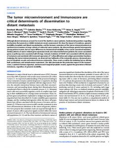
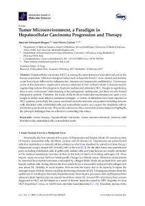
Figure 1. Stromal Cav-1 can be used to stratify human breast cancer patients into two transcriptionally distinct patient populations. The transcriptional profiles of Cav-1-positive (+) tumor stroma (N = 4) versus Cav-1-negative (–) tumor stroma (N = 7) were compared. We identified 238 gene transcripts that were upregulated and 232 gene transcripts that were downregulated in the stroma of tumors showing a loss of Cav-1 expression (Sup. Table 1). Note that the two patient populations are transcriptionally different. One-way ANOVA was setup to extract differentially expressed genes between Cav-1 positive and Cav-1 negative samples. The resultant p-values were further adjusted by multi-test correction (MTC) method of FDR step-up. The standardized intensity data from the stringent gene list (p-value ≤ 0.01 and fold change ≥1.5) were used in generating the hierarchical clustering HeatMap.
sections) in which the status of their stromal Cav-1 levels (positive or negative) was first determined by immuno-histochemical staining. After laser-capture, the stromal material isolated from Cav-1 (+) and Cav-1 (-) patients was then subjected to genomewide transcriptional profiling, to mechanistically unravel which signaling pathway(s) that are activated in the microenvironment of patients which are deficient in stromal Cav-1. We show that the new transcriptional gene signature(s) that we have defined can effectively be used to cleanly separate patients based on their stromal Cav-1 status, into Cav-1 (+) and Cav-1 (-) sub-groups, greatly facilitating the prediction of their prognosis. Our findings provide novel mechanistic insights into how the presence or absence of Cav-1 in the stroma regulates tumor progression and breast cancer metastasis. Results Transcriptional profiling of a Cav-1 deficient tumor microenvironment. Loss of stromal Cav-1 in human breast cancer(s)
www.landesbioscience.com
is associated with tumor recurrence, metastasis and drug-resistance, conferring poor clinical outcome.1-5 To mechanistically understand the “lethality” of a Cav-1 negative tumor microenvironment, we performed laser capture microdissection on the tumor stroma of patients that were pre-classified as stromal Cav-1-positive(+) (N = 4) and stromal Cav-1-negative(-) (N = 7), based on immuno-histochemical (IHC) staining. Then, the RNA extracted from these samples was subjected to genomewide transcriptional profiling. Based on this approach, we identified 238 gene transcripts that were specifically upregulated and 232 gene transcripts that were downregulated in the stroma of tumors showing a loss of Cav-1 expression [p ≤ 0.01 and fold-change (f.c.) ≥1.5] (Sup. Table 1). Using these stringent criteria, we were able to transcriptionally separate these two patient populations, in accordance with their IHC stromal Cav-1 status (negative versus positive). A HeatMap of the total patient cohort is shown in Figure 1, demonstrating that these two patient populations appear transcriptionally and “genetically” distinct.
Cell Cycle
1795
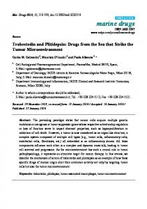
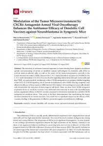
Figure 2. HeatMaps of gene transcripts associated with myofibroblast differentiation, autophagy, lysosomal degradation and glycolysis. Note that Cav-1-deficient stroma shows the upregulation of myofibroblast differentiation (15 transcripts), autophagy (22 transcripts), lysosomal proteases (5 transcripts), lysosomal proteins (29 transcripts) and glycolysis/pyruvate metabolism (15 transcripts). See Supplemental Tables 4, 5 and 10.
Gene set enrichment analysis (GSEA) of a Cav-1 deficient tumor microenvironment. To understand what cellular processes are characteristic of a Cav-1-deficient tumor microenvironment, we next performed gene set enrichment analysis (GSEA), by comparison with other gene signatures available in various public databases. For this purpose, we focused on a wider list of gene transcripts that were upregulated in Cav-1-deficient stroma [5,424 transcripts encoding 3,459 unique genes; p ≤ 0.1 and foldchange (f.c.) ≥1.15] (Sup. Table 2), as is normally recommended for GSEA. Table 1 shows the results of this detailed analysis. Note that we see the upregulation of cellular processes normally associated with “stemness,” inflammation, DNA damage, aging, oxidative stress, hypoxia, apoptotic signaling, autophagy and mitochondrial dysfunction in the tumor stroma of patients lacking stromal Cav-1. Individual HeatMaps [based on 1,819 transcripts encoding 1,297 unique genes; p ≤ 0.05 and fold-change (f.c.) ≥1.15] (Sup. Table 3) for key cellular processes are shown in Figures
1796
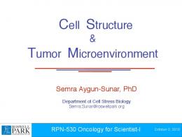
Figure 3. HeatMaps of gene transcripts associated with the response to hypoxia and mitochondria. Note that Cav-1-deficient stroma shows the upregulation of hypoxia target genes (65 transcripts) and mitochondrial-associated proteins (41 transcripts). See Supplemental Tables 10 and 12.
2–8. These HeatMaps illustrate the upregulation of gene transcripts associated with myofibroblast differentiation, autophagy, lysosomal degradation, glycolysis, hypoxia, mitochondria, inflammation and redox signaling, DNA damage and repair, aging, BRCA1-mutation positive and ER-negative breast cancer patients, apoptosis and neural stem cells (See Sup. Tables 4–14). The observed association between a Cav-1-deficient tumor stroma and neural stem cells may reflect increased stromal “neurogenesis,” which has previously been implicated in an aggressive reactive stromal phenotype in a subset of prostate cancers.7,8 Similarly, “neurogenesis” was associated with increased DNA damage in the tumor stroma.7,8 In addition, the upregulation of gene transcripts or gene sets associated with GOLPH3, NRF1 (nuclear respiratory factor 1), PRKDC (protein kinase, DNA-activated, catalytic polypeptide) or arsenic treatment is highly suggestive of mitochondrial
Cell Cycle
Volume 10 Issue 11
Table 1. Cellular processes that are associated with a lethal tumor micro-environment in human breast cancer (Cav-1 deficient stroma) Data Set
p-value
Detailed Description
STEMCELL_NEURAL_UP
1.45E -17
Enriched in mouse neural stem cells, compared to differentiated brain and bone marrow cells
MORF_BMI1
3.74E -10
Neighborhood of BMI1 B lymphoma Mo-MLV insertion region (mouse) in the MORF expression compendium
STEMCELL_EMBRYONIC_UP
6.51E -09
Enriched in mouse embryonic stem cells, compared to differentiated brain and bone marrow cells
HSC_LATEPROGENITORS_SHARED
8.03E -09
Upregulated in mouse hematopoietic late progenitors from both adult bone marrow and fetal liver (Cluster VI, Late Progenitors Shared)
STEMCELL_HEMATOPOIETIC_UP
8.50E -09
Enriched in mouse hematopoietic stem cells, compared to differentiated brain and bone marrow cells
HSC_LATEPROGENITORS_FETAL
2.72E -08
Up regulated in mouse hematopoietic late progenitors from fetal liver (Late Progenitors Shared + Fetal)
RUTELLA_HEPATGFSNDCS_UP
6.88E -06
Genes upregulated by HGF treatments
MORF_TPR
1.59E -05
Neighborhood of TPR translocated promoter region (to activated MET oncogene) in the MORF expression compendium
WIELAND_HEPATITIS_B_INDUCED
5.92E-12
Genes induced in the liver during hepatitis B viral clearance in chimpanzees
REOVIRUS_HEK293_UP
1.56E-06
Upregulated at any timepoint up to 24 hours following infection of HEK293 cells with reovirus strain T3Abney
DEFENSE_RESPONSE
1.73E-06
Genes annotated by the GO term GO:0006952. Reactions, triggered in response to the presence of a foreign body or the occurrence of an injury, which result in restriction of damage to the organism attacked or prevention/recovery from the infection caused by the attack
REJECTION_UP
3.80E-06
Genes upreglated in acute rejection transplanted kidney biopsies relative to well functioning transplanted kidney biopsies from stable, immunosuppressed, recipients (median FDR