fracture site immobilized with a Hoffman- Vidal external fixator. A dial gage was ... similar fractures and subjective X-ray interpretation. External fixators .... (10, 20 and 30 Ib). ..... 12 Anon., du Pont de Nemours and Co., The Language oj Rubber,.
Reprinted from May 1983, Vol. 105, Journal of Biomechanical Engineering
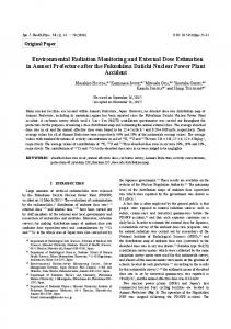
G. S. Beaupre W. C. Hayes
M. H. Jofe A. A. White, III Orthopaedic Biomechanics Laboratory, Charles A. Dana Research Institute, Beth Israel Hospital and Harvard Medical School. Boston, Mass. 02215
Monitoring Fracture Site Properties With External Fixation
1
An in-vitro system has been devised to monitor the properties of an idealized fracture site immobilized with a Hoffman- Vidal external fixator. A dial gage was used to measure the relative pin displacements under controlled axial loading. The displacement measurements were then used in conjunction with a finite element model to predict the modulus of an idealized fracture site. Five fracture sites made of neoprene disks of different mechanical properties were monitored in order to simulate the increasing modulus of a healing fracture. Good agreement was observed between directly measured mechanical properties of the neoprene and those inferred from the combinedfinite element and pin displacement tests.
Introduction The use of external skeletal fixators for the treatment of fractures, nonunions and joint fusions is enjoying a new popularity as improvements in materials and techniques have made the approach more effective [1, 2, 3]. In spite of this resurgence in use, few quantitative biomechanical data exist to determine the optimal treatment period or to evaluate the progress of fracture healing with this method [4]. The potential for pin tract infection dictates that the external fixator should, as a rule, be removed as soon as is safely possible [5]. At present, however, treatment times are most often based upon a combination of previous experience with similar fractures and subjective X-ray interpretation. External fixators offer an ideal opportunity to monitor the characteristics of healing fractures using noninvasive techniques. The direct coupling to bone provided by the transfixing pins permits accurate measurement of quantities which may be correlated with the degree of healing. Chao, et al. [6] and Briggs and Chao [7], using a Hoffman device applied to a synthetic bone model of rectangular cross section, measured the stiffness response to bending, torsional, compressive and tensile loadings. In addition, a finite element model was created to compare with the experimental results. Burny [8] used strain gages attached to the fixator frame to measure bending deformation as a function of healing time for a given load. He suggested that there were eight types of fracture healing based on the form of the deformationhealing-time curve. Jorgensen [9] generated similar in-vivo healing curves and suggested that the information from these curves could be used to develop a gradually increasing loading program for the patient. (The loading program would begin with partial weight bearing when the fracture had stiffened to a predetermined value and progress to full weight bearing and fixator removal as the fracture stiffness increased.) The structural rigidity of a composite external fixation system includes contributions from the rigidity of the device 1 This work was supported by NIH RCDA 5 K04 AM00368 and GM07806. Contributed by the Bioengineering Division for publication in the JOURNAL received by the Bioengineering Division, June II, 1982; revised manuscript received October 31, 1982.
OF BIOMECHANICAL ENGINeERING. Manu~cript
1 20 I V 0 I. 105, MAY 1983
itself and from the rigidity of the healing bone. Contributions to the bone rigidity are made both by changes in fracture site geometry and by changes in the mechanical properties of the tissues participating in the healing process. Contributions to the rigidity of the fixation device are made by its geometry, its method of application and its material properties. Wolf, et al. [10] demonstrated that the rigidity of fixation of a fracture can markedly influence the time course, histological patterns and mechanical properties of healing fractures. We assume, therefore, that fracture site rigidity is: I) a potentially important variable controlling fracture healing, and 2) an important variable to monitor since it can be related most directly to the structural behavior (i.e., stiffness and strength) of a healing fracture. The purpose of this study is to show that we can determine the fracture site modulus based on pin displacement measurements and a parallel finite element analysis. In companion studies in our laboratory, we have generated healing curves using human subjects beginning as early as two weeks postoperatively. We are also conducting similar tests using both sheep and rabbits as animal models. These animal studies permit the determination of both strength and stiffness data, since these are the biomechanical properties of the fracture site of ultimate concern.
Methods and Materials We analyzed a Hoffmann-Vidal (H-V) fixator in two complimentary ways. First, experiments were conducted to determine the response of the H-V system under controlled axial loading. Second, finite element models were generated to determine the precision of mathematical predictions of the fracture site modulus. In Fig. 1, we show the double-frame H- V apparatus in the standard six-pin quadrilateral configuration that was used exclusively for in-vitro testing. The H- V device was applied to a hollow plexiglass tube in which a central aluminumneoprene "fracture site" was introduced. The plexiglass tube was chosen over actual or synthetic bone because of its regular geometry and because its material properties are well known.
Transactions of the ASME
Although the modulus of plexiglass (2.4 GPa) is much less than that of compact bone (15-25 GPa), it was used in this preliminary study because of its availability and ease of machining. The question of whether to use real bone or a substitute material is an important consideration. Kempson and Cambell [II] have tested a variety of external fixation devices connected to both human tibiae and cylindrical steel tubes. Although they found that the overall compressive stiffness of the two systems was different, they state that in some cases the pins became loose in the bone and thus some of the deformation was a result of the movement of the pin relative to the bone. In addition, the pins were brazed into the steel tube which may also result in a stiffer response. The variability in shape and material properties of real bones make their choice impractical for this preliminary study, where a testing methodology is being developed and inter-test consistency is important. The representative "fracture site" consisted of a neoprene disk securely bonded between two aluminum fixtures. The aluminum fixtures slipped into the ends of the hollow plexiglass tube and served to secure and locate the neoprene disk. These fixtures also acted as part of the gripping system for subsequent material characterization tests of the neoprene. In all, five sets of aluminum-neoprene composites were constructed using neoprene of various hardnesses ranging from durometer 20 to durometer 90. This range of hardness allowed the modulus of the fracture siteto be varied in order to simulate a healing fracture, though not necessarily to span the entire range of properties of healing bone or callous. The experimental protocol consisted of suspending known weights from the lowest transfixing pin while supporting the frame from a cable threaded through a hole in the upper segment of the plexiglass tube. The relative displacement between the two pins nearest the fracture site was measured using a dial gage sensitive to 20 !Lm (0.0005 in.). In all cases, the H- V frame was applied to the plexiglass tube with a fixed amount of precompression. The precompression was necessary both to prevent debonding of the glued aluminumneoprene interfaces and to ensure that even in the presence of the hanging weights, the fracture site remained in compression. A rough attempt was made to maintain the same level of precompression for each test by adjusting the thumb screws at the center of each side bar until the bending in the transfixing pins was approximately equal. For each hardness of neoprene, displacement values were recorded as a function of time for each of three weights: 44.5N, 89N and 133.5N (10, 20 and 30 Ib). Displacement-time histories were recorded since neoprene behaves in a viscoelastic manner. The final part of the experimental investigation was to characterize the material properties of each neoprene disk. Typically, rubberlike materials are characterized according to hardness using durometer values [12]. For our purposes, hardness values were not such a useful parameter and material charact.erization using an Instron electro-hydraulic materials testing machine was necessary in order to obtain information about the compressive modulus of the neoprene. The modulus was calculated from load-displacement curves at deformation rates spanning four decades. In conjunction with the experimental testing, a series of finite element models were also generated. The first model was similar to one used by Chao [6] with the addition of two elements to simulate the aluminum-neoprene fracture site. For this initial study, a two-dimensional model was assumed sufficient to describe the behavior of the H-V system under the experimental loading conditions. In addition, it was necessary to model only one half of the structure since symmetry existed for both the geometry and loads. A finite
Fig.1
Double·frame Hoffmann·Vidal fixator
Fig. 2 Finite element model used for preliminary analYSis. The lower end of the elements representing the side bars and the element representing the fracture site were constrained to have no rotation or translation due to symmetry conditions.
element software package, GIFTS [13] was used for model generation, analysis and results display. Figure 2 shows the initial model in the undeformed configuration. Preliminary results from this model indicated that the nodal points at the ends of the transfixing pins underwent displacements at least two orders of magnitude less than the next largest displacement. Therefore, for subsequent models, the elements representing the side bars were ontitled and the translational degrees of freedom for the nodes situated at the pin ends were eliminated. Rotational rigidity was maintained by the addition of torsional springs to these nodes. Egker, et MAY 1983, Vol. 105/121
al. [14] showed that the pin-side bar connections behaved neither as rigidly as the clamped case nor as flexibly as the hinged case. Instead, the rotational rigidity provided by the side bars lies somewhere between the two extremes. With torsional springs the spring constant could be adjusted to supply any degree of torsional constraint. We conducted a simple experiment to determine the appropriate value of the spring constant. An H-V frame was assembled with transfixing pins, but without the plexiglass tube. A known weight was suspended from the center of one of the pins and the displacement was'measured using a dial gage, The measured value was found to lie between the theoretical predictions for a clamped-clamped beam and a hinged-hinged beam (see Appendix), In conjunction with this experiment, a finite element model was generated consisting of a beam made up of two elements with torsional springs at the ends. By adjusting the spring constant, it was possible to match the displacement obtained experimentally. This value of the spring constant ( - 288 N-m/rad) was then used for the complete H- V finite element model. Another input parameter which had to be determined experimentally was the effective length of the transfixing pins. Due to the finite width of the plexiglass tube, a portion of the center section of each pin underwent less bending than it would have if the tube were infinitely thin. This in turn resulted in less displacement at other points along the pin length. As a result, the effective length of each pin was slightly less than the distance between side bars. The effective pin length was found by conducting another simple experiment. This time, a transfixing pin attached to the plexiglass tube was loaded at its ends by equal weights. Again using a dial gage, the displacement near the pin end was measured. Using simple beam theory, the displacement was expressed as a function of the beam dimensions and material properties (see Appendix): The effective length was determined graphically by equating the experimental and theoretical displacements. The results of this experiment indicated that the effective pin length is equal to the free length of each pin (the distance from the side bars to the tube outer diameter) plus an additional length of 0.953 cm. The results of these two simple experiments together with the dimensions and material properties of the H- V plexiglass tube system completed the finite element input phase. The finite element model then consisted of 15 nodal points, 14 two-dimensional beam elements and 6 torsional spring elements (Fig. 3). The two-dimensional beam elements permitted two translational degrees of freedom (x,y) and one rotational degree of freedom (OJ at each node. Table 1 lists the geometric and material properties for each element. Two additional finite-element models were used to examine the effects of changes in both the pin end boundary conditions and the effective pin length. For the first model, representing
Table 1 Structure
the least rigid configuration, we assumed the pin ends were hinged and the full length of the pins was available for bending. For the second model, representing the most rigid configuration, we assumed the pin ends were clamped and the effective length of the pins was reduced by the full thickness of the tube. The loading for all models consisted of a concentrated axially distractive force applied to the upper plexiglass tube element. A series of computer runs was made for each model to determine the effect of changing the modulus of the element representing the neoprene.
Results Because of the viscoelastic nature of the neoprene, we recorded displacement-time histories as each weight was suspended from the frame. Figure 4 is a typical set of displacement histories with durometer 60 neoprene installed in the central region. Each curve displays an initial elastic response followed by a period of creep reaching an asymptotic displacement. For each weight, each curve represents the results from two or three tests which have been averaged to yield a single curve. The error bars indicate the range of displacements from the different tests. The limiting displacement for each weight was taken as a measure of the response of the system, The composite modulus of each neoprene-aluminum composite fixture was then determined by testing in the Instron materials testing machine. Displacement-rate-controlled
15 ~========7========F==t========~8=========16 19
=====~=====~12~~~~13~==~14~====
20
4
~5
C::i:;!6
Fig. 3 Finite element model used for final analysis. Elements 15 through 20 are torsional spring elements. The lower end of element 6 was constrained to have no rolation or translation due to symmetry conditions.
Length (mm) 103.
Area (mm 2 ) 285.0
Plexiglass
2,3
21.6
285.0
Plexiglass
4
63.6
285.0
6.35 3.18 107.0 69.0 38.0
794.0 794.0 12.9 12.9 12.9
122/VoI.105, MAY 1983
e=======~9========~~========~IO~=======18
Geometric and material properties for finite element model
Element no.
Plexiglass
Aluminum Neoprene Pins Pins Pins Torsional springs
17
5 6 7-10 11,14 12,13 15-20
K,orsional =
Diameter (mm) 31.8 OD 25.4 ID 31.8 OD 25.4 ID 31.8 OD 25.4 ID 31.8 31.8 4.06 4.06 4.06
E (GPa)
2.40
0.20
2.40
0.20
2.40
0.20
71.0 variable 203.0 203.0 203.0
0.33 0.45 0.29 0,29 0.29
N.m 282 rad
Transactions of the ASME
1000
.---,----,.---.---,.----r---,
6 900
5
........
..
800
E: E:
4
700
........
'~