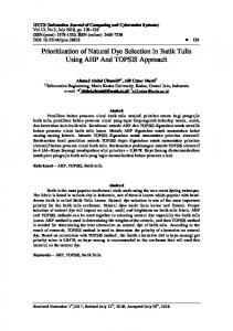
Jurnal Perlindungan Tanaman Indonesia, Vol. 21, No. 2, 2017: 72–79 DOI: 10.22146/jpti.25469
Research Article
Morphological and Molecular Characterization of Rhizoctonia solani Isolates from Two Different Rice Varieties Karakterisasi Morfologi dan Molekuler Isolat Rhizoctonia solani dari Dua Varietas Padi Berbeda
Aisyah Surya Bintang1)*, Arif Wibowo1), Achmadi Priyatmojo1), & Siti Subandiyah1) 1)
Department of Crop Protection, Faculty of Agriculture Universitas Gadjah Mada Jln. Flora 1, Bulaksumur, Sleman, Yogyakarta 55281 *Corresponding author. E-mail:
[email protected] Submitted May 30, 2017; accepted September 19, 2017
ABSTRACT
Six isolates of Rhizoctonia solani, i.e. two isolates collected from infected rice plants and four isolates from laboratory collection were studied by using morphological characters and molecular analysis. Un-weighted pair group method with arithmetic mean dendogram constructed based on cluster analysis showed that these isolates were grouped into three clusters at the 0.77 similarity coefficient. Cluster I consisted of BA, BNJ, and NBR isolates with 100% similarity and indicated that those were from AG 1 IA sub group, cluster II consisted of BND, and cluster III consisted of SL1 and SL2. Mycelium was very light brown or whitish with few and moderate sclerotia except SL1 and SL2. Molecular characterization showed that BA, BNJ, and NBR were amplified at 140 bp using Rs1F/Rs2R specific primer for R. solani AG1 IA. All isolates were amplified between 350−400 bp using Rhsp1 primer, meanwhile SL1 and SL2 were not amplified using AG2sp and AG22sp2 primers. Based on Maximum Likelihood tree analysis showed that SL1 and SL2 had high similarity based on ITS sequence data. Keywords: character, fungal, molecular, morphology, Rhizoctonia solani
INTISARI
Enam isolat Rhizoctonia solani yang berasal dari tanaman padi bergejala dan koleksi laboratorium diuji secara morfologi dan molekuler. Analisis UPGMA dengan koefisien persamaan 0,77 menunjukkan bahwa enam isolat tersebut terbagi atas tiga klaster. Klaster I terdiri atas isolat BA, BNJ, dan NBR dengan kesamaan 100% dan menunjukkan bahwa isolat tersebut berasal dari subgrup AG 1 IA , klaster II yakni isolat BND, dan klaster III terdiri atas isolat SL1 dan SL2. Miselium berwarna putih hingga cokelat muda dengan jumlah sklerotia sedang, kecuali isolat SL1 dan SL2. Uji keragaman secara molekuler menunjukkan bahwa isolat BA, BNJ, dan NBR teramplifikasi pada kisaran 140 bp dengan menggunakan primer Rs1F/Rs2R yang merupakan primer spesifik dari R. solani AG1 IA. Seluruh isolat teramplifikasi pada kisaran 350−400 bp dengan menggunakan primer Rhsp1, sedangkan isolat SL1 dan SL2 keduanya tidak teramplifikasi oleh primer AG2sp dan AG22sp2. Analisis Maximum Likelihood tree berdasar data sekuen ITS menunjukkan bahwa isolat SL1 dan SL2 memiliki tingkat kesamaan yang tinggi. Kata kunci: jamur, karakter, morfologi, molekuler, Rhizoctonia solani
INTRODUCTION
Rice (Oryza sativa) is main food for almost peoples in Indonesia. However, in some areas the rice production was decreased. Plant disease is a factor that can decrease rice productivity in Indonesia. Rice sheath blight caused by Rhizoctonia solani is one of the most important plant diseases on rice. R. solani can infect rice and lost due to this pathogen is up to 30%. Each year, the blight causes up to 50% decrease in the rice yield under favorable condition around the world (Zheng et al., 2013).
The fungi generally grouped as Rhizoctonia solani types occur in all parts of the world and are probably indigenous to uncultivated areas. R. solani is capable of attacking a tremendous range of host plants (Menzies, 1970). R. solani is one of the necrotrophic pathogen that difficult to control, based on their longevity in the soil, ability to outgrow or evade plant defenses and the logistics, cost and efficacy of fungicide applications. In many cases, the pathogen causes disease on more than one host species (Okubara, 2014). Based on Aye & Matsumoto (2012), humidity
Bintang et al.: Morphological and Molecular Characterization of Rhizoctonia solani
is an important criterion after inoculation. The disease level cannot be obtained in humidity under 70%. R. solani is one of the most important rice diseases in tropical areas. R. solani is the causal agent of rice sheath blight, which has become a major constraint to rice production during the last two decades (Kobayashi et al., 1997). Rhizoctonia solani can infect crops of nearly 50 species, including barley, lettuce, tomato, sorghum, maize, and rice (Jones & Belmar, 1989; Zhang et al., 2009). Rice varieties in Indonesia are generally susceptible to R. solani. Different rice variety showed different respond to the pathogen. Cisadane rice variety showed lower disease intensity rather than Ciherang variety and mostly affected lowland area (Nuryanto, 2011). Variability of R. solani has also been reported by Sayler and Yang (2007) using specific primers for R. solani from rice, and many attempts have been made to organize isolate into groups based on morphology, physiology, and pathology of this pathogen. Despite of the morphological and physiological character is useful and still acceptable, it is just a laboratory scale and need longer time. To understand about genetic variability of R. solani species, DNA-based analysis has been reported all around the world, substantial information is not available on morphological characterization along with molecular analysis in Indonesia. Molecular techniques, such as using specific primers for species identification has quicker result rather than conventional methods (Moni et al., 2016). Molecular characterization gives a real time result and can distinguish isolates from each host and even from different geographic area (Lopez-Olmoset al., 2005; Lübek, 2004; Sneh et al., 1991).This study was undertaken to assess morphological and molecular characters of some R. solani isolates using several different primers.
MATERIALS AND METHODS
Collection and Isolation of Rhizoctonia solani Isolates
Isolates of R. solani were collected from infected rice field from Sleman of Yogyakarta Special Region. The pathogens were isolated and purified using potato dextrose agar (PDA) medium. Others R. solani isolates from another host were collected from the collection of laboratory of Plant Health Clinic, Faculty of Agriculture, Universitas Gadjah Mada. The basic information of isolates were listed in the Table 1.
73
Table 1. List of Rhizoctonia solani isolates with the origins Code Host (AG) SL1 SL2 BA1 NBR BNJ BND
Rice Rice Rice (AG 1-IA) Corn (AG 1-IA) Corn (AG 1-IA) Potato (AG 3)
Origin
Sleman Sleman Bantul Blora Binjai Bandungan
Note
Ciherang variety IR64 variety Lab. Collection Lab. Collection Lab. Collection Lab. Collection
Molecular Identification Using Specific Primers
DNA isolation. Actively growing mycelial was put into 50 ml potato dextrose broth (PDB) and placed on electric shaker at room temperature for 3 until 7 days. Mycelia were separated from the broth and DNA was isolated using CTAB 2% method. Isolated DNA was quantified on 0.8% agarose gel. DNA amplification using specific primers. The DNA was amplified on the basis of PCR. A mastermix of 12.5 µl reaction was prepared with PCR components with the total volume 25 µl (mastermix 12.5 µl; ddH2O or RNase free water 9.5 µl; primer forward and reverse each 1 µl; and 1 µl genomic DNA). Specific primer and PCR programs for these isolates were listed in the Table 2. Morphological Characterization
Mycelial disc of 0.5 cm diameter from margin of 3 days old colonies were placed aseptically to potato dextrose agar plates and incubated at room temperature for 12 days. Twenty characters (Moni et al., 2016) were used for morphological characterization. Morphological data were recorded according to the character stage and analyzed by NTSYSpc 2.0 using cluster analysis. Molecular Characterization Using Universal Primers
The DNA was amplified on the basis of PCR. A master-mix of 12.5 µl reaction was prepared with PCR components with the total volume 25 µl (KAPA 2G Fast ReadyMix PCR Kit 12.5 µl; ddH2O or RNase free water 9.5 µl; primer forward (ITS1) and reverse (ITS4) each 1 µl; and 1 µl genomic DNA). DNA amplification from denaturation until extension was proceed at 34 cycles using temperature (Nadarajah et al., 2014). Amplification of isolated R. solani DNA was performed under the following condition: Pre-denaturation 94ºC for 5 min; then 34 cycles of denaturation 94ºC for 30 s, annealing 58ºC for 30 s, extension 72 ºC for 1 min; post-extension 72 ºC for 10 min and hold at 4 ºC.
Vol. 21 No. 2 Jurnal Perlindungan Tanaman Indonesia 74
GTTCAAAGAYTCGATTCAC
AACAAGGTTTCCGTAGGTG
Sequence
Pre-Denaturation for 2,5 min at 94ºC then 40 cycles of 94ºC for 15 s 59ºC for 30 s 72ºC for 1,5 min 72ºC for 10 min and hold at 4ºC.
PCR Program
Table 2. List of specific primers for identification of Rhizoctonia solani isolate Primer
Rhsp 1 5,8SKhotR Rs1F
Pre-Denaturation for 2,5 min at 94ºC then 40 cycles of 94ºC for 15 s, 62ºC for 30 s 72ºC for 1,5 min . 72ºC for 10 min and hold at 4ºC
Pre-Denaturation for 2,5 min at 94ºC then 40 cycles of 94ºC for 15 s, 50ºC for 30 s 72ºC for 1,5 min 72ºC for 10 min and hold at 4ºC.
GCCTTTTCTACCTTAATTTGGCAG Pre-Denaturation 95 ºC for 15 min then 40 cycles of 95ºC for 15 s, 52ºC for 30 s, GTGTGTAAATTAAGTAGACAGCAAATG 72ºC for 15 s 72ºC for 5 min and hold at 4ºC. ATTATTGAATTTAAACAAAG
Rs2R AG2sp
TAGCTGGATCCATTAGTTTG
GTTCAAAGAYTCGATTCAC
AG22sp2 GTTCAAAGAYTCGATTCAC
5,8SKhotR
5,8SKhotR
137−140 (Syler & Yang, 2007)
350−400 (Salazar et al., 2010)
Specific primer for Rhizoctonia solani sub group AG2
Specific primer for Rhizoctonia solani sub group AG1-IA
Specific primer for Rhizoctonia solani species
Target (bp)
322−330 (Salazar et al., 2010)
Specific primer for Rhizoctonia solani sub group AG2 2 IIIB
(Fredricks et al., 2010) 322−330 (Salazar et al., 2010)
(Fredricks et al., 2010)
Bintang et al.: Morphological and Molecular Characterization of Rhizoctonia solani
Electrophoresis and Visualization
DNA fragment was separated using 1% of agarose gel under TBE 1X solution by electrophoresis machine for 50 minutes on 50 V. Gel was stained by Ethidium Bromide (EtBr) solution for 15 minutes. The stained gel was rinsed with distilled water for distaining, illuminated on UV transilluminator and photographed by gel documentation unit for measuring the bands of amplified DNA fragments. DNA sequencing and Molecular Identification
Samples were sent to 1st BASE Malaysia. Sequence results were analyzed based on GenBank’s data at National Center for Biotechnology International (NCBI) using BLAST (Basic Local Alignment Tools) program and phylogeny analysis constructed by MEGA 6.0 software. RESULTS AND DISCUSSION
Molecular Identification Using Specific Primers
Three specific primers were used to identify collected isolates. Each primer was capable of amplifying DNA fragment. The sizes of the amplified DNA products varied for each primer. The DNA of all isolates amplified with Rhsp1 primer which is specific primer for R. solani species (Figure 1), SL1, SL2, and BND amplified at ± 350 bp and another samples amplified at ± 400 bp. DNAs of three isolates (BA, BNJ, NBR) were amplified at ±140 bp using Rs1F/Rs2R. Rs1F/ Rs2R are specific primers for R. solani AG1-IA (Figure 4). Two primers set (AG2sp and AG22sp2)
500 bp
300 bp
Figure 1. DNA amplification of Rhizoctonia solani isolates with primerRhsp1/5,8KhotR; 100 bp DNA ladder (M)
75
were used to amplify DNA of SL1 and SL2, but both primers showed that no DNA fragments were amplified. AG2sp and AG22sp2 are specific primer to identify R. solani AG 2 and AG 2 2 IIIB (Figure 2). Morphological Characterization
After 12 days of incubation on potato dextrose agar, isolates of R. solani showed diversity in their growth (Figure 3). Isolates BA, BNJ, and NBR sclerotia tended to be brown to dark brown. Some isolates produced a moderate quantity of aerial mycelium on the colony surface as well as on the lid and produced at least some sclerotia on the lid. Combined morphological data set of 20 characters with UPGMA resulted 3 clusters with 0.77 similarity coefficient using NTSYSpc 2.0 (Figure 4). Three isolates (BA, BNJ, NBR) were constellated in cluster I with 100% similarity, which indicate that those were the same isolates. The isolate BND was grouped in cluster II, the isolates SL1 and SL2 were in cluster III. Mycelium was usually very light brown or whitish, with moderate aerial mycelia. The number of sclerotia was few to abundant except for SL1 and SL2. Molecular Characterization Using Universal Primers
Universal primers(ITS1/ITS4) were used for SL1 and SL2 (Figure 5). From PCR amplification, each sample was amplified at 600 bp. Analysis of sequence data showed that SL1 and SL2 had high statistical similarity (100%). ML phylogenies generated in this study also indicated that there was a close phylogenetic association between SL1 and SL2 with R. solani AG3 and R. solani AG 1 IA in a monophyletic clade (Figure 6). Variation between isolates from different hosts and geographical regions has previously been studied for R. solani for many years. It is already recognized that R. solani consists of a lot of races, groups or sub-groups, morphology in culture and even the sclerotial distribution (Bonman, 1992). The result of this study also showed that R. solani isolates showed morphological from rice had a variation among the isolates. Two isolates from rice (SL1 and SL2) were genetically similar based on sequence analysis and its showed difference between another rice isolate (BA). R. solani AGs 1, 2, 3, and 4 are the biggest pathogen groups, meanwhile group 5 to 13 are less important as plant disease agent (Lehtonen, 2009). Evolution analysis of sequenced Agaricomycete fungi including R. solani AG 1 IA provides important evidence to molecular characterization and confirms the divergent
Jurnal Perlindungan Tanaman Indonesia
76
Vol. 21 No. 2
500 bp 500 bp
140 bp
B
A
Figure 2. DNA amplification of Rhizoctonia solani isolates with primer: Rs1F/Rs2R (A); AG2sp/5,8KhotR and AG22sp2/5,8KhotR (B); 100 bp DNA ladder (M)
BA
NBR
BNJ
BND
SL1
SL2
Figure 3. Morphological variation of Rhizoctonia solani isolates on PDA
Figure 4. Un-weighted pair group method with arithmetic mean dendogram of Rhizoctonia solani isolates constructed with NTSYSpc 2.0 using cluster analysis based on morphological characters
Bintang et al.: Morphological and Molecular Characterization of Rhizoctonia solani
600 bp
Figure 5. DNA amplification of Rhizoctonia solani isolates with primer ITS1/ITS4; 100 bp DNA ladder (M)
placement of R. solani AG 1 IA. As only sequenced plant fungal pathogen in the subphylum, the genome will undoubtedly provide valuable information about the genetic features that distinguish pathogenic and saprophytic lifestyles among Basidiomycetes. In addition, comprehensive comparisons with forthcoming genome data from other fungal taxa may important clues about the origins and influences on other organisms. Based on this study, three isolates (BA, BNJ, NBR) collected from different locations and hosts. BA isolate was collected from rice in Yogyakarta,
77
BNJ was collected from corn in Binjai, and NBR was collected from corn in Blora. Molecular identification showed that isolates come from same sub-group and morphological characterization showed that isolates had high similarity. Toda et al. (1999) reported that the isolates originated from the same geographical or host plants were not always genetically related. Two isolates from rice (SL1 and SL2) were genetically similar based on sequence analysis and it showed difference between another rice isolate (BA). R. solani AGs 1, 2, 3, and 4 are the biggest pathogen groups, meanwhile group 5 to 13 are less important as plant disease agent (Lehtonen, 2009). Evolution analysis of sequenced Agaricomycete fungi including R. solani AG 1 IA provides important evidence to molecular characterization and confirms the divergent placement of R. solani AG 1 IA. As only sequenced plant fungal pathogen in the subphylum, the genome will undoubtedly provide valuable information about the genetic features that distinguish pathogenic and saphrophytic lifestyles among Basidiomycetes. In addition, comprehensive comparisons with forthcoming genome data from another fungal taxa may important clues about the origins and influences on other organisms. It complex multiple AGs and its multi-nuclear nature have made understanding R. solani evolution previously very difficult (Zheng et al., 2013). Rhizoctonia solani AG 3 commonly infected potatoes, but has been found in several crops and weed hosts worldwide (Tsror, 2010). Based on pathogenicity tests, maize has been found to be susceptible to AG3 infection along with other cereal crops such as oat
Figure 6. Maximum Likelihood tree of ITS sequences analysis to show relationship of SL1 and SL2 among another sub-group of Rhizoctonia solani; bootstrap values higher than or equal to 50% (1000 replicates) are shown at each branches
78
Jurnal Perlindungan Tanaman Indonesia
and wheat (Carling et al., 1986), meanwhile R. solani AG 1 IA infected rice and also maize crops (GonzalezVera et al., 2010). This study showed that SL1 and SL2 had closer relationship to AG3 and AG 1 IA based on ITS sequence, it means that R. solani had diverse genetic compound and also broad range of host, the characteristics of high genetic diversity, high gene flow, and the mixed reproductive system place hosts infecting populations of R. solani among pathogens with a very high evolutionary potential. For these pathogens, major resistance genes and fungicides that target a single protein or biochemical pathway should be used with caution. Measures to minimize gene flow among populations (e.g. by reducing the spread of sclerotia via shared irrigation systems or contaminated machinery) are also strongly recommended. The impact of molecular characterization on fungal classification and identification has been profound. Indeed phylogenies based on the sequence of ribosomal RNA genes contributed significantly to the diversity of unknown R. solani. There have been concerns about the utility of the ITS regions of the rDNA as a phylogenetic marker in molecular systematic studies. Despite all intricacies of the ribosomal DNA gene, it is still quite reliable in assessing generic placement of many unidentified fungal species (Jeewon et al., 2013). The quickly developing area of host and genomics of pathogen continues to provide insights into the processes of pathogenesis, host defense responses, and changes to genome structure and composition associated with diversifying host-pathogen interactions. Genome-based information for phytopathogenic Rhizoctonia is pivotal identification of candidate genes, pathways and molecular markers for phenotypes and pathosystems of interest. Advances in sequencing and sequence assembly technology have enabled comparative genomics at the individual laboratory as well as broader level (Okubara et al., 2014).
CONCLUSIONS
The morphological character and molecular analysis of R. solani from rice and another hosts using few specific primers was studied. Morphological characters showed diversity between isolates from different hosts. Molecular analysis showed that each isolate from rice, corn, and potato come from different sub-
Vol. 21 No. 2
group based on specific primers. These results suggested the presence of different sub-group within the same host. Our finding may be helpful for the molecular identification and phylogenetic classification of this complex species and may provide knowledge about R. solani isolates. For better understanding of the pathogen population of this important pathogen, further study is needed with more number of isolates and another primer for the diversity. ACKNOWLEDGEMENT
This research was funded by Koppert Biosystem Netherlands in collaboration with Central Study for Biotechnology Universitas Gadjah Mada and Wageningen University & Research. LITERATURE CITED
Aye, S.S & M. Matsumoto. 2012. Characterization of Antagonistic Soil Microbes against Rhizoctonia spp. and Sclerotium hydrophilum. Archives of Phytopathology and Plant Protection 20: 2465−2473.
Bonman, J.M. 1992. Durable Resistance to Rice Blast Disease: Environmental Influences. Euphytica 63: 115−123. Carling, D.E., R.H. Leiner, & K.M. Kebler. 1986. Characterisation of Rhizoctonia solani and Binucleate Rhizoctonia-like Fungi Collected from Alaskan Soils with Varied Crop Histories. Canadian Journal of Plant Pathology 8: 305−310.
Gonzalez-Vera, A.D., J. Bernardes-de-Assis, M. Zala, B.A. McDonald, F. Correa-Victoria, J. GraterolMatute, & P.C. Ceresini. 2010. Divergence between Sympatric Rice- and Maize-infecting Populations of Rhizoctonia solani AG 1 IA from Latin America. American Phytopathological Society 100: 172−182.
Jones, R.K. & S.B. Belmar. 1989. Characterization and Pathogenicity of Rhizoctonia spp. Isolated from Rice, Soybean, and Other Crops Grown in Rotation with Rice in Texas. Plant Disease 73: 1004−1010. Lehtonen, M.J. 2009. Rhizoctonia solani as a Potato Pathogen Variation Isolates in Finland and Host Response. Thesis. University of Helsinki, Helsinki. 18 p.
Lopez-Olmos, K., S. Delgado-Hernandez, & N. PerezMayek. 2005. AFLP Fingerprinting for Identification of Anastomosis Groups of Rhizoctonia solani Isolates from Common Bean (Phaseolus vulgaris L.) in Mexico. Revista Mexicana de Fitopatologia 23: 147−151.
Bintang et al.: Morphological and Molecular Characterization of Rhizoctonia solani
Lübeck, M. 2004. Molecular Characterization of Rhizoctonia solani. Applied Mycology and Biotechnology 4: 205−224.
Menzies, J.D. 1970. Survival of Microbial Plant Pathogens in Soil. Botany 29: 79−122.
Moni, Z.R., Ali, M. Salam, M.A. Rahman, M.R. Bhuiyan, M.S. Mian, K.M. Ifterkharuddaula, M.A. Latif, & M.A.I. Khan. 2016. Morphological and Genetic Variability among Rhizoctonia solani Isolates Causing Sheath Blight Disease of Rice. Rice Science 23: 42−50.
79
Sayler, R.J. & Y. Yang. 2007. Detection and Quantification of Rhizoctonia solani AG1-IA, the Rice Sheath Blight Pathogen, in Rice Using Real-Time PCR. Plant Disease 12: 1663−1668. Sneh, B., L. Burpee, & A. Ogoshi. 1991. Identification of Rhizoctonia Species. The Journal of Agricultural Science 120: 273−278.
Suparyono & Sudir. 1999. The Role of Sclerotia and Other Propagules of Rhizoctonia solani as the Primary Inoculums of Rice Sheath Blight. Entomology and Plant Pathology Journal 1: 7−12.
Nadarajah, K., N.S. Omar, M.M. Rosli, & S.T. Ong. 2014. Molecular Characterization and Screening for Sheath Blight Resistance Using Malaysian Isolates of Rhizoctonia solani. BioMed Research International 2014: 1−18.
Toda, T., M. Hyakumachi, & D.K. Arora. 1999. Genetic Relatedness among and within Different Rhizoctonia solani Anastomosis Groups as Assessed by RAPD, ERIC and REP-PCR. Microbiological Research 154: 247−258.
Okubara, P.A., M.B. Dickman, & A.E. Blechi. 2014. Molecular and Genetic Aspects of Controlling the Soilborne Necrotrophic Pathogens Rhizoctonia and Phytium. Plant Science 228: 61−70.
Zhang, C.Q., Y.H. Liu, X.Y. Ma, Z. Feng, & Z.H. Ma. 2009. Characterization of Sensitivity of Rhizoctonia solani, Causing Rice Sheath Blight to Mepronil and Boscalid. Crop Protection 28: 381−386.
Nuryanto, B. 2011. Varietas, Kompos, dan Cara Pengairan sebagai Komponen Pengendali Penyakit Hawar Upih. Disertasi Program Pascasarjana, Universitas Gadjah Mada, Yogyakarta. 113 p.
Salazar, O., M.C. Julian, & V. Rubio. 2000. Primers Based on Specific Rdna-ITS Sequences for PCR Detection Rhizoctonia solani, R. solani AG 2 Subgroups and Ecological Types, Binucleate Rhizoctonia. Mycology Research 104: 281−285.
Tsror, L.J. 2010. Biology, Epidemiology and Management of Rhizoctonia solani on Potato. Journal of Phytopathology 158: 649−658.
Zheng, A., R. Lin, D. Zhang, P. Qin, L. Xu, L. Ding, Y. Wang, Y. Chen, Y. Liu, Z. Sun, H. Feng, X. Liang, R. Fu, C. Tang, Q. Li, J. Zhang, Q. Deng, S. Li, S. Wang, J. Zhu, L. Wang, H. Liu, & P. Li. 2013. The Evolution and Pathogenic Mechanisms of the Rice Sheath Blight Pathogen. Nature Communication 4: 1−10.