Acanthococcus (type species: A. antarcticus J.D. Hooker et Harvey), from southern South America and the ...... Emani Mefiez and Frank Ferrari of the Smith.
Phycologia (1992) Volume 31 (1), 101-118
Morphology and systematics of Acanthococcus (Cystocloniaceae, Rhodophyta) S.
FREDERIcQ,'
M.H.
HOMMERS
antarcticus
AND ' AND G.L. LEISTER2
'Department of Biology, University of North Carolina, Chapel Hill, NC 27599-3280, USA 2Carolina Biological Supply Company, Burlington, NC 27215, USA S. FREDERICQ, M.H. HOMMERS AND AND G.L. LEISTER. 1992. Morphology and systematics of Acan thococcus antarcticus (Cystocloniaceae, Rhodophyta). Phycologia 31: 101-118. Acanthococcus (type species: A. antarcticus J.D. Hooker et Harvey), from southern South America and the Falkland Islands, is shown to be correctly placed in the Cystocloniaceae. Geographically remote from other members ofthe family, Acanthococcus is readily distinguished anatomically from the other genera. Successive axial cells each initiate a single cortical filament in an alternate-distichous arrangement. Periaxial and inner cortical cells first produce ascending rhizoids that form a bundle around the central axis. At a greater distance from the apex, the rhizoids may branch extensively between inflated medullary and inner cortical cells. Carpogonial branches are straight, 3-celled, with hypogynous cells initially broader than the basal cells. The supporting cell elongates after fertilization, cortical cells distal to the auxiliary cell are transformed into a nutritive tissue, and short files of nutritive filaments produced secondarily from vegetative cells surround the supporting cell. Terminal cells of these filaments fuse directly on to the supporting cell, depositing their nuclei and leaving behind secondary pit connections. The supporting cell, auxiliary cell and inner gonimoblast cells unite to form a central fusion cell bearing chains of carposporangia. Spermatangia develop on special filaments in shallow pits, and tetrasporangia with zonately arranged spores are embedded amongst cortical filaments. On the basis of type material from the Falkland Islands, the taxa known as Cystoclonium obtusangulum (J.D. Hooker et Harvey) Kiitzing or Acanthococcus spinuligerus (J. Agardh) J. Agardh are found to be conspecific with A. antarcticus.
INTRODUCTION
The family Cystocloniaceae Kiitzing 1843 (for merly known as the Rhodophyllidaceae Schmitz 1892; see Guiry 1978), currently contains 10 gen era (Wynne & Kraft 1981), five of which are endemic to the southern hemisphere. Kylin (1932, 1956) recognized the following characters as be ing diagnostic of the Cystocloniaceae (as the Rhodophyllidaceae): thallus uniaxial; axial cells each typically bearing two periaxial cells; cortex compact, composed of large inner and small out er cells; carpogonial branches inwardly directed and typically 3-celled; female gametophytes pro carpic, the auxiliary cell being an ordinary cor tical cell borne directly on the supporting cell and cutting off a single, inwardly directed goni moblast initial; carposporangia in chains; car posporophyte lacking both an enveloping sheath and an ostiole; and tetrasporangia zonately cleaved. Kylin (1956) identified two types of cys tocarp in the Cystocloniaceae: one in which a large central fusion cell is surrounded by car-
posporangia-bearing filaments (as found in the genera Cystoclonium, Rhodophyllis, and Fim briJolium), and the other in which gonimoblasts are secondarily connected to a small-celled nu tritive tissue situated in the floor of the cystocarp (as found in the genera Calliblepharis and Cras pedocarpus). Acanthococcus J.D. Hooker et Harvey (1845, p. 261) is known from southern South America and the Falkland Islands (Pujals 1963, 1977; Pa penfuss 1964). According to Hariot (1889, p. 79), A. antarcticus J.D. Hooker et Harvey is one of the most common subtidal red algae in Tierra del Fuego. The record of A. antarcticus from iles Kerguelen (Reinbold 1908) on almost the op posite site of the globe requires confirmation. Acanthococcus antarcticus was originally de scribed by J.D. Hooker & Harvey (1845, p. 261) on the basis of cystocarpic specimens from Cape Horn and the Falkland Islands. Acanthococcus, named for its spiny cystocarps, was further char acterized as having a cellular medulla surround ing a central bundle of rhizoids (as 'tubes'). The 101
102
Phycologia, Vol. 31 (1), 1992
habit, external view of a cystocarp, and cross section of a vegetative axis were illustrated in Part II of Flora Antarctica (J.D. Hooker 1847, pI. 181). Hooker & Harvey (1845) and Hooker (1847) were unsure about the taxonomic position of Acanthococcus, first placing it in the Delesserieae next to Plocamium, and subsequently in the Sphaerococceae next to Hypnea. J. Agardh (1852, p. 434) first placed Acanthococcus in his 'Ordo' Hypneaceae next to Hypnea and later (J. Agardh 1876, p. 349) in his 'Ordo' Rhodymeniaceae next to Plocamium. When Schmitz (1892, p. 1 9) erected the Rhodophyllidaceae (=Cystocloni aceae), he assigned Acanthococcus, together with Rhodophyllis, to his new 'subgroup' Rhodo phyllideae. Acanthococcus was retained in the Rhodophyllidaceae by Kylin & Skottsberg (1919, p. 16) without the addition of further morpho logical information. Subsequently, Kylin (1932, p. 39, 1956) mentioned the presence of a large post-fertilization fusion cell in the cystocarp of Acanthococcus. Joly et al. (1964) described and illustrated aspects of the vegetative and repro ductive morphology of a robust form of A. ant arcticus from Puerto Deseado, Provo Santa Cruz, Argentina. Additional morphological data that we have been able to provide as a result of recent collections contribute significantly to our under standing of Acanthococcus and its position in the Cystocloniaceae. At present, we recognize only one species in Acanthococcus, A. antarcticus, in which we in clude plants reported as Cystoclonium obtusan gulum (J.D. Hooker et Harvey) Kiitzing or Acanthococcus spinuligerus (J. Agardh) J. Agardh from southern South America and the Falkland Islands.
medium : water (Hommersand & Fredericq 1988), or was stained with 1% aniline blue and mounted in glycerine. Herbarium abbreviations follow those of Holmgren et al. (1981).
OBSERVATIONS
Acanthococcus J.D. Hooker et Harvey (1845: 261) DESCRIPTION: Plants uniaxial, with successive axial cells each cutting off a single periaxial cell on opposite sides, producing cortical filaments in an alternate-distichous arrangement; carpo gonial branch ascending, 3-celled, with all cells uninucleate and with the second cell initially broader than the first; carpogonium with straight trichogyne; procarpic, auxiliary cell distal to sup porting cell; supporting cell elongating after fer tilization, and cortical cells distal to auxiliary cell enlarging and developing into a nutritive tissue; additional nutritive filaments produced second arily around supporting cell, the terminal cells fusing with it and thereby establishing secondary pit connections; gonimoblast initial single, cut off more inwardly from auxiliary cell; fusion cell formed by fusion of supporting cell, auxiliary cell and inner gonimoblast cells; carposporangia in branched chains; sterile gonimoblasts lacking; pericarp weakly developed, ostiole lacking; cys tocarps sessile, hemispherical to subglobose, with or without spiny outgrowths; spermatangia borne on special filaments in shallow pits; tetrasporan gia formed terminally, zonately cleaved, embed ded amongst cortical cells. E SPECIES: Acanthococcus antarcticus J.D. TYP Hooker et Harvey (1845, p. 261)
Acanthococcus antarcticus J.D. Hooker et Harvey (1845: 261) MATERIALS AND METHODS
The specimens examined in this study were col lected by R.B. Searles, G.L. Leister and J. F. Brauner during the 1972 and 1973 NSF R/V Hero cruises to southern Argentina and Chile (Fig. 1). Liquid-preserved material in 5% Formalin/ seawater was hand-sectioned with a platinum chrome double-edged razor blade. For longitu dinal sections, branchlets were held with forceps and split into two halves. Material was stained with aceto-iron-haematoxylin-chloral hydrate (Wittmann 1965) and mounted in 1 : 1 Hoyer's
Figs 2-43 DESCRIPTION: Thalli up to 18 cm tall, erect, attached by a fibrous holdfast; main axes com pressed to subcylindrical, 0.5 to 2(-3) mm wide; branching up to 5(-6) orders, dense to sparse or denuded, repeatedly alternate-distichous to ir regularly subdichotomous, sometimes secund; branches of any but the final order may bear slender determinate branchlets up to 2 mm long; ultimate branchlets short and tapering abruptly, or long and tapering gradually; branch tips acute to acuminate; axial cells elongated, typically sur rounded by a bundle of rhizoids that are often
Fredericq et al.: Acanthococcus antarcticus
42 "
69 "
72"
75"
100 I
+
I 200
103
66"
2 00 I
mi 400km
ARGENTINA 44"
+
4 4"
+
4 6"
4 6"
+
48"
+
50"
+
52"
+
54"
56"
+
+
+
+
+
1 78"
4 8"
52 "
54"
5 6" 75 "
7 2"
69 "
66"
63"
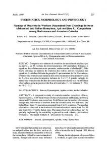
Fig. I. Distribution map of Acanthococcus antarcticus showing R/V Hero cruise collections (circles) and presumed east and west coast distribution limits (triangles). Records from the Falkland Islands are not shown.
104
Phyco!ogia, Vol. 31 (1), 1992
3cm
2
"
100pm
7
9
Figs 2-9. Acanthococcus antarcticus. Fig. 2. Habit of robust cystocarpic specimen of Acanthococcus antarcticus, Searles, Leister & Brauner 73-37-3,
Punta ValparaISo, Tierra del Fuego, Chile, NCU. Fig. 3. Upper left portion of lectotype of Acanthococcus antarcticus showing sessile, protuberant cystocarps. Fig. 4. Lectotype, Acanthococcus antarcticus J.D. Hooker et Harvey (Capt. Crozier, 1 842, Port Williams, E. Falkland Island, BM). Fig. S. Syntype, Graci/aria? obtusangula var. a (J.D. Hooker et Harvey, Falkland Islands, J.D.S., BM). Fig. 6. Holotype, Sphaerococcus subulatus var. nigrescens C. Agardh (Freycinet, LD-28225, Falkland Islands, LD). Fig. 7. Cross-section close to apex of fourth-order branch through specimen in Figs 3-4. Fig. 8. Cross-section close to apex of fourth-order branch through specimen in Fig. 5. Fig. 9. Cross-section close to apex of fourth-order branch through specimen in Fig. 6.
Fredericq et al.: Acanthococcus antarcticus
indistinguishable from them in lower cross-sec tions; medulla composed of 1-3 layers of large, inflated cells reaching 300 x 400 �m; cortex of 1-3 layers of progressively smaller cells; surface layer uniformly distributed, composed of rect angular to irregularly polygonal cells averaging 6 x 10 �m. Rhizoids at first ascending, later also descending, confined to central core in distal branches, or sparsely distributed between med ullary and cortical cells, later branching second arily, becoming abundant between medullary and inner cortical cells in main axes or towards the base; cystocarps intercalary or subterminal on lateral branches, naked or bearing up to six spiny outgrowths; carposporangia up to 50 �m in di ameter; branched carposporangial chains 200250 �m long; spermatangial pits averaging 50 �m in diameter; tetrasporangia up to 20-50 x 50100 �m, immersed in modified cortex. LEcrO TYP E: We have chosen as the lectotype a cystocarpic specimen that corresponds closely to the reverse image of the left-hand side of the habit drawing in J.D. Hooker i847, pI. 181, Port Williams, E. Falkland I., Capt. Crozier, 1842, BM (Figs 3-4, 7). No material was found that corresponded to the base and sterile part Of the habit drawing (right-hand side) in this plate. NOMENCLA TURAL SYNO MYM : Callophyllis ant artica (J.D. Hooker et Harvey) Kiitzing (1849, p. 747). TAXONOMIC SYNO NYM S: Gracilaria? obtusan gula J.D. Hooker et Harvey (1845, p. 261). Cys toclonium ? obtusangulum (J.D. Hooker et Har vey) Kiitzing (1849, p. 757). Sphaerococcus subulatus var. nigrescens C. Agardh (1822, p. 329). Graci/aria ? nigrescens J.D. Hooker et Har vey in J.D. Hooker (1847, p. 477). Graci/aria? nigrescens var. tenuior J.D. Hooker et Harvey in J.D. Hooker (1847, p. 477). Cystoclonium spin guligerum J. Agardh (1849, p. 87). Sphaero coccus nigrescens (J.D. Hooker et Harvey) Kiitz ing (1849, p. 777). Sphaerococcus nigrescens (J.D. Hooker et Harvey) var. tenuior Kiitzing (1849, p. 777). Acanthococcus spinuligerus (J. Agardh) J. Agardh (1852, p. 437). Acanthococcus spinu ligerus (J. Agardh) J. Agardh var. tenuior (J.D. Hooker et Harvey) Hariot (1889, p. 80). Acan thococcus spinuliger (J. Agardh) J. Agardh as cit ed in De Toni (1897, p. 351). Gigartina spinifera Kiitzing (1849, p. 750). The literature contains numerous references to a narrow, filiform species usually cited as Cys toclonium obtusangulum based on Gracilaria ob tusangula J.D. Hooker et Harvey or Acantho-
105
coccus spinuliger based on Sphaerococcus subulatus var. nigrescens C. Agardh (Pujals 1963, 1977; Papenfuss 1964). Gigartina spinifera Kiitz ing was placed in synonymy under Acanthococ cus spinuligerus by Hariot (1889, p. 80). Type material, presumably labeled 'Ad ins. Maloui nas: Delise' was not found. It could be a syntype of Sphaerococcus subulatus var. nigrescens. We have concluded after examining numerous spec imens from South America and the Falkland Is lands that the narrow, subcylindrical forms fall within the range of variation of Acanthococcus antarcticus and are conspecific with it. All col lections of these forms examined have proved to be sterile. Records of Acanthococcus spinuliger from the South Orkney Islands (Gepp & Gepp 1905) and Cystoclonium obtusangulum from the South Shetland Islands (Moe & DeLaca 1976), localities where A. antarticus has not been re ported, require further investigation. HISTORICAL SPECIMENS EXAMINED: Lectotype, Port Williams, E. Falkland Island (Capt. Crozier, 1842, BM, Figs 3-4, 7); syntype, Port Williams, E. Falkland Island (Capt. Crozier, vii. 1842, L, published as Callophyllis antarctica (J.D. Hook er et Harvey) Kiitzing 1849, 1867, pI. 93); NW Bay, Hermite Island, Cape Horn (spermatangial, undated, BM; tetrasporangial, undated, TCD; undated LD-28224, LD); St Martin's Cove, Her mite Island, Cape Horn (Capt. Crozier, Septem ber-October 1842, BM); outer coast Cape Pem broke, Falkland Islands (undated, TCD); Ancud, Chiloe, Chile (Lechler, undated, C); Falkland Is lands (det. Lenormand, undated, S; C); Detroit de Magellan (undated, ex. Herb. Lenormand, UPS); Slogget Bay, Fuegia (c. Skottsberg, 16.iii.1909, GB); Insula Maclov (undated, Herb. Kjellman, UPS); lIe de Tova, Patagonie (undat ed, Herb. Kiitzing, L). As Cystoclonium obtu sangulum var. a: Falkland Islands (JDH, BM, Figs 5, 8); undated, no location (BM). As Cysto clonium obtusangulum var. f3: NW Bay, Hermite Island, Cape Horn (J.D.H., undated, BM); Cape Horn (undated, L-938 92 194, L, illustrated by Kiitzing 1868, pI. 17); Berkeley Sound, Falkland Islands (D. Lyall, as DL), v. 1842, TCD; PC); St Salvador Bay, E. Falkland Island (iv. 1842, BM); St Salvador Bay, Falkland Islands (J.D. H. , un dated, BM); Falkland Islands (undated, BM); Fret. Magellan (Hohenacker 486, undated, L-938 92 128, L). As Acanthococcus spinuligerus: holo type, Falkland Islands (Freycinet LD-28225, Sphaerococcus subulatus var. nigrescens C. Agardh, Figs 6, 9); Syntype, Falkland Islands
106
Phycologia, Vol. 31 (1), 1992
(Gaudichaud #130, x. 1822, ex Herb. J. Gay, BM); Falkland Islands (#233 ex Herb. J. Ag., undated, BM). NEW RECORDS: RlV Hero cruises to southern Argentina and Chile: exposed island at entrance to Puerto Alert, Chile, 49°53.6' S, 75°12.5'W, sublittoral fringe to 6 m (Searles, Leister& Brau ner 72-19-70, tetrasporangial, 31.x.1972, NCU); Isla San Pedro, Gulfo de Peiias, Chile, 47°43.2' S, 74°53.3' W, low water to 3 m (Searles, Leister & Brauner 72-32-52, 6.xi.1972, NCU); Kelp bed, Punta Conway, Isla de los Estados, Argentina, 54°43.8' S, 64°13.9' W, 9-12 m (Searles, Leister & Brauner 73-1-29, cystocarpic, 4.v.1973, NCU); outer fringe of kelp bed, Bahia Colnett, Isla de los Estados, Argentina, 54°42.6' S, 64°20.3' W, 14 m (Searles, Leister & Brauner 73-6-5, cys tocarpic, 5.v.1973, NCU); very exposed, Puerto Vancouver, Isla de los Estados, Argentina, 54°47.4' S, 64°04.35' W, 5-25 m (Searles, Leister & Brauner 73-18-24, cystocarpic, 1 0.v.1 973, NCU); small cove, behind Islet Alexander, Isla de los Estados, Argentina, 54°50.5' S, 64°23.8' W, 20 m (Searles, Leister& Brauner 73-22-20,
cystocarpic, 1O.v.1973, NCU); outer edge of kelp bed, Bahia Crossley, Isla de los Estados, Argen tina, 54°47.5' S, 64°42.1' W, 14 m (Searles, Leis ter& Brauner 73-33-11, 1 4.v.1973, NCU); very sheltered, Cta Awaia Kirrh, Canal Beagle, Chile, 55°0.0' S, 69°02.2' W, 0-2 m (Searles, Leister& Brauner 73-35-50 cysto carp ic& male, 16.v.1973, NCU); Punta ValparaIso, Tierra del Fuego, Chile, 54°22.2' S, 71°21. 7' W (Searles, Leister& Brau ner 73-37-3 cystocarpic, 17.v.1973, NCU); Puerto Alert, Canal Trinidad, Chile, 49°53.6' S, 75°12.7' W, 5-18 m (Searles, Leister & Brauner 73-42-11, cystocarpic, 20.v.1973, NCU). DISTRIBUTION: Distributional records of Acan thococcus are given by Papenfuss (1964), Pujals (1963, 1977) and Moe& DeLaca (1976). Robust, compressed forms and narrow, spinuligerous forms of Acanthococcus antarcticus were found at many of the same localities in Tierra del Fuego and the Falkland Islands in the expeditions of the last century. The most northerly records of A. antarcticus are from Ancud, Chiloe, Chile, and Puerto Deseado, Argentina (Fig. 1). We confirm that the Lechler specimen at Copenhagen (C) from
Figs 10-13. Acanthococcus antarcticus. Searles, Leister & Brauner 73-35-50. Vegetative morphology. Fig. 10. Side branch showing uniaxial tips, surface cells, and rhizoids beneath. Fig. 11. Optical section through spine on cystocarp showing apical cell, uniaxial construction and branching of
cortical filaments (aniline-blue stained). Each axial cell (arrowhead) has cut off a periaxial cell bearing a cortical filament. Fig. 12. Longitudinal section through tip showing apical cell (a), axial cells (arrowheads) and apical cells of corticating filaments (arrows). Periaxial cells and inner cortical cells below have produced rhizoids (r). Fig. 13. Optical section of tip showing rhizoids (arrows) ascending from periaxial cells and inner cortical cells. Darkly staining spherical bodies are visible in some cortical cells (arrowheads). Figs 14-19. Acanthococcus antarcticus. Searles, Leister & Brauner 73-35-50. Vegetative morphology. Fig. 14. Axial cells (ax) and periaxial cells (p) linked by enlarged pit connections (arrows). Fig. 15. Axial cells (ax) and periaxal cell (p) bearing cortical filament (co). Rhizoids (r) arising from periaxial and cortical cells form numerous secondary pit connections (arrows). A conjunctor cell from a rhizoid is shown on the left-hand side (arrowhead). Fig. 16. Longitudinal section through upper part of third-order branch showing rhizoids primarily restricted to central core. Fig. 17. Longitudinal section through stipe showing abundant rhizoids in thick, central bundle and between surrounding medullary and cortical cells. Fig. 18. Cross-section through third-order branch showing few central and interspersed (arrowhead) rhizoids. Fig. 19. Cross-section through stipe showing abundant central and interspersed rhizoids. Figs 20-24. Acanthococcus antarcticus. Searles, Leister & Brauner 73-37-3. Female reproductive system. Fig. 20. Longitudinal section of branchlet tip showing position of carpogonial branches (arrows) and auxiliary cells (ac). Fig. 21. Supporting cell (sc) bearing 3-celled carpogonial branch terminated by carpogonium (arrow); auxiliary cell (ac) is pit-connected (arrowhead) to supporting cell. Fig. 22. Post-fertilization stage. Nutritive filaments (nf), supporting cell (sc), first and second cells of carpogonial branch (cbl, cb2) and auxiliary cell (ac) containing a central haploid (hn) and proximal diploid (dn) nucleus. Cells are slightly displaced due to squashing. Fig. 23. Elongated supporting cell (sc) and auxiliary cell (ac) with gonimoblast initial (gi) and surrounded by nutritive filaments (nf) and modified terminal cortical filaments (mcf). Fig. 24. Detail of central region shown in Fig. 20. Supporting cell (sc) is uninucleate, except where a conjunctor cell derived from a nutritive filament has fused with it, depositing a nucleus and leaving a small process (arrow). Auxiliary cell (ac) distal to supporting cell has cut off a gonimoblast initial (gi).
Fredericq et al.: Acanthococcus antarcticus
200pm
10 25pm
107
25pm
108
Phyc% gia, Vol. 31 (1), 1992
Fredericq et al.: Acanthococcus antarcticus
109
110
Phyc% gia, Vol. 31 (1), 1992
20 pm
Fredericq et al.: Acanthococcus antarcticus
Ancud, Chiloe, is A. antarcticus, but its geo graphical origin requires reinvestigation. HABITAT: The larger, more robust specimens come from wave-exposed habitats in the vicinity of Cape Hom (Fig. 2), and from Puerto Deseado, Santa Cruz, Argentina (Joly et al. 1 964). Plants from more northerly latitudes along the coast of Chile and from the Falkland Islands are usually narrower and less robust. Vegetative morphology
Plants are uniaxial, with a single wedge-shaped apical cell that protrudes slightly at the apex (Figs 1 0-1 2). An apical cell has two oblique faces from which it alternately cuts off axial segments by concavo-convex septa at angles of 50-60°. Suc cessive divisions (Figs I I , 1 2) alternate to one side and then the other, generating a file of axial cells in a zig-zag pattern. Each axial cell cuts off a single periaxial cell from its upper side in an alternate-distichous arrangement (Figs 1 1 , 1 2, 1 4). A periaxial cell first produces the initial of the leading determinate filament (Fig. 1 2). When this filament is 2-3 cells long, laterals are initi ated obliquely on opposite sides that also de velop into ascending, branched cortical fila ments. Branching of the cortical filaments is initially irregularly trichotomous or subdichotomous. Approximately 6-7 cells behind the apex, inner cells expand into isodiametric shapes, except for the basal periaxial cell, which first expands but later becomes elongated (Figs 1 2, 1 4). Subcor tical cells may generate as many as five or six lateral filaments as the thallus elongates and ex pands, forming an outer cortex two or three lay ers thick and composed of progressively smaller, predominantly uninucleate or binucleate, sub ovoid to subspherical cortical cells (Figs 1 6, 1 8). Surface cortical cells are of nearly uniform size
III
(Fig. 1 0), and contain a single, parietal, lobed chloroplast. Both medullary and cortical cells typically include one to few spherical globules, separate from the chloroplasts, that stain darkly with aniline blue (Fig. 1 3). Rhizoidal initials issue from periaxial cells ap proximately 1 0 segments below the apex (Figs 1 2, 1 3). Close to the apex they arise singly, pri marily from the upper sides of periaxial cells, and produce ascending, septate rhizoids (Figs 1 3, 1 5), although they may also originate in lesser numbers from lower sides and form descending rhizoidal filaments. Lower down the axis, addi tional rhizoidal filaments are produced on peri axial cells and inner cortical cells, which are ei ther added to the central core surrounding the axial filament, or grow between the medullary and inner cortical cells (Figs 1 6-1 9). Rhizoids form numerous secondary pit connections with axial cells, inner cortical cells and each other (Fig. 1 5). As a result, most cells in the interior of the thallus become multinucleate. Rhizoids may be localized primarily in a central bundle with only a few filaments distributed between medullary and cortical cells (Figs 7-9, 1 6, 1 8; Kiitzing 1 868, pI. 1 7, 'Cystoclonium obtusangulum ,), or may branch secondarily, becoming abundant between medullary and inner cortical cells in the main axes or towards the base (Figs 1 7, 1 9; Hooker 1 847, pI. 1 81 , fig. 3; Joly et al. 1 964, figs 4-7, 1 0, 1 1 ). Axial cells start to elongate within a few cells of the apices in vegetative tips (Fig. 1 2), becom ing up to 1 20 ILm long (Fig. 1 4). Pit connections linking axial cells progressively enlarge up to 1 0 ILm in diameter (Fig. 1 4). Cortical filaments as cend at a steep angle (> 60°) alongside two or more successive axial segments (Figs 1 1 , 1 2). As a result, the inflated cells surrounding any given axial cell, together with the core of rhizoidal cells seen in cross-section (Figs 18, 1 9), are derived
Figs 25-29. Acanthococcus antarcticus. Searles, Leister & Brauner 73-1-29. Female reproductive system. Fig. 25. Detail of multinucleate supporting cell (sc) showing pit connection (arrow) to distal auxiliary cell (ac)
and 2-celled gonimoblast (g). Fig. 26. Auxiliary cell (ac) fused (arrow) to supporting cell (sc) and bearing a 2-celled gonimoblast (g). Fig. 27. Remnant of pit plug (arrow) between fused auxiliary cell (ac) and supporting cell (sc). Nutritive filaments
have become linked to supporting cell, which now contains many nuclei. Fig. 28. Gonimoblasts (g) arising from fusion cell and surrounded by nutritive filaments (nf). Gonimoblast cells are uninucleate; cells of nutritive filaments are mostly multinucleate. Fig. 29. Close-up of nutritive filaments (nf) linked by secondary pit connections to multinucleate fusion cell (fu).
112
Phycologia, Vol. 31 (1), 1992
Figs 30-35. Acanthococcus antarcticus. Searles, Leister & Brauner 73-33 - 1 1 . Female reproductive system. Fig. 30. Cross-section showing early gonimoblasts (g) surrounding fusion cell (fu). Fig. 31. Gonimoblast filaments cutting off conjunctor cells (arrows) that are fusing on to the fusion cell (fu). Fig. 32. Innermost gonimoblast cells that have divided anticlinally (between arrowheads). One conjunctor cell is visible (arrow). Fig. 33. Branched gonimoblast filaments directed outwardly, and linked by secondary pit connections to fusion cell.
Fredericq et al.: Acanthococcus antarcticus
from two or more files of overlapping cortical filaments on each side. Female reproduction
Carpogonial branches are formed near the apices of ultimate branchlets. The supporting cell is an intercalary cortical cell borne on a periaxial cell. It can either be the suprabasal cell of the leading cortical filament or the corresponding cell of a lateral filament. Occasionally, more than one carpogonial branch is produced in adjacent branches of the same filament system. The car pogonial branch initial is cut off adaxially from the supporting cell and develops into a 3-celled carpogonial branch directed towards the thallus apex (Fig. 20). The supporting cell elongates, be coming crescent-shaped at maturity (Figs 20, 21). Prior to fertilization, all cells of the carpogonial branch, the supporting cell, and the auxiliary cell are uninucleate (Fig. 21). The hypogynous cell is the largest cell of the young carpogonial branch (Fig. 20), although at maturity the basal cell reaches the same size as the hypogynous cell (Fig. 21). The carpogonium is more or less conical, remains small, and bears a straight to slightly curved trichogyne directed towards the thallus surface (Fig. 20). There are no sterile cells on any of the carpogonial branch cells. The auxiliary cell lies just distal to the sup porting cell and is linked to it by a primary pit connection (Fig. 21). Prior to fertilization, it is barely distinguishable in shape from the sur rounding cortical cells (Figs 20, 21). The fertil ization nucleus is transferred to the auxiliary cell, probably as a result of direct fusion of the car pogonium with the auxiliary cell. The diploid ized auxiliary cell is binucleate, containing a cen tral haploid nucleus and a proximal diploid nucleus (Fig. 22). The basal and hypogynous cells of the carpogonial branch are still visible at this stage, but they soon disintegrate. Carposporopbyte development
Following presumed fertilization, cortical cells distal to the auxiliary cell become densely stain ing and their nuclei replicate and enlarge. Con currently, the supporting cell extends in length
1 13
parallel to the central axis and short, densely staining, branched nutritive filaments develop from the surrounding rhizoidal and medullary cells and grow towards the supporting cell (Figs 22-24). In contrast to the darker staining distal filaments, which are transformed from pre-ex isting cortical cells, the nutritive filaments below are produced secondarily. The auxiliary cell cuts offa single gonimoblast initial towards the thallus interior by an oblique, concavo-convex division (Figs 23, 24). Pit con nections between the auxiliary cell and the mod ified distal cortical cells enlarge (Fig. 23), and the secondary nutritive filaments become linked to the supporting cell. Terminal cells of nutritive filaments fuse directly on to the supporting cell, depositing their nuclei (Fig. 24) and leaving be hind secondary pit connections. By the time the gonimoblast is 2-celled, the supporting cell al ready contains several nuclei derived from nu tritive cells (Fig. 25). Ultimately, all of the nu tritive filaments become linked to the supporting cell, with the deposition of many nuclei (Figs 2729). The auxiliary cell fuses with the supporting cell (Fig. 26) around the primary pit connection (Fig. 27), and inner gonimoblast cells also fuse with it (Figs 26, 29). Young gonimoblast cells at the anterior end of the fusion cell are readily distinguished because they are uninucleate (Fig. 28). The nutritive cells linked to the supporting cell are mostly multinucleate, stain darkly at this stage, and their pit connections broaden (Figs 28, 29). Ultimately, the outline of the supporting cell, auxiliary cell and fused inner gonimoblast cells becomes indistinct, and the fusion cell rounds up. Gonimoblast filaments that were initially lo calized at the anterior end of the supporting cell (Fig. 28) continue to divide until the carpospo rophyte completely surrounds the fusion cell (Figs 30, 33). Apical gonimoblast cells adjacent to the fusion cell divide anticlinally as the gonimoblasts spread, followed by periclinal divisions (Fig. 32) forming branched chains up to six cells in length that radiate outwardly (Figs 30, 33). The inner most gonimoblast cells cut off conjunctor cells (Figs 31, 32) basally that unite with the fusion cell, leaving behind secondary pit connections
;Fig. 34. Fusion cell bearing chains of carposporangia and str�tched, remnant cortical filament (arrow i rmg the Fig. 35. Cross-section of mature cystocarp with central fusIOn cell and chams of carposporangIa
cystocarp cavity.
J
114
Phyc% gia, Vol. 31 (1), 1992
50pm
Figs 36-43. Acanthococcus antarcticus. Male (Searles, Leister & Brauner 73-35-50) and tetrasporangial (Searles, Leister & Brauner 72-1 9-70) reproductive systems. Fig. 36. Cross-section of mature male reproductive structures organized in shallow, superficial sori. Fig. 37. Spermatangial parent-cell initial (arrowhead). Fig. 38. Two-celled spermatangial filament (arrowheads). Fig. 39. Right, 3-celled spermatangial filament (arrowheads); left, sorus of branched spermatangial filaments
bearing terminal spermatangia (arrows). Fig. 40. Sorus of spermatangial filaments bearing terminal spermatangia (arrows). Fig. 41. Terminal tetrasporocyte (arrowhead). Fig. 42. Centre, tetrasporocyte laterally pit-connected (arrow) to its bearing cell; right, tetrasporocyte (arrowhead) after first division. Fig. 43. Cross-section of mature tetrasporangia surrounded by elongated cortical cells and outer covering of cortical filaments.
Fredericq et al.: Acanthococcus antarcticus
115
(Fig. 33). Both cortical and secondary nutritive
of the distended initial provides a reliable indi
filaments lose their stainable contents and be
cation of the previous position of this pit con
come indistinct in old cystocarps. All gonimo
nection (Fig. 42).
blast cells that are not incorporated into the fu
Cortical cells adjacent to a tetrasporocyte elon
sion cell become carposporangia (Figs 34, 35),
gate, becoming stretched and thin as the initial
there being no sterile gonimoblast cells in the
expands. Surface cells continue to divide, cov
mature cystocarp.
Except for an occasional
ering the mature tetrasporangia (Fig. 43). Tetra
stretched inner cortical filament (Fig. 34), the
sporangia cleave successively twice in the same
entire space between the fusion cell and the sur
plane, resulting in the formation of regularly zon
rounding cortex becomes filled with branched
ately arranged tetraspores. Tetrasporangia are 50-
chains of carposporangia up to six cells long (Figs
100 ILm long and 20-50 ILm wide at maturity
34, 35). A strongly developed multilayered peri
(Fig. 43).
carp and ostiole are lacking (Fig. 35). Cystocarps are sessile, hemispherical, subglobose, wider than the subtending branch (Figs 2, 3), and may bear
DISCUSSION
up to six spiny outgrowths (Hooker 1847, pI. 181).
Acanthococcus possesses all the familial char acters that Kylin (1932, 1956) considered to be
Male reproduction
Male reproductive structures occur on separate plants in shallow sori (Fig. 36) covering the thal lus surface on the higher-order- branches. Sorus development begins with the transformation of a terminal cortical cell into the primordium of a spermatangial filament. This ceif loses its plastid, stains uniformly with haematoxylin, and its nu cleus enlarges and stains darkly (Fig. 37). The cell divides obliquely, first to one side (Fig. 38) and then to the other (Fig. 39). Each of the re sulting three cells may function directly as a sper� matangial parent cell, or may undergo transverse and oblique divisions that extend the sperma tangial filaments (Figs 39, 40). Terminal cells cut off a single, hyaline, uninucleate spermat�mgium in which the nucleus is displaced towards the apex (Figs 39, 40). Cortical cells adjacent to sper matangial filaments elongate to form the borders of the soral cavity (Figs 36, 39, 40). Tetrasporangial reproduction
diagnostic of the Cystocloniaceae. The distin guishing generic characters of Acanthococcus within the family are: (I) axial cells each bearing one periaxial cell; (2) 3-celled carpogonial branch, with the hypogynous cell initially broader than the basal cell; (3) nutritive filaments produced after diploidization of the auxiliary cell, becom ing linked by secondary pit connections to the elongated supporting cell largely before fusion cell formation; (4) spermatangia in sori borne on spermatangial filaments that develop from sur face primordial cells. Thallus growth is initiated by a wedge-shaped apical cell with two oblique cutting faces throughout the Cystocloniaceae. In other genera, each axial cell cuts off two periaxial cells at an angle of 90° in an alternating sequence (Kylin 1932, 1956; Min-Thein & Womersley 1976). Acanthococcus appears to be unique among the genera of the Cystocloniaceae in that each axial cell cuts off only one periaxial cell. Comparable single periaxials per axial cell are encountered in
Mature tetrasporangia are scattered over the sur
the uniaxial genera Rhabdonia, Areschougia and Melanema of the Solieriaceae (Min-Thein &
face of higher order branches. They are zonately
Womersley 1976, as the Rhabdoniaceae). We in
cleaved (Fig. 43) and embedded amongst the cor
terpret the single periaxial cell of Acanthococcus
tical filaments. Tetrasporocytes (Figs 41, 42) are
as an advanced character that evolved in com
terminal cortical cells that may be distinguished
bination with other changes in filament ontogeny
from neighbouring vegetative cells by the pres
at the thallus apex and independently from sim
ence of an enlarged nucleus. At first a tetraspo
ilar arrangements in solieriaceous genera. The
rocyte initial is connected to its bearing cell by
potential loss of structural components in the
a basal pit connection, but as the initial enlarges,
axes that might result from halving the number
increasing in length from its inner side, the pit
of cortical filaments per axial cell is achieved in
connection comes to lie laterally at mid-level.
two ways in Acanthococcus: ( I) each leading cor
Although pit connections are often difficult to
tical filament extends alongside two axial cells
see and persist for only a short time, the shape
above the bearing cell on one side of the thallus,
116
Phyc% gia, Vol. 31 (1), 1992
thus filling in the gaps; and (2) inner cortical cells produce up to four secondary filaments (5-6 fil aments in total), which fill in the cortex as the thallus elongates and expands. Acanthococcus antarcticus is characterized by ascending and descending rhizoidal filaments that form a central bundle around the axial filament surrounded by one or two layers of swollen med ullary cells, later extending between the medul lary and cortical cells to just beneath the thallus surface. Descending rhizoids are abundant in Cystoclonium (Kylin 1923, 1956), and are usu ally present to some degree in other members of the Cystocloniaceae and in the Solieriaceae, where they are often generated in species-specific pat terns. The initially ascending rhizoids that char acterize Acanthococcus antarcticus appear to be unique among the genera of the Cystocloniaceae, although ascending rhizoids have been reported in species of Agardhiella (Solieriaceae; Gabriel son & Hommersand 1982b). Enlarged inner cells that are separated from the central axis and each other by a network of bridging filaments are char acteristic of Mychodea of the Mychodeaceae (Kraft 1978), several species of which had pre viously been placed in Acanthococcus. The early production of rhizoidal filaments from periaxial and inner cortical cells and their tendency to form numerous secondary pit connections with adja cent cells of all types rapidly obscures the basic uniaxial organization of the thallus. The male reproductive system of Acanthococ cus differs from that typically encountered in the Cystocloniaceae, where spermatangial parent cells are cut off outwardly, either from untransformed terminal surface cells or from surface cells in a rosette configuration (Kylin 1956; Min-Thein & Womersley 1976; Hansen 1980). In Acantho coccus, terminal cortical cells are first modified into spermatangial filament primordia, which then divide to form chains of spermatangial par ent cells that cut off single spermatangia to the outside. The resulting spermatangial sori of Acanthococcus bear a resemblance to the 'pits' or 'conceptacles' that are found in species of Graci/aria in which spermatangial parent cells are also generated in filaments (Fredericq & Hommersand 1989). Further observations may reveal that this type of spermatangial sorus is more widespread in the Cystocloniaceae than ex isting studies have indicated. In Acanthococcus no lateral filaments are borne on the carpogonial branch cells, such as occur in Cystoclonium (Kylin 1923). The basal and hy-
pogynous cells are laterally expanded and con tain an enlarged nucleus. Although the carpo gonial branch resembles that of other members of the Cystocloniaceae or Hypnea (Hypneaceae; Kylin 1956), in none of these genera are the prox imal cells of the carpogonial branch as enlarged as in Acanthococcus. Cortical filaments distal to the auxiliary cell enlarge and become densely staining after fertil ization, and either before or immediately follow ing diploidization of the auxiliary cell in Acan thococcus. The resulting structure resembles the auxiliary cell complex formed prior to diploid ization of the auxiliary cell in Solieria and Agardhiella of the Solieriaceae (Gabrielson & Hommersand 1982a, 1982b). Cortical filaments distal to the auxiliary cell are transformed into a nutritive tissue in other genera belonging to the Cystocloniaceae and Hypneaceae, but it is un clear, at present, whether cells proximal to the auxiliary cell are similarly modified. However, in no instance has the supporting cell been shown to elongate after fertilization in the manner ob served in Acanthococcus. The suite of characters listed by Gabrielson & Kraft (1984) as being diagnostic of the Solieri aceae includes files of cells cut off around the auxiliary cell following diploidization, but prior to gonimoblast initiation. These files may or may not consolidate to form a distinct pericarp. This feature is also found in most members of the Cystocloniaceae and Hypneaceae. Such files of cells probably function as nutritive filaments that become linked in some chhracteristic way, either directly with the gonimoblasts or with a fusion cell. Details vary in different genera, and are un known in many instances. Acanthococcus ap pears to be unique in that the nutritive filaments formed after fertilization become directly linked to the elongated supporting cell through fusion of terminal cells prior to the formation of a cen tral fusion cell. Examples of cases where gonimoblast cells link with vegetative nutritive cells, either through di rect fusion or by means of secondary pit con nections, are widespread among the Rhodophyta (Hommersand & Fredericq 1990). However, the opposite behaviour, in which secondary nutri tive filaments initiate the fusion process, is less common and, to our knowledge, only occurs in some members of the Cystocloniaceae and Hyp neaceae. In Cystoc/onium, for example, cells at the ends of nutritive filaments elongate and fuse directly on to the highly ramified gonimoblast
Fredericq et al.: Acanthococcus antarcticus
fusion cell in the vicinity of the carposporangial chains (Thrainsson 1986). At present, direct link age of nutritive filaments to the supporting cell or fusion cell is known only in Cystoclonium purpureum (Hudson) Batters and Acanthococcus antarcticus, but it may well occur in other cys tocloniaceous genera. In Acanthococcus, as in other members of the Cystocloniaceae, outer gonimoblast filaments are converted into chains of carposporangia that abut against the stretched inner cells of the pericarp. Some thickening of the pericarp takes place in Acanthococcus due to resumption of outward growth of the surface cortical filaments, but a massive pericarp is not produced, nor do sterile gonimoblast filaments form extensions or pro cesses that link up with cells in the outer pericarp, as in some species of Rhodophyllis, Craspedo carpus, and Calliblepharis from Australia (Min Thein & Womersley 1976), and Hypnea mus cijormis from North Carolina (Hommersand & Fredericq 1990). Ostiole-like structures are sometimes formed in these genera, but not in Acanthococcus. Tetrasporangia are generally described as be ing scattered or localized in sori, either on the thallus surface or on proliferations in the Cys tocloniaceae. The somewhat nemathecial char acter of the tetrasporangial sori in Acanthococ cus, in which the neighbouring sterile cells are markedly elongated and the surface cells divide and branch to form a cover above the tetraspo rangia, may be a specialized feature of this genus within the Cystocloniaceae. The procarpial family Cystocloniaceae is prov ing to be as diverse an assemblage and as rich in genera and species as the non-procarpial family Solieriaceae. It is represented by some very dis tinct genera in boreal and antiboreal waters, as well as in the warm-temperate regions of the At lantic and Indian oceans (Hommersand 1990). New morphological studies of the other genera of the Cystocloniaceae are required to elucidate the phylogeny and biogeography of the entities referred to this family.
AC
KN OWLEDGEMENTS
This study was supported by the National Sci ence Foundation under contract number DPP7413988 to the Smithsonian Institution. We thank Emani Mefiez and Frank Ferrari of the Smith sonian Oceanographic Sorting Center for their
117
encouragement and for the loan of specimens. Liquid-preserved collections from the 1972-1973 NSF R/V Hero cruises (Grants GV-31766, Di vision of Polar Program, to Duke University and BMS-75-21500 to G.L.) formed the basis for this research. We thank G.T. Kraft, D.J. Garbary and M.D. Guiry for their critical reviews of the manuscript, and Paul Silva for advice on no menclatural problems. REFERENCES
AG ARD H C.A. 1 822. Species Algarum . . . Vol. 1. Mauritius, Greifswald, 1 89 pp. AGARDH J.G. 1 849. Algologiska bidrag. Ofversight af Forhandlingar: Kongelige Svenska Vetenskaps-Aka demien 6: 79-89. AGARDH J.G. 1 852. Species Genera et Ordines Al garum . . . Vol. 2(2}, pp. 337-786. C. W. K. Gleerup, Lund. AGARDH J.G. 1 876. Species genera et ordines alga rum . . . Vol. 3(1}, Weigel, Leipzig, vii + 724 pp. DE TONI, G.B. 1 897. Sylloge algarum . . . Vol. 4 (Florideae). Privately published, Padua, xx + Ixi + 386 pp. FREDERICQ S. & HOMMERSAND M.H. 1 989. Proposal of the Gracilariales ord. nov. (Rhodophyta) based on an analysis of the reproductive development of Gracilaria verrucosa. Journal of Phycology 25: 2 1 3227. GABRIELSON P.W. & HOMMERSAND M.H. 1 982a. The Atlantic species of Solieria (Gigartinales, Rhodoph yta). Their morphology, distribution and affinities. Journal of Phycology 18: 3 1-45. GABRIELSON P.W. & HOMMERS AND M.H. 1 982b. The morphology ofAgardhiella subulata representing the Agardhielleae, a new tribe in the Solieriaceae (Gig artinales, Rhodophyta). Journal ofPhycology 18: 4658. GABRIELSON P.W. & KRAFT G.T. 1 984. The marine algae of Lord Howe Island (N.S.W.): the family So lieriaceae (Gigartinales, Rhodophyta). Brunonia 7: 2 1 7-25 1 . GEPP A. & GEPP E.S. 1 905. Antarctic algae. Journal of Botany 43: 1 0 5- 1 09, pI. 470. GUIRY M.D. 1 978. Notes on some family names of Florideophyceae (Rhodophyta). Taxon 27: 1 9 1-1 95. HANSEN G.I. 1 980. A morphological study of Fim brifolium, a new genus in the Cystocloniaceae (Gig artinales, Rhodophyta). Journal ofPhycology 16: 2072 1 7. HARlOT P.A. 1 889. Algues. In: Mission Scientifique du Cap Horn, 1882-1883. Vol. 5, pp. 3-1 1 9, pI. 1 0. Botanique. Gauthier-Villars et fils, Paris. HOLMGREN P.K., KEUKEN W. & SCHOFIELD E.K. 1 98 1 . Index Herbariorum, I. The Herbaria of the World, 7th edn. Bohn, Scheltema & Holkema, Utrecht, 452 pp. [Regnum Vegetabile vol. 1 06]. HOMMERS AND M. H . 1 990. Biogeography of the ma rine red algae of the North Atlantic Ocean. In: Evo lutionary Biogeography of the Marine Algae of the North Atlantic (Ed. by D.J. Garbary & G.R. South), pp. 349-4 1 0. NATO Advanced Science Institutes
118
Phycologia, Vol. 31 (1), 1992
Series G: Ecological Sciences, Vol. 22. Springer-Ver lag, Berlin. HOMMERsAND M.H. & FREDERICQ S. 1 98 8 . An in vestigation of cystocarp development in Gelidium pteridifolium with a revised description ofthe Gelid iales (Rhodophyta). Phycologia 27: 254-272. M.H. & FREDERICQ S. 1 990. Sexual HOMMERS AND reproduction and cystocarp development. In: Biol ogy of the Red Algae (Ed. by K.M. Cole & R.G. Sheath), pp. 305-345. Cambridge University Press, New York. HOOKER J.D. 1 847. Algae. In: The Botany ofthe Ant arctic Voyage of H.M. Discovery ships 'Erebus' and Terror' in the Years 1839-1843 . . . Vol. 1. Flora antarctica . . . Part 11. Botany of Fuegia, the Falk lands, Kerguelen 's Land, etc. (1845-1847), pp. 209574. Reeve, London. HooKER J.D. & HARVEY W.H. 1 845. Algae antarc ticae . . . London Journal of Botany 4: 249-276. GUISHI N. 1 964. La JOLY A.B., CORDEIRO M . & Y AMA estructura y reproduccion de Acanthococcus antarc ticus Hooker et Harvey. Boletin del Instituto de Bio logia Marina (Mar del Plata) 5: 1-9. G.T. 1 9 7 8 . Studies of marine algae in the KRAFr lesser-known families of the Gigartinales (Rhodoph yta). III. Mychodeaceae and Mychodeophyllaceae. A ustralian Journal of Botany 26: 5 1 5-6 1 0. KUTZING F. 1 843. Phycologia generalis . . . Brock haus, Leipzig, xxxii + 458 pp. KUTZING F. 1 849. Species Algarum . . . Brockhaus, Leipzig, vi + 922 pp. KUTZING F. 1 867. Tabulae phycologicae . . . Vol. 1 7. Privately published, Nordhausen, 30 pp. + 1 00 pis. ZING F. 1 868. Tabulaephycologicae . . . Vol. 1 8, Kih Privately published, Nordhausen, 35 pp. + 1 00 pis. KYLIN H. 1 923. Studien iiber die Entwicklungsge schichte der Florideen. Kongelige Svenska Vetensk apsakademiens Handlingar 63 ( 1 1 ) : 1 - 1 39. KYLIN H. 1 9 32. Die Florideenordnung Gigartinales. Lunds Universitets Arsskriji, N.F., Avd. 2 28(8): 1 8 8 pp., 28 pis. KYLIN H. 1 956. Die Gattungen der Rhodophyceen. C.W.K. Gleerup, Lund, xv + 673 pp. KYLIN H. & SKOTTSBERG c. 1 9 1 9. Zur Kenntnis der subantarktischen und antarktischen Meeresalgen. II. Rhodophyceen. In: Wissenschaftliche Ergebnisse der
schwedischen sud-polar Expedition 1 901-1 903, Vol. 4 (Ed. by O. NordenskjOld). Lithographisches Insti tut des General stabs, Stockholm. 88 pp, I pI. MIN-THEIN U. & WOMERSLEY H.B.S. 1 976. Studies on Australian taxa of Solieriaceae, Rhabdoniaceae, and Rhodophyllidaceae (Rhodophyta). A ustralian Journal of Botany 24: 1 - 1 66. MOE R.L. & DELAcA T.E. 1 976. Occurrence of mac roscopic algae along the Antarctic Peninsula. Ant arctic Journal of the United States 11: 20-24. PAPENFUSS G .F. 1 964. Catalogue and bibliography of Antarctic and subantarctic benthic marine algae. A ntarctic Research Series, Washington 1: 1-76. PUJALS C. 1 963. Caulogo de Rhodophyta citadas para la Argentina. Revista del Museo Argentino de Ciencias Naturales 'Bernardino Rivadavia' (Botiini cay 3: 1 - 1 39. PuJALS C. 1 977. Adiciones y correcciones al 'CataJo go de Rhodophyta citadas para la Argentina.' Revista del Museo Argentino de Ciencias Naturales 'Bernar dino Rivadavia' (Botiinica) 5: 1 23- 1 63. REINBOLD T. 1 908. Die Meeresalgen der deutchen Siidpolar-Expedition 1 90 1- 1 903. In: Deutsche Siid polar Expedition 1 901-1 903, Vol. 8 (Ed. by E. Von Drygalski), pp. 1 77-202. Botanik, Berlin. SCHMITZ [C.J.] F. 1 892. [6. Klasse Rhodophyceae] 2. Unterklasse Florideae. In: Syllabus der Vorlesungen uber specielle und medicinische-pharmaceutische Botanik. Grosse A usgabe (Ed. by A. Engler), pp. 1 623. Gebriider Bomtraeger, Berlin. SSON S.A. 1 986. Morphological studies on TH RAIN some members of the family Cystocioniaceae (Gig artinales, Rhodophyta). Unpublished MA thesis, University of North Carolina at Chapel Hill, 1 0 1 pp. W. 1 965. Aceto-iron-haematoxylin-chlo MANN WITT ral hydrate for chromosome staining. Stain Tech nology 40: 1 6 1 - 1 64. G.T. 1 9 8 1 . [Appendix]. Clas KRAFr E M.J. & W YNN sification summary. In: The Biology of Seaweeds, Botanical Monographs Vol. 1 7 (Ed. by C.S. Lobban & M. J . Wynne), pp. 743-750. Blackwell Scientific Publications, Oxford.
Accepted 17 July 1991