Journal of the International Neuropsychological Society (2005), 11, 482–487. Copyright © 2005 INS. Published by Cambridge University Press. Printed in the USA. DOI: 10.10170S1355617705050587
BRIEF COMMUNICATION
Motor and non-motor sequence learning in children and adolescents with cerebellar damage
ANDREA BERGER,1 MICHELLE SADEH,2,3 GABRIEL TZUR,1 AVINOAM SHUPER,2,4 LIORA KORNREICH,2,4 DOV INBAR,2,4 IAN J. COHEN,2,4 SHALOM MICHOWIZ,2,4 ISAAC YANIV,2,4 SHLOMI CONSTANTINI,5 and ELI VAKIL 6 1 Ben-Gurion
University of the Negev, Beer Sheva, Israel Children’s Medical Center, Petach Tikva, Israel 3 Israel Cancer Association, Givataim, Israel 4 Sackler Faculty of Medicine, Tel Aviv University, Tel Aviv, Israel 5 Dana Hospital, Sackler Faculty of Medicine, Tel Aviv University, Tel Aviv, Israel 6 Bar-Ilan University, Ramat Gan, Israel 2 Schneider
(Received August 25, 2004; Revised December 28, 2004; Accepted March 25, 2005)
Abstract Cerebellar involvement in motor and non-motor sequence learning was examined with serial reaction time tasks (SRT). Our sample consisted of 8 children and adolescents who had undergone surgical removal of a benign posterior fossa tumor (PFT) during childhood. None of them had undergone chemotherapy or cranial radiation therapy (CRT). Ages ranged from 1–11 years at surgery and 9–17 years at testing. The children were tested not earlier than 2.5 years after surgery (M 5 5.9 years), enabling brain plasticity and recovery of functions. Their performance was compared with a matched control sample. The PFT group was not impaired in the implicit learning of sequences, as reflected in their performance in blocks with a repeated sequence, both before and after a random block. However, in the perceptual task, their performance deteriorated more than that of the control group when a random block was introduced, suggesting that it was more difficult for the patients to respond flexibly or change their response set when encountering changing task demands. These results are in line with another study by our group on task switching with the same patients. (JINS, 2005, 11, 482–487.) Keywords: Serial reaction time, Procedural learning, Motor learning, Perceptual learning, Cerebellum, Posterior fossa tumor
recognition of the sequence. At this point, changes in the stimuli order, such as switching to a random sequence, cause increases in reaction times and errors, which decrease again if the original sequence is reinstated. Implicit learning of the sequence occurs also in the perceptual0non-motor version of the task, where the participants follow the sequence with their eyes instead of their fingers (Vakil et al., 2000). Although the exact brain network involved in procedural skill learning is still not fully understood, there is consistent evidence relating the basal ganglia to this kind of learning (Pascual-Leone et al., 1993; Vakil et al., 2000), and increasing evidence for the involvement of the cerebellum (Doyon, 1997; Laforce & Doyon, 2001; Friston et al., 1992; Grafton et al., 1995; Hallet & Grafman, 1997; Pascual-Leone et al., 1993; Seidler et al., 2002; Seitz et al., 1990). Imaging stud-
INTRODUCTION Procedural learning refers to the acquisition of ability through practice. This acquisition can be purely implicit because it is reflected in improved performance, even without conscious recognition or retrieval (Squire, 1992). In the SRT paradigm, procedural implicit learning is usually studied in terms of the learning of a repeated sequence of finger movements (Nissen & Bullemer, 1987). With practice, participants usually learn the sequence implicitly, and responses become faster and more accurate even without conscious
Reprint requests to: Andrea Berger, Ph.D., Department of Behavioral Sciences, Ben-Gurion University of the Negev, Beer Sheva 84105 Israel. E-mail:
[email protected]
482
SRT in posterior fossa patients
483
ies and studies of patients with cerebellar damage have provided evidence of cerebellar involvement both during implicit learning (Doyon et al., 1996) and during the practice of a motor sequence of which patients have complete explicit knowledge (Seitz et al., 1990). However, more recent findings seem to indicate that the cerebellum is not critical for the early stages of skill acquisition, but only for the later stages in which it becomes automatic. This has been found in a heterogeneous sampling of patients with cerebellar damage, including those with infarcts and atrophies (Doyon, 1997). On the other hand, other imaging data (Seidler et al., 2002) suggests that the cerebellum is not involved in the sequence-encoding phase, but rather in the modification of performance resulting from learning. In the present study we focused on the performance in the SRT task of human patients with focal damage to the cerebellum. We were interested in the long-term sequelae of damage not originating from degenerative processes. In previous reports, we described a group of children and adolescents who underwent surgical removal of a benign posterior fossa tumor during childhood. The sample was homogeneous in terms of the nonmalignant nature of the tumors such that none of the participants underwent chemotherapy or cranial radiation therapy. In recent years there have been several general neuropsychological follow-ups on PFT patients. These follow-ups showed that (1) the severity of long-term cognitive sequelae is affected by the amount of additional post-surgical treatment (Grill et al., 1999); (2) a cognitive affective cerebellar syndrome, characterized by deficits in executive functions, visual-spatial functions, expressive language, verbal memory and regulation of affect, can be found even in cases of surgery without any additional treatment (Levisohn et al., 2000); (3) in some of the samples, cognitive deficits are found in a wide range of domains without a clear profile (Aarsen et al., 2004; Steinlin et al., 2003); (4) vermis involvement increases the likelihood of long-term neuropsychological and psychiatric problems (Levisohn et al., 2000; Steinlin et al., 2003), and of transient mutism after surgery (Pollack, 1997); and (5) patients usually recover from most of the
symptoms (e.g., mutism) over a few weeks or months (Pollack, 1997). Moreover, PFT children have been found to be impaired in perception and estimation, at least regarding short-duration intervals, regardless of their exact pathology and treatment (Hetherington et al., 2000).
METHODS Research Participants The participant population consisted of two groups, a PFT group and a normal control group. The PFT group consisted of eight children and adolescents (5 boys and 3 girls). The tumor type was either astrocytoma grade I, astrocytoma grade II, pilocytic astrocytoma, or non-malignant cystic. None of the participants underwent chemotherapy or CRT. The children’s ages ranged from 1–11 years at surgery (Mdn 5 7.2), and 9–17 years at testing (Mdn 5 13.55). These children were tested at a minimum of 2.5 years after surgery (enabling brain plasticity and recovery of functions), with the average time being 5.9 years after surgery (SD 5 2.76). Three of these patients underwent more than one operation (see Table 1). For these 3 patients the age at surgery was based on the date of the first surgery, while the time for testing after surgery was calculated from the date of the last surgery. All the patients learned in regular schools and developed intelligence within the normal range. Neurological examination on entry to the study revealed only 1 case of gait ataxia and 1 case of clumsiness; there were no other neurological abnormalities. Neuropsychological assessment yielded three major findings: (1) intelligence of the PFT patients was within the average range in all cases (VIQ 5110, PIQ 5119); (2) the PFT patients’ performance in all cognitive domains, excluding verbal memory, did not differ from their matched controls. The differences were seen in verbal memory, in both immediate and delayed memory for words and stories, and in verbal working memory (Sadeh et al., 2005); and (3) no clear signs of frontal deficits were found. None of our patients suffered from mutism after surgery, even though the vermis was damaged in most of the cases.
Table 1. Clinical data on the PFT group
Pt
Sex
VIQ
PIQ
Age at Surg I
Age at Surg II
Age at Surg III
Age at Surg IV
Age at testing
Years since last surgery
Neuro signs
1 2 3 4 5 6 7 8
M M M M M F F F
108 116 114 101 110 125 105 101
118 122 123 112 128 134 100 118
10.8 8.9 3.9 6.5 10.0 2.5 1.0 7.9
None None None 6.6 None 2.6 5.8 None
None None None None None 3.5 None None
None None None None None 4.5 None None
16.8 12.9 14.9 9.1 16.8 8.9 14.2 12.1
6.0 4.0 11.0 2.5 6.8 4.4 8.4 4.2
2 2 1 2 2 2 1 2
Note. Ages presented in years. Pt 5 patient; VIQ 5 verbal IQ, estimation based on four WISC–R subtests; PIQ 5 performance IQ, estimation based on 4 WISC–R subtests; Neuro signs 5 presence of any type of neurological sign at the time of testing.
484
A. Berger et al. Table 2. Extent of damage to cerebellum in PFT group Damage to cerebellar hemisphere
Damage to specific structures
Pt.
Vermis
Rt
Lt
Den
1 2 3 4 5 6 7 8
2 1 2 3 1
3 3 2 2 1
2 2 2 2 2
1 2 2 1 2
2 3
3 2
1 2
1 2
Corpus med
Estimated size of damage (cm) I Ped
1 2 2 2 2 2 1 1 2 2 MRI unavailable 1 2 2 2
AP axis
ML axis
CC axis
2.0 1.2 1.0 0.6 2.6
1.5 1.0 0.8 2.5 0.8
2.0 2.5 0.5 1.8 2.5
3.5 1.0
3.0 1.2
3.5 1.2
Note. Pt. 5 patient; Rt 5 right; Lt 5 left; Den 5 dentate; med 5 medullaris; I Ped 5 inferior peduncle; AP 5 anterior–posterior; ML 5 medio–lateral; CC 5 cranio–caudal; Damage: 1 5 less than 50%, 2 5 more than 50%, 3 5 diffuse atrophy, 2 5 no damage, 1 5 damage.
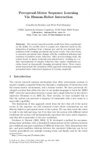
Patients were scanned using magnetic resonance imaging (MRI) before and after surgery, in order to evaluate the damage in the posterior fossa caused by the tumor and its removal. Every MRI was independently evaluated for the study by an expert neuroradiologist, except for that of one participant, whose MRI were unavailable due to family relocation (Participant 6). A detailed description of the extent of damage to the cerebellum in the different patients is presented in Table 2. An illustration of the typical localization of the damage can be found in Figure 1. The control group consisted of a sample of 8 healthy children with no neurological history. This group was individually matched to the PFT group according to sex, age, education, intellectual ability according to the Wechsler Intelligence Scale for Children–Revised (WISC–R; Wechsler, 1974; VIQ 5 116, PIQ 5 121), socio-economic back-
ground, and level. All members of both the PFT and the control groups studied in regular schools.
Apparatus and Procedure Testing was preformed using an IBM PC with a Pentium I processor running the MS-DOS operating system. Participants performed the two versions of SRT mentioned above: the motor standard version (SRTm) and the perceptual version (SRTnm), following the methodology used by Vakil (Vakil et al., 2000, 2002). In both tasks, the stimuli consisted of four squares (3.3 3 3.3 cm) arranged adjacent to each other horizontally on the computer screen, with a red light appearing in a sequence in the four squares. In both tasks there were repeated sequence blocks in which the red light positions followed a specific sequence of which the
Fig. 1. Representative coronal MRI slice from a patient with a posterior fossa tumor before (a) and after (b) surgery.
SRT in posterior fossa patients participants were not explicitly informed about (i.e., 2131431241 in the SRT motor task and 1434312413 in the SRT non-motor task). Participants were presented with six blocks with a 1-min rest period between blocks. The first four blocks and the last one (i.e., Blocks 1– 4 and 6) were repeated sequence blocks, while the fifth block was a random block in which the red light positions were pseudorandomly determined. Each sequence was composed of 10 trials, each trial presenting the red light in a different position. Each block was composed of 100 trials (10 repetitions of a 10-trial sequence). In the SRT motor task, participants were asked to press the key corresponding to each light as it appeared in the array. The red light changed positions following the sequence. In this task, the red light moved to the following position only after there was a finger press, therefore the stimuli presentation pace was under the participant’s control. In the SRT non-motor task also, the red light changed positions following the sequence. However, it changed positions continuously. Participants were asked to respond only whenever the red light appeared in the second square from the left (target position) by pressing the space-bar with their dominant hand. Therefore in this task, they followed the sequence visually and not with finger movements, and sequence presentation was paced by the computer. Task order was counter balanced. Keyboard time resolution0 accuracy was 10 ms.
485 included the general mastering of the task as well as the implicit learning of the sequence.
Deterioration in performance when a random block is introduced The difference between the fourth block (repeated sequence block) and the fifth block (random block) reflected the implicit learning of the sequence.
Preservation of learning Similar performance in the fourth block (which was the last repeated sequence block before the random block) and the sixth block (which was again a repeated sequence block) reflected the preservation of the sequence learning after the disruption introduced by the random block.
RESULTS SRTnm RTs Learning There was a gradual improvement in performance within the first four blocks [F(3,42) 5 7.18, p , .001], which did not interact with group (F , 1). Moreover, there was no group main effect (F , 1).
Analysis
Deterioration in performance when a random block is introduced
Separate analyses were run for the mean reaction time (RT) and the sum of errors of each of the tasks. The analyses were carried out using a repeated measures ANOVA with group (PFT, Control) as a between-subjects variable, and block as a within-subjects variable, looking for the following effects:
There was a significant drop in performance [F(1,14) 5 111.60, p , .001], which interacted with group [F(1,14) 5 5.67, p , .05]. Both groups were slower in the random block as compared with the repeated Sequence 1 (see Figure 2a), but this difference was greater for the PFT group. There was no group main effect (F , 1).
General learning
Preservation of learning
The mean RTs in Blocks 1– 4 were expected to become gradually shorter, showing a learning curve. This learning
There was some loss of learning, as the RT in the repeated sequence block after the random block was slower than in
(a)
(b)
Fig. 2. Mean group performance and standard errors of (a) reaction times and (b) accuracy levels, for the PFT and Control groups in the different blocks of the SRTnm task.
486 the repeated sequence block before the random one [F(1,14) 5 10.83, p , .01]. This pattern was not significantly different between the groups (F , 1).
SRTnm Errors The only effect found in the analysis of errors was in the deterioration in performance when a random block is introduced: More errors were made in the random block [F(1,14) 5 6.31, p , .05]. The group main effect and the interaction between Group 3 Block were both marginal [F(1,14) 5 3.27, p , .09; and F(1,14) 5 3.55, p , .08, respectively], meaning both groups made more errors in the random block as compared with the repeated sequence block, with a trend suggesting that the PFT group did worse than the control group, especially in the random block (see Figure 2b).
SRTm RTs Learning There was a marginal improvement in performance within the first four blocks [F(3,42) 5 2.50, p , .0723], which did not interact with group (F , 1). Moreover, there was no group main effect (F , 1).
Deterioration in performance when a random block is introduced For both groups there was a significant drop in performance [F(1,14) 5 18.20, p , .001], which interacted with group [F(1,14) 5 5.67, p , .05]. There was no group main effect ( p 5 .3124), and no interaction between Group 3 Block ( p 5 .2562).
Preservation of learning None of the effects reached significance and therefore no loss of learning was found after the random sequence in any of the groups.
SRTm Errors None of the effects reached significance.
DISCUSSION Our study is the first report regarding implicit sequence learning in children following resection of a posterior fossa tumor. The results show that the implicit learning of a sequence by the PFT patients was like that of the control group, since they learned the sequence at the same rate as controls. No speed–accuracy trade-off was observed. Patients were not slower than controls and their retention of the learning was the same as controls. The preservation of the learning was also similar between the groups. This is consistent with imaging evidence indicating that there is no involvement of the cerebellum in the learning phase of a
A. Berger et al. sequence (Seidler et al., 2002), and evidence that children with cerebellar dysmorphology do not exhibit implicit learning problems (Colvin et al., 2003). However, our results markedly contradict what has been reported about patients suffering from cerebellar degeneration (Pascual-Leone et al., 1993), for whom the learning curve is flat. There are several possible explanations for this discrepancy: (1) cerebellar degeneration is not restricted to the cerebellum itself but spreads to afferent connections and pathways essential for sequence learning; (2) when the cerebellar lesion is focal and occurs at a young age, plasticity processes can provide alternative pathways to the cerebellar circuitry required for sequence learning; and, (3) the relatively small amount of participant training in our study may have limited the degree of cerebellar involvement. The length of our experiment was limited due to considerations of the overall length of our assessment battery and previous reports by Vakil et al. (2000, 2002) that even such small scale SRT brings out differences between patients with brain lesions and controls. We can not rule out the possibility that there was some explicit learning involved, since our paradigm did not include a check for this kind of learning. However, we doubt this possibility in light of previous studies that employed exactly the same task, such as Vakil et al. (2000, 2002). In these studies, the performance of the control group in the “generate” phase was about chance level. In our study we did not systematically ask our participants to generate the sequence after the sixth block. Informal conversation with them after the testing indicated that they noticed that there was “some kind of sequence,” however, they were unable to reproduce it. The performance of the PFT group was not identical to that of the controls in all aspects. Their impairment arose when the sequence was changed to random. In this case, the performance of the patients was much more disrupted than the performance of the controls. They became markedly slower and also made more errors. This pattern was observed for each participant within the PFT group. Their pattern of results might be interpreted as reflecting over-strictness or rigidity, that is, over-commitment to the task currently performed. This would suggest that they compensate by overengaging the task, a strategy that seems to succeed in bringing performance to a normal level. Overall, the PFT patient group seems to have overcome their brain lesions, developing normal levels for the majority of cognitive abilities. Still, a sequela of their cerebellar injury seems to be elicited when confronting rapid changes, such a change in the sequence. In this case, they seem to have difficulty disengaging from an established response set and therefore, tend to carry out previously learned responses. This is one plausible interpretation of our results. It is compatible with the fact that their deficit arose only in the perceptual SRT and not in the motor SRT, since these tasks differ in the stimuli pace, which is under the participant’s control in the motor version but out of his hands in the perceptual version. Moreover, it is compatible with the fact that their performance in
SRT in posterior fossa patients the last block, which returns to the predictable sequence, was comparable to normals. In addition, this interpretation is strengthened by an additional study with the same patients that yielded similar results (Berger et al., in press). In that study, seven of the patients were tested with computerized task switching. In that paradigm, again, results indicated normal learning of the task and a deficit when rapid behavioral adjustment was required: the PFT group had an enhanced switching cost when the preparation time was short, but did not differ from controls when the preparation time was long.
REFERENCES Aarsen, F.K., Van Dongen, H.R., Paquier, P.F., Van Mourik, M., & Catsman-Berrevoets, C.E. (2004). Long-term sequelae in children after cerebellar astrocytoma surgery. Neurology, 62, 1311–1316. Berger, A., Sadeh, M., Tzur, G., Shuper, A., Kornreich, L., Inbar, D., Cohen, I.J., Michowiz, Sh., Yaniv, I., Constantini, Sh., Kessler, Y., & Meiran, N. (2005). Task switching after cerebellar damage. Neuropsychology. In press. Colvin, A.N., Yeates, K.O., Enrile, B.G., & Coury, D.L. (2003). Motor adaptation in children with myelomeningocele: Comparison to children with ADHD and healthy siblings. Journal of the International Neuropsychological Society, 9, 642– 652. Doyon, J. (1997). Skill learning. In R. Bradley, R. Harris & P. Jenner (Series Eds.) & J.D. Schamahmann (Vol. Ed.), International review of neurobiology: Vol. 41, The cerebellum and cognition (pp. 273–294). San Diego, CA: Academic Press. Doyon, J., Owen, A.M., Petrides, M., Szkilas, V., & Evans, A.C. (1996). Functional anatomy of visuomotor skill learning in human participants examined with positron emission tomography. European Journal of Neuroscience, 8, 637– 48. Friston, K.J., Frith, C.D., Passingham, R.E., Liddle, P.F., & Frackowiak, R.S.J. (1992). Motor practice and neuropsychological adaptation in the cerebellum: A positron tomography study. Proceedings of the Royal Society of London. Series B Biological Sciences, 248, 223–228. Grafton, S.T., Hazeltine, E., & Ivry, R.E. (1995). Functional mapping of sequence learning in normal humans. Journal of Cognitive Neuroscience, 7, 497–510. Grill, J., Renaux, V.K., Bulteau, C., Viguier, D., Levy-Piebois, C., Sainte-Rose, C., Dellatolas, G., Raquin, M.A., Jambaque, I., & Kalifa, C. (1999). Long-term intellectual outcome in children with posterior fossa tumors according to radiation doses and volumes. International Journal of Radiation, Oncology, Biology, Physics, 45, 137–145. Hallet, M. & Grafman, J. (1997). Executive function and motor skill learning. In R. Bradley, R. Harris, & P. Jenner (Series Eds.) & J.D. Schamahmann (Vol. Ed.), International Review of Neurobiology: Vol. 41, The cerebellum and cognition (pp. 297–323). San Diego, CA: Academic Press.
487 Hetherington, R., Dennis, M., & Spiegler, B. (2000). Perception and estimation of time in long-term survivors of childhood posterior fossa tumors. Journal of the International Neuropsychological Society, 6, 682– 692. Laforce, R. & Doyon, J. (2001). Distinct contribution of the striatum and cerebellum to motor learning. Brain and Cognition, 45, 189–211. Levisohn, L., Cronin-Golomb, A., & Schamahmann, J.D. (2000). Neuropsychological consequences of cerebellar tumor resection in children: Cerebellar cognitive affective syndrome in a paediatric population. Brain, 123, 1041–1050. Nissen, M.J. & Bullemer, P. (1987). Attentional requirements of learning: Evidence from performance measures. Cognitive Psychology, 19, 1–32. Pascual-Leone, A., Grafman, J., Clark, K., Stewart, M., Massaqoui, S., Lou, J.S., & Hallett, M. (1993). Procedural learning in Parkinson’s disease and cerebellar degeneration. Annals of Neurology, 34, 594– 602. Pollack, I.F. (1997). Posterior fossa syndrome. In R. Bradley, R. Harris, & P. Jenner (Series Eds.) & J.D. Schamahmann (Vol. Ed.), International Review of Neurobiology: Vol 41, The cerebellum and cognition (pp. 411– 432). San Diego, CA: Academic Press. Sadeh, M., Berger, A., Tzur, G., Shuper, A., Kornreich, L., Constantini, S., Friedman, O., Michowiz, S., Cohen, I.J., Yaniv, I., & Inbar, D. (2005). Overcoming early cerebellar injury: Neuropsychological long-term sequela of posterior fossa tumors in childhood. Manuscript submitted for publication. Seidler, R.D., Purushotham, A., Kim, S.G., Ugurbil, K., Willigham, D., & Ashe, J. (2002). Cerebellum activation associated with performance change but not motor learning. Science, 296, 2043–2046. Seitz, R.J., Roland, E., Bohm, C., Greitz, T., & Stone-Elander, S. (1990). Motor learning in man: A positron emission tomographic study. NeuroReport, 1, 57– 60. Squire, L.R. (1992). Declarative and nondeclarative memory: Multiple brain systems supporting learning and memory. Journal of Cognitive Neuroscience, 4, 232–243. Steinlin, M., Imfeld, S., Zulauf, P., Boltshauser, E., Lovblad, K.-O., Luthy, A.R., Perrig, W., & Kaufmann, F. (2003). Neuropsychological long-term sequelae after posterior fossa tumor resection during childhood. Brain, 126, 1998–2008. Vakil, E., Kahan, S., Huberman, M., & Osimani. A. (2000). Motor and non-motor sequence learning in patients with basal ganglia lesions: The case of serial reaction time (SRT). Neuropsychologia, 38, 1–10. Vakil, E., Kraus, A., Bor, B., & Groswasser, Z. (2002). Impaired skill learning in patients with severe close-head injury as demonstrated by the serial reaction time (SRT) task. Brain and Cognition, 50, 304–315. Wechsler, D. (1974). Manual for the Wechsler Intelligence Scale for Children–Revised. New York: The Psychological Corporation.