SUBMITTED TO IEEE JBHI SPECIAL ISSUE ON SKIN LESION IMAGE ANALYSIS FOR MELANOMA DETECTION
1
Multi-class Semantic Segmentation of Skin Lesions via Fully Convolutional Networks
arXiv:1711.10449v1 [cs.CV] 28 Nov 2017
Manu Goyal, Student Member, IEEE, and Moi Hoon Yap, Member, IEEE,
Abstract—Early detection of skin cancer, particularly melanoma, is crucial to enable advanced treatment. Due to the rapid growth of skin cancers, there is a growing need of computerized analysis for skin lesions. These processes including detection, classification, and segmentation. There are three main types of skin lesions in common that are benign nevi, melanoma, and seborrhoeic keratoses which have huge intra-class variations in terms of color, size, place and appearance for each class and high inter-class visual similarities in dermoscopic images. The majority of current research is focusing on melanoma segmentation, but it is also very important to segment the seborrhoeic keratoses and benign nevi lesions as these regions potentially indicate the pre-cancer stage. We propose a multiclass semantic segmentation for these three classes from publicly available ISBI-2017 challenge dataset which consists of 2750 dermoscopic images. We propose an end-to-end solution using fully convolutional networks (FCNs) for multi-class semantic segmentation, which will automatically segment the melanoma, keratoses and benign lesions. To overcome the issue of data deficiency, we propose a transfer learning approach which uses both partial transfer learning and full transfer learning to train FCNs for multi-class semantic segmentation of skin lesions. The results are presented in Dice Similarity Coefficient (Dice) to compare the performance of the deep learning segmentation methods on the dataset with 5-fold cross-validation. The results showed that the two-tier level transfer learning FCN-8s achieved the overall best result with Dice score of 0.785 in a benign category, 0.653 in melanoma segmentation, and 0.557 in seborrhoeic keratoses. Index Terms—Skin lesion, Melanoma, Fully convolutional networks, Transfer learning, Semantic segmentation
I. I NTRODUCTION KIN cancer is the most common cancer among all other cancers [1]. The malignant skin lesions consist of the melanocytic lesion, i.e. melanoma, and non-melanocytic lesion, i.e. basal cell carcinoma. Although melanoma is the least common type of skin cancer, it is the most aggressive and deadly cancer [2]. In addition, it has the high capacity to invade tissues and other organs (cite). Hence, it is important to have early detection to save a life. According to the prediction of Melanoma Foundation [3], the estimated new cases of melanoma in the United States is 87,110 (200% increased since 1973) with 9,730 predicted deaths. In current medical practice, skin cancer specialists primarily examine the patients on visual inspection with manual measurements tools called dermoscopy assessment to determine
S
M. Goyal and M.H. Yap are with the School of Computing, Mathematics and Digital Technology, Manchester Metropolitan University, Manchester, UK. E-mail:
[email protected] Manuscript received xxxxx xx, 2017; revised xxx xx, 201x.
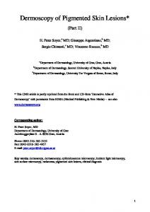
the skin lesions. Relying on self-vigilance and medical inspection by human vision risk life and survival rate as it is difficult to identify the type of lesions by naked eyes. Hence, over the years, different image modalities were used to inspect the skin, including macroscopy (clinical) and microscopy (also known as Dermoscopy). Dermoscopy is a non-invasive imaging that allows visualisation of skin surface by the light magnifying device and immersion fluid [4]. It is one of the most widely used imaging techniques in dermatology and it has increased the diagnosis rate [5]. In dermoscopic imagery, melanoma starts as minuscule and mole-like [1], with a gradual change in size and color. Like other cancer malignancies, the appearance of melanoma is Irregular diffuse pigmentation as illustrated in Figure 1(a), compared to benign, more regular pigment as illustrated in Figure 1(b). Braun et al. [6] studied the morphology of keratoses using dermoscopic imaging, where they described that the common characteristics are comedolike openings and milialike cyst(round, whitish and yellow), as illustrated in Figure 1(c). The majority of the state-of-the-art computer-aided diagnosis on dermoscopy images composed of multi-stages, which include image pre-processing, image segmentation, features extraction and classification [7], [1]. Using hand-crafted feature descriptors, the dermatologists are able to differentiate benign lesions based on their shape features as they normally have small dimensions and more circular, as illustrated in Figure 1(b). Other feature descriptors used in previous works including asymmetry features, color features and texture features. Pattern analysis was widely used to describe the appearance of skin lesions, this including the melanocytic algorithm elaborated by Argenziano et al. [8]. Using the features descriptors and pattern analysis, machine learning algorithms were used to classify the lesions types. There are many research in developing computerized methods based on image processing and conventional machine learning approaches. These research were presented in two survey papers where the majority of the approaches were using hand-crafted features to classify or segment the lesions, the earlier review was in 2012 reviewed by Korotkov et al. [7] and the later was conducted by Pathan et al. [1]. Korotkov et al. [7] concluded that there is a large discrepancy in previous research and the computer-aided diagnosis systems were not ready for implementation. The other issue was the lack of benchmark dataset which makes it harder to assess the algorithms. Pathan et al. [1] concluded that the CAD systems worked for experimental settings but subject to rigorous validation in real-world clinical settings. With the rapid growth of deep learning approaches, many researchers [9], [10], [11] have proposed Deep Convolutional
SUBMITTED TO IEEE JBHI SPECIAL ISSUE ON SKIN LESION IMAGE ANALYSIS FOR MELANOMA DETECTION
(a)
(b)
(c)
Fig. 1. Courtesy images from ISBI 2017 Challenge. Original images without artifacts (first row) and Original images with artifacts (second row). The skin lesion diagnosis from left to right: (a) melanoma, (b) benign and (c) keratoses.
Neural Networks for melanoma detection and segmentation. However, there is no previous research for multi-class semantic segmentation to detect all types of skin cancer lesions. The idea of big data and data science is to encourage data sharing and development of data-driven approaches to better diagnostics in clinical applications, this will improve the performance of learning algorithms. Ackerman and Mones [12] claimed that solar keratoses is malignant lesion (squamous cell carcinoma (SCC)), whereas Fuchs and Ellen [13] defined it as with potential to progress to SCC. These research findings show the importance of monitoring keratoses lesions. To help the dermatologists to evaluate and understand the development of keratoses, we have included keratoses as one of the classes in our semantic segmentation. Previous work [9], [10], [11] has combined keratoses with other benign lesions as non-melanoma classes and working on two-class semantic segmentation. The contributions of this paper are: 1) To provide a review on current state-of-the-art deep learning approaches for skin cancer lesion and melanoma segmentation. 2) To propose a multi-class semantic segmentation for melanoma, keratoses and benign lesions on the ISBI2017 challenge dataset rather than using only one class for segmentation task. 3) To review the performance of state-of-the-art deep learning algorithms on this proposed multi-class segmentation task given that there is high visual intra-class in each class and inter-class similarities between these skin lesion classes. 4) To use both types of transfer learning called two-tier transfer learning to overcome the data deficiency to train the FCNs. II. D EEP L EARNING FOR M ELANOMA S EGMENTATION Deep learning has gained popularity in medical imaging research including Magnetic Resonance Imaging (MRI) on brain [14], breast ultrasound cancer detection [15] and diabetic foot ulcer segmentation [16]. A popular deep learning approach in biomedical imaging research is U-Net, proposed by Ronneberger et al. [17]. U-Net enables the use of data
2
augmentation, including the use of non-rigid deformations, to make full use of the available annotated sample images to train the model. These aspects suggest that the U-Net could potentially provide satisfactory results with the limited size of the biomedical datasets currently available. An upto-date review of conventional machine learning methods is presented in [1]. This section reviews the state-of-the-art deep learning approaches for segmentation for skin lesions. To date the majority of existing research focuses on skin lesions segmentation, but not on semantic segmentation. Yu et al. [10] proposed very deep residual networks of more than 50 layers for two-stage framework of skin lesions segmentation followed by classification. They claimed that the deeper networks produce richer and more discriminative features for recognition. By validating their methods on ISBI 2016 Skin Lesion Analysis Towards Melanoma Detection Challenge dataset [18], they reported that their method ranked first in classification when compared to 16-layer VGG-16, 22-layer GoogleNet and other 25 teams in the competition. However, in segmentation stage, they ranked second in segmentation among the 28 teams. Although the work showed promising results, but the two-stage framework and very deep networks are computationally expensive. Bi et al. [11] proposed a multi-stage fully convolutional networks (FCNs)for skin lesions segmentation. The multistage involved localised coarse appearance learning in the early stage and detailed boundaries characteristics learning in the later stage. Further, they implemented a parallel integration approach to enable fusion of the result that they claimed that this has enhanced the detection. Their method outperformed others in PH2 dataset [19] of 90.66% but achieved marginal improvement if compared to Team ExB in ISIB 2016 competition with 91.18%. Yuan et al. [9] proposed an end-to-end fully automatic method for skin lesions segmentation by leveraging 19-layer DCNN. They introduced a loss function using Jaccard Distance as the measurement. They compared the results using different parameters such as input size, optimisation methods, augmented strategies, and loss function. To fine tune the hyperparameters, 5-fold cross-validation with ISBI training dataset was used to determine the best performer. Similar to Bi et al. [11], they evaluated their results on ISBI 2016 and PH2 dataset. The results were outperformed the state-of-the-art methods but they suggested that the method achieved poor results in some challenging cases including images with low contrast. The research showed that deep learning achieved promising results for skin lesions segmentation and classification. However, these methods did not make their codes available and not validated on the ISBI 2017 dataset, which has 2000 images compared to 900 in ISBI 2016 dataset. In addition, there is no existing work in semantic segmentation for the benign lesion, keratoses lesion, and melanoma lesion. Even though there are datasets available for skin lesions, but the scale of combined datasets is relatively smaller than the general field, e.g. ImageNet [20]. To overcome the data deficiency in skin lesions segmentation and to enable fully automated of skin lesions segmentation, we propose a transfer learning approach
SUBMITTED TO IEEE JBHI SPECIAL ISSUE ON SKIN LESION IMAGE ANALYSIS FOR MELANOMA DETECTION
3
TABLE I C LASSIFICATION OF SKIN LESIONS IN ISBI CHALLENGE DATASET
Training Data
Benign Nevi
Melanoma
Seborrhoeic Keratoses
Total
1372
374
254
2000
Validation Data
78
30
42
150
Test Data
393
117
90
600
Total
1843
521
386
2750
(a)
(b)
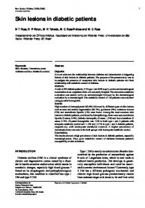
Fig. 2. The sample input RGB image used in PASCAL-VOC 2012 dataset and its corresponding labeling in PASCAL-VOC format.
for multi-class semantic segmentation. The following section detailed our proposed method. III. M ETHODOLOGY This section discusses the ISBI skin cancer dataset, the preparation of the ground truth labeling, the two-tier transfer learning approach and the type of performance metrics to validate our segmentation results. A. Datasets We solely used publicly available ISBI 2017 skin cancer challenge dataset for training the fully convolutional deep learning models. RGB colorspace is used to represent all the images in this dataset. It includes 3 important skin lesion classes on the dermoscopy images that are benign nevi, melanoma and seborrhoeic keratoses. The segmentation task on these dermoscopy images based on these 3 classes is very challenging due to high inter-class similarity between these 3 classes. This challenge dataset consists of training set of 2000 dermoscopy images in which 1372 dermoscopy images are classified as benign nevi, 374 as melanoma and 274 as seborrhoeic keratoses. There are 150 images in validation set and 600 images in the test set. The classification of the whole dataset of 2750 images is shown in Table I. In this dataset, the size of images vary between 540 × 722 and 4499 × 6748. For the training of FCNs, we resized all the images to 500 × 375 to improve the performance and reduce the computational costs. In the competition, the segmentation challenge was only lesion boundaries from dermoscopic images. Hence, there was only one class for the segmentation task. Whereas, in our segmentation task, we divided the whole dataset on the basis of 3 classes i.e. benign nevi, melanoma and seborrhoeic keratoses. B. Ground Truth Format Since, the performance of convolutional neural networks for semantic segmentation are widely tested for the PASCALVOC 2012 dataset [21], [22]. In this dataset, the input images are all defined in RGB colorspace and 8-bit paletted images are used for representing the ground truth for input images i.e. a sample input image showing the person riding motorbike with ground truth in Figure 2. The ISBI dermoscopy dataset has input images in the same format i.e. RGB colorspace. As mentioned above, the segmentation challenge was only for one class, hence binary mask was used for representing the ground truth as illustrated in Figure 3. The Pascal VOC
(a)
(b)
Fig. 3. The sample input RGB image and binary mask used in original ISBI2017 segmentation challenge
dataset has originally 21 classes where as in our task, we have 3 classes to represent benign nevi, melanoma and seborrhoeic keratoses. Figure 4 illustrates the dermoscopic images with the corresponding ground truth labeling in PASCAL-VOC format, with index 1 indicates benign, index 2 indicates melanoma and index 3 represents seborrhoeic keratoses. We did not use the index 255 to represent the boundary of object as this index is ignored by the deep learning methods.
(a)
(b)
(c)
Fig. 4. Original images (first row) and PASCAL-VOC format (second row). The skin lesion diagnosis from left to right: (a) benign, (b) melanoma and (c) seborrhoeic keratoses.
C. Fully Convolutional Networks for Skin Cancer Segmentation The convolutional neural networks proved to be breakthrough in the computer vision tasks and relatively match the human accuracy. These networks are very computational intense to extract the hierarchies to features to detect the objects in images. The state-of-the-art CNN networks are trained on huge datasets and used to classify the number of classes of different objects by providing the score for each class.
SUBMITTED TO IEEE JBHI SPECIAL ISSUE ON SKIN LESION IMAGE ANALYSIS FOR MELANOMA DETECTION
The problem with these networks, they actually have single prediction for each image. If there are multiple objects in single image, these networks detect the most prominent object in an image. The advanced convolutional neural networks such as FCNs and encoder-decoder CNNs which can detect the multiple objects as well localize the objects by using pixelwise prediction. The pixel-wise prediction enables to learn which pixel of an image belongs to which class of object. Recently, the FCNs have become the state-of-the-art methods to do the segmentation tasks for both non-medical and medical imaging and proved their superiority over the conventional machine learning and other deep learning methods. We used the four different variants of FCNs that are FCN-AlexNet, FCN-32s, FCN-16s, and FCN-8s to perform the skin cancer segmentation task. The first variant FCN-AlexNet is a modified version of original state-of-the-art classification model called AlexNet which won ImageNet ILSVRC-2012 competition in classification category [23], [24]. The FCN-AlexNet enables the pixelwise prediction by using the deconvolutional layers which upsample the features learned by the earlier convolutional layers. We have trained the FCN-AlexNet on the Caffe deep learning framework [25]. The input and ground truth images are both 500×375. We have fine-tuned the network parameters to allow the method more time to learn the features from dermoscopy images by using 100 epochs, stochastic gradient descent with a learning rate of 0.0001. The other FCNs variant FCN-32s, FCN-16s and FCN-8s are based on the another state-of-the-art classification network called which won localization challenge and made second position in classification challenge in the ImageNet ILSVRC2014 competition [26], [23]. The difference between these models are the up-sampling layers with different pixel stride. As the name suggested by these FCNs variants, in FCN-32s, up-sampling is performed with the help of 32-pixel stride where as 16-pixel stride for FCN-16s and 8-pixel stride for FCN-8s. With the small pixel stride, the models were able to predict finer-grained analysis of the objects. Similarly, the same network parameters are used as of FCN-AlexNet to train these models. D. The Two-tier Transfer Learning Approach The convolutional neural networks generally requires a huge dataset to learn the features to get the positive results for detection of objects in images [27]. Since, we have RGB images in dermoscopic images, it is good to use two-tier transfer learning from huge datasets in non-medical backgrounds such as ImageNet and Pascal-VOC dataset to converge the weights associated with each convolutional layers of networks [28], [22], [16]. The main reason for using two-tier transfer learning is because, the medical imaging datasets are very limited, hence, when convolutional neural networks are trained from the scratch on these datasets, they don’t produce effective results due to non-convergence of weights associated with each convolutional layer with limited medical imaging datasets. When we use deep learning in medical imaging, there is a big concern regarding the size of the datasets [29].
4
Hence, when we train convolutional neural networks on these limited medical datasets, it is very important to use transfer learning by training models on huge non-medical datasets to produce better results. The transfer learning transfers the feature learned by previous models on huge non-medical datasets to medical image datasets. There are two types of transfer learning i.e. partial transfer learning in which only the features from few convolutional layers are transferred and full transfer learning in which features are transferred from all the layers of previous pre-trained models. In two-tier transfer learning, we have used both types of transfer learning, as in first tier, we have used the partial transfer learning by transferring the features only from the convolutional layers trained on huge classification dataset called ImageNet which consists of more than 1.5 million images with 1000 classes and full transfer learning to transfer the features from model trained on semantic segmentation dataset called Pascal-VOC that consists of more than 2000 images with 21 classes. Hence, we used the two-tier transfer learning technique to use the pretrained model for all corresponding FCNs in our skin cancer segmentation task in Fig. 5. E. Performance Metrics In medical imaging, Sensitivity and Specificity are the standard evaluation metrics and where as for segmentation evaluation, Dice Similarity Coefficient (Dice) is popularly used by researchers [30], [31]. We report our findings in Dice, Sensitivity, Specificity, and Matthew Correlation Coefficient (MCC) [32] as our evaluation metrics for segmentation. Sensitivity =
TP TP + FN
(1)
Specif icity =
TN FP + TN
(2)
Dice =
M CC = p
2 ∗ TP (2 ∗ T P + F P + F N )
(3)
TP ∗ TN − FP ∗ FN (T P + F P )(T P + F N )(T N + F P )(T N + F N ) (4)
Sensitivity is defined in eq (1), where TP is True Positives and FN is False Negatives. A high Sensitivity (close to 1.0) indicates good performance in segmentation which implies all the lesions were segmented successfully. On the other hand, Specificity (as in eq. (2)) indicates the proportion of True Negatives (TN) of the non-lesions. A high Specificity indicates the capability of a method in not segmenting the non-lesions. Dice Similarity Index (Dice) is a measure of how similar both prediction and ground truth are, by measuring of how many TP found and penalising for the FP that the method found, as in eq. (3). MCC has a range of -1 (completely wrong binary classifier) to 1 (completely right binary classifier). This is a suitable measurement for the performance assessment of our segmentation algorithms based on binary classification (lesion versus non-lesions), as in eq. (4).
SUBMITTED TO IEEE JBHI SPECIAL ISSUE ON SKIN LESION IMAGE ANALYSIS FOR MELANOMA DETECTION
5
Fig. 5. The two-tier transfer learning from big datasets to produce more effective segmentation [16]
TABLE II D ISTRUBUTION OF I MAGES FOR SEGMENTATION TASK
Training Set
Benign Nevi
Melanoma
Seborrhoeic Keratoses
Total
1290
365
271
1926
Validation Data
184
52
104
275
Test Data
369
104
76
549
Total
1843
521
386
2750
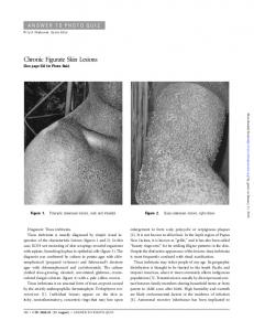
IV. R ESULT AND DISCUSSION There are total 2750 dermoscopy images in the ISBI challenge dataset. For 5-fold validation test, we have divided the images with 70% data in training set, 10% in validation and 20% in test according to the Table II. A. Quantitative Performance Measure We tested four state-of-the-art fully convolutional networks for our proposed segmentation task as described above. We train the models with input-size of 500×375 using stochastic gradient descent with a learning rate of 0.0001, 60 epochs with a dropout rate of 33%. In Table III, we report Dice Similarity Coefficient (Dice), Sensitivity, Specificity, Matthews Correlation Coefficient (MCC) as metrics for performance evaluation of multi-class segmentation of skin cancer lesions. In performance measure for multi-class segmentation, FCNs performed best in the class of benign nevi, that’s because, we have more data of this class in the dataset available rather than melanoma and seborrhoeic keratoses. Due to high intraclass and inter-class visual similarities, performance for both
classes, melanoma and seborrhoeic keratoses suffer due to less available data in the dataset. The total melanoma images are only approx. 37% and keratoses images are just approx. 22% of total benign images in the dataset. In Table III, we report the performance evaluation of fully convolutional networks for multi-class segmentation on 5-fold cross validation data. In benign class category, all FCNs achieved good segmentation results, but FCN-AlexNet achieved the best results with Dice score of 0.819, MCC score of 0.814, and Senstivity is 0.798. In this category, FCN-8s performed 2nd best with Dice score of 0.779 and MCC score of 0.779. In melanoma and keratoses category, FCN-8s has performed best with Dice score of 0.653 and 0.557 respectively and also, in terms of Senstivity, Specificity and MCC. With few data available in these both categories, due to finer grained analysis of the objects, the FCN-8s performed best among all other FCN-8s. Overall, the FCN-8s and FCN-Alexnet worked well for our proposed multi-class segmentation. B. Visual Comparison In Fig. 6, we reported the accurate results predicated by every FCN models score in each category whereas in Fig. 7, we reported the inaccurate results. Due to visual similarities, there are many overlaps between the classes of skin lesion from Fig. 7, row 1 and row 3 particularly show there is overlap between benign and keratoses. V. C ONCLUSION In this work, we propose multi-class semantic segmentation to segment the melanoma, benign and seborrhoeic keratoses in the combined dataset. It is a very challenging segmentation
SUBMITTED TO IEEE JBHI SPECIAL ISSUE ON SKIN LESION IMAGE ANALYSIS FOR MELANOMA DETECTION
6
TABLE III C OMPARISON OF DIFFERENT FCN S ARCHITECTURES ON S KIN C ANCER DATASET (SK DENOTES S EBORRHOEIC K ERATOSES ) Dice
Method
Specificity
Sensitivity
MCC
Benign
Melanoma
SK
Benign
Melanoma
SK
Benign
Melanoma
SK
Benign
Melanoma
SK
FCN-AlexNet
0.819
0.609
0.488
0.989
0.982
0.987
0.798
0.4864
0.456
0.814
0.541
0.484
FCN-32s
0.779
0.549
0.484
0.991
0.977
0.968
0.751
0.430
0.478
0.775
0.484
0.463
FCN-16s
0.761
0.590
0.506
0.988
0.979
0.978
0.706
0.471
0.466
0.764
0.528
0.501
FCN-8s
0.785
0.653
0.557
0.990
0.984
0.988
0.747
0.527
0.509
0.779
0.582
0.5683
Ground truth
FCN-AlexNet
FCN-32s
FCN-16s
FCN8s
Fig. 6. The segmentation results to visually compare the performance of FCNs on multi-class segmentation. First row is benign, Second row is melanoma and Third and last row is Keratoses The first column is the ground truth delineation, the second column is the results of FCN-AlexNet, the third column is the FCN-32s, the fourth column is the FCN-16s and the last column is the FCN-8s.
task as there are high intra-class and inter-class similarities in terms of visual, size, color and appearance in these classes. There was only one class used in the recent literature on the skin lesion segmentation. For computer vision algorithms, it is comparatively easy to segment one class of skin cancer lesions from the normal healthy skin to predict the segmentation for one class. But, when it comes to differentiating the different skin lesions, it stands a major challenge to achieve good multi-class segmentation results for each category. We tested this segmentation problem with state-of-the-art segmentation algorithms known as fully convolutional networks. We get the best results for benign category and good results in melanoma and keratoses categories. It is mainly because of unbalanced data of each category in the dataset. With the more data especially for melanoma and keratoses categories, can further improve the performance of fully convolutional networks.
[2]
[3]
[4]
[5]
[6]
[7]
R EFERENCES
[8]
[1] S. Pathan, K. G. Prabhu, and P. Siddalingaswamy, “Techniques and algorithms for computer aided diagnosis of pigmented skin lesionsa
[9]
review,” Biomedical Signal Processing and Control, vol. 39, pp. 237– 262, 2018. National Cancer Institute, “Cancer stat facts: Melanoma of the skin,” 2017, last access: 26/10/17. [Online]. Available: https://seer.cancer.gov/ statfacts/html/melan.html Melanoma Foundation (AIM), “Melanoma stats, facts and figures,” 2017, last access: 27/10/2017. [Online]. Available: https://www. aimatmelanoma.org/about-melanoma/melanoma-stats-facts-and-figures/ G. Pellacani and S. Seidenari, “Comparison between morphological parameters in pigmented skin lesion images acquired by means of epiluminescence surface microscopy and polarized-light videomicroscopy,” Clinics in dermatology, vol. 20, no. 3, pp. 222–227, 2002. J. Mayer, “Systematic review of the diagnostic accuracy of dermatoscopy in detecting malignant melanoma.” The Medical Journal of Australia, vol. 167, no. 4, pp. 206–210, 1997. R. P. Braun, H. S. Rabinovitz, J. Krischer, J. Kreusch, M. Oliviero, L. Naldi, A. W. Kopf, and J. H. Saurat, “Dermoscopy of pigmented seborrheic keratosis: a morphological study,” Archives of dermatology, vol. 138, no. 12, pp. 1556–1560, 2002. K. Korotkov and R. Garcia, “Computerized analysis of pigmented skin lesions: a review,” Artificial intelligence in medicine, vol. 56, no. 2, pp. 69–90, 2012. G. Argenziano, H. P. Soyer, V. De Giorgio, D. Piccolo, P. Carli, M. Delfino, A. Ferrari, R. Hofmann-Wellenhof, D. Massi, G. Mazzocchetti et al., “Interactive atlas of dermoscopy,” 2000. Y. Yuan, M. Chao, and Y.-C. Lo, “Automatic skin lesion segmentation
SUBMITTED TO IEEE JBHI SPECIAL ISSUE ON SKIN LESION IMAGE ANALYSIS FOR MELANOMA DETECTION
Ground truth
FCN-AlexNet
FCN-32s
FCN-16s
7
FCN8s
Fig. 7. The inaacurate segmentation results to visually compare the performance of FCNs on multi-class segmentation. First row is benign, Second row is melanoma and Third and last row is Keratoses The first column is the ground truth delineation, the second column is the results of FCN-AlexNet, the third column is the FCN-32s, the fourth column is the FCN-16s and the last column is the FCN-8s.
[10]
[11]
[12]
[13]
[14]
[15]
[16]
[17]
[18]
[19]
using deep fully convolutional networks with jaccard distance,” IEEE Transactions on Medical Imaging, 2017. L. Yu, H. Chen, Q. Dou, J. Qin, and P.-A. Heng, “Automated melanoma recognition in dermoscopy images via very deep residual networks,” IEEE transactions on medical imaging, vol. 36, no. 4, pp. 994–1004, 2017. L. Bi, J. Kim, E. Ahn, A. Kumar, M. Fulham, and D. Feng, “Dermoscopic image segmentation via multi-stage fully convolutional networks,” IEEE Transactions on Biomedical Engineering, 2017. A. Ackerman and J. Mones, “Solar (actinic) keratosis is squamous cell carcinoma,” British Journal of Dermatology, vol. 155, no. 1, pp. 9–22, 2006. A. Fuchs and E. Marmur, “The kinetics of skin cancer: progression of actinic keratosis to squamous cell carcinoma,” Dermatologic Surgery, vol. 33, no. 9, pp. 1099–1101, 2007. W. Zhang, R. Li, H. Deng, L. Wang, W. Lin, S. Ji, and D. Shen, “Deep convolutional neural networks for multi-modality isointense infant brain image segmentation,” NeuroImage, vol. 108, pp. 214–224, 2015. M. H. Yap, G. Pons, J. Mart´ı, S. Ganau, M. Sent´ıs, R. Zwiggelaar, A. K. Davison, and R. Mart´ı, “Automated breast ultrasound lesions detection using convolutional neural networks,” IEEE Journal of Biomedical and Health Informatics, 2017. M. Goyal, N. D. Reeves, S. Rajbhandari, J. Spragg, and M. H. Yap, “Fully convolutional networks for diabetic foot ulcer segmentation,” arXiv preprint arXiv:1708.01928, 2017. O. Ronneberger, P. Fischer, and T. Brox, “U-net: Convolutional networks for biomedical image segmentation,” in International Conference on Medical Image Computing and Computer-Assisted Intervention. Springer, 2015, pp. 234–241. D. Gutman, N. C. Codella, E. Celebi, B. Helba, M. Marchetti, N. Mishra, and A. Halpern, “Skin lesion analysis toward melanoma detection: A challenge at the international symposium on biomedical imaging (isbi) 2016, hosted by the international skin imaging collaboration (isic),” arXiv preprint arXiv:1605.01397, 2016. T. Mendonc¸a, P. M. Ferreira, J. S. Marques, A. R. Marcal, and J. Rozeira, “Ph 2-a dermoscopic image database for research and benchmarking,” in Engineering in Medicine and Biology Society (EMBC), 2013 35th Annual International Conference of the IEEE. IEEE, 2013, pp. 5437– 5440.
[20] J. Deng, W. Dong, R. Socher, L.-J. Li, K. Li, and L. Fei-Fei, “Imagenet: A large-scale hierarchical image database,” in Computer Vision and Pattern Recognition, 2009. CVPR 2009. IEEE Conference on. IEEE, 2009, pp. 248–255. [21] A. Garcia-Garcia, S. Orts-Escolano, S. Oprea, V. Villena-Martinez, and J. Garcia-Rodriguez, “A review on deep learning techniques applied to semantic segmentation,” arXiv preprint arXiv:1704.06857, 2017. [22] M. Everingham, S. M. A. Eslami, L. Van Gool, C. K. I. Williams, J. Winn, and A. Zisserman, “The pascal visual object classes challenge: A retrospective,” International Journal of Computer Vision, vol. 111, no. 1, pp. 98–136, Jan. 2015. [23] J. Long, E. Shelhamer, and T. Darrell, “Fully convolutional networks for semantic segmentation,” in Proceedings of the IEEE Conference on Computer Vision and Pattern Recognition, 2015, pp. 3431–3440. [24] A. Krizhevsky, I. Sutskever, and G. E. Hinton, “Imagenet classification with deep convolutional neural networks,” in Advances in neural information processing systems, 2012, pp. 1097–1105. [25] Y. Jia, E. Shelhamer, J. Donahue, S. Karayev, J. Long, R. Girshick, S. Guadarrama, and T. Darrell, “Caffe: Convolutional architecture for fast feature embedding,” in Proceedings of the 22nd ACM international conference on Multimedia. ACM, 2014, pp. 675–678. [26] K. Simonyan and A. Zisserman, “Very deep convolutional networks for large-scale image recognition,” arXiv preprint arXiv:1409.1556, 2014. [27] Y. LeCun, Y. Bengio, and G. Hinton, “Deep learning,” Nature, vol. 521, no. 7553, pp. 436–444, 2015. [28] O. Russakovsky, J. Deng, H. Su, J. Krause, S. Satheesh, S. Ma, Z. Huang, A. Karpathy, A. Khosla, M. Bernstein et al., “Imagenet large scale visual recognition challenge,” arXiv preprint arXiv:1409.0575, 2014. [29] A. Van Opbroek, M. A. Ikram, M. W. Vernooij, and M. De Bruijne, “Transfer learning improves supervised image segmentation across imaging protocols,” IEEE transactions on medical imaging, vol. 34, no. 5, pp. 1018–1030, 2015. [30] D. Zikic, Y. Ioannou, M. Brown, and A. Criminisi, “Segmentation of brain tumor tissues with convolutional neural networks,” Proceedings MICCAI-BRATS, pp. 36–39, 2014. [31] S. Pereira, A. Pinto, V. Alves, and C. A. Silva, “Brain tumor segmentation using convolutional neural networks in mri images,” IEEE transactions on medical imaging, vol. 35, no. 5, pp. 1240–1251, 2016.
SUBMITTED TO IEEE JBHI SPECIAL ISSUE ON SKIN LESION IMAGE ANALYSIS FOR MELANOMA DETECTION
[32] D. M. Powers, “Evaluation: from precision, recall and f-measure to roc, informedness, markedness and correlation,” 2011.
8