probe, Lucifer Yellow, into single MNGCs, initially only diffuse fluorescence .... Viable cells were incubated in the presence of either acridine orange. (10 µg/ml ...
Journal of Cell Science 108, 3233-3241 (1995) Printed in Great Britain © The Company of Biologists Limited 1995 JCS9380
3233
Multinucleated cells can continuously generate mononucleated cells in the absence of mitosis: a study of cells of the avian osteoclast lineage F. Solari1, C. Domenget1, V. Gire2, C. Woods3, E. Lazarides3, B. Rousset2 and P. Jurdic1,* 1Département de Biologie Moléculaire et Cellulaire, Ecole Normale Supérieure, 46 Allée d’Italie, 69364 2INSERM U.369, Faculté de Médecine A. Carrel, 8 rue G. Paradin, 69372 Lyon Cedex 08, France 3Alliance Pharmaceutical Corporation, 3040 Science Park Road, San Diego, CA 92121, USA
Lyon Cedex 07, France
*Author for correspondence
SUMMARY The multinucleated bone-resorbing osteoclast has a hematopoietic origin. We have demonstrated previously that osteoclasts are derived from the monocytic lineage by fusion of mononuclear macrophage precursors. Using an in vitro osteoclast differentiation model derived from pure populations of chick macrophage cultures, osteoclast-like multinucleated giant cells (MNGCs) can be formed by fusion following an active proliferation phase. However, after reaching a peak with 70% of the culture being MNGCs, a new round of expansion of the mononuclear cells is observed. The following experiments suggest that these mononuclear cells were derived directly from the MNGCs by a budding process, selectively from the central zone of the apical surface. After microinjection of the membrane-impermeable probe, Lucifer Yellow, into single MNGCs, initially only diffuse fluorescence, limited to the whole MNGC injected, was observed. However, after 24-48 hours fluorescent mononuclear cells were observed adjacent but distinct from the injected MNGC. To confirm that these mononuclear cells were indeed derived from a parent MNGC, single MNGCs were cloned into single wells. Within a week, the MNGC was surrounded by mononuclear cells, which eventually populated the entire well. These mononuclear cells could
then give rise to a second generation of MNGCs following a three-week period of culture. To determine whether this process required mitosis, MNGCs were cultured for three days in the presence of the mitotic inhibitor, Ara-C, prior to microinjection with Lucifer Yellow. Fluorescent mononuclear cells were still seen to arise from a single injected MNGC under these conditions. Detailed observations by scanning electron microscopy and confocal microscopy indicated that these mononuclear cells arise by a budding process from the central zone of the apical cell surface. A continuum of nuclei was observed to exist in MNGCs, with a distinct and characteristic spatial localization of nuclei. Nuclei located at the basal surface were uniformly oval and regular in shape, being clustered in a central location in a single plane at the base of the MNGCs. Nuclei at the apical surface, in contrast, were clustered and irregular, and twisted in shape. Taken together, our data provide the first evidence that mononuclear cells can be generated from a multinucleated ‘differentiated’ cell type, by a budding process that is independent of mitosis.
INTRODUCTION
calcitonin receptor is an important marker but has not yet been demonstrated in avian osteoclasts (Gay, 1991). Osteoclasts are hematopoietic cells in origin and considerable evidence has been obtained in support of osteoclasts being derived from monocytic precursors (Mundy, 1995; Nijweide and de Grooth, 1992). It is generally accepted that fully mature multinucleated osteoclasts are postmitotic cells generated by fusion of mononucleated precursors without endoploidy (Jotereau and Le Douarin, 1978; Takahashi et al., 1994). Recently, we described an in vitro culture system consisting of chicken macrophages without contamination from any other cell type. Following a period of proliferation up to a threshold characterized by a high density, macrophages aggregate and then fuse to give rise to MNGCs that bear many of the hallmarks of immature osteoclast cells (Alvarez et al., 1991; Woods et al., 1995). Clonal analysis indicated that the majority
Mature osteoclasts, the cells responsible for bone resorption, are large, multinucleated and highly polarized cells. The basal surface of these cells is characterized by a ruffled, highly motile plasma membrane border encompassing a ‘sealing zone’, the tight cell-substratum adhesion zone that seals off the subosteoclastic resorption pit (Baron, 1993). This membrane surface expresses a high concentration of electrogenic proton ATPases, which appear to be responsible for the pit acidification. The apical surface in contrast expresses high concentrations of Na+,K+-ATPase (for a review see Sims et al., 1992). Apart from their morphology, osteoclasts are characterized by the expression of tartrate-resistant acidic phosphatase (TRAP), high levels of carbonic anhydrase II and c-src protooncogene, as well as the αvβ3 vitronectin receptor (Mundy, 1995). The
Key words: macrophage, multinucleated cell, osteoclast, cell generation, cell fusion
3234 F. Solari and others of individual macrophages were competent to give rise to these immature osteoclast-like MNGCs (Woods et al., 1995). We demonstrate here that the immature osteoclast-like MNGCs obtained in vitro by macrophage fusion, are able to generate mononucleated cells. Our results indicate that the generation of mononucleated cells from large multinucleated cells is independent of mitosis. We propose that the mononuclear cells are generated from MNGCs by a budding mechanism taking place within the central apical zone of the MNGC where nuclei exhibit morphological and staining properties that are different from those of nuclei located closer to the basal zone. MATERIALS AND METHODS Cells Heparinized blood was collected from the wing vein of a 3-monthold SPAFAS chicken maintained in our animal facility. Leucocytes were isolated on top of Lymphocyte Separation Medium (LSM, Organon Teknika, USA), washed twice in BT88 complete medium and seeded at 106 cells per 100 mm in tissue culture-treated Petri dishes. Non-adherent cells were removed 2 days later. Adherent cells were macrophages. These cells were used in secondary cultures after trypsinization of primary macrophages in trypsin (0.5%)/EDTA (0.2%) (Boehringer).
Macrophage cultures were performed as described by Woods et al. (1995). Briefly, they were cultured in a Dulbecco-derived medium (Gibco, Formula #78-544 OEA) containing 5% fetal bovine serum (Boehringer Mannheim, Germany), 5% chicken serum (Eurobio, France), 10% tryptose phosphate broth (Difco), and antibiotics. This medium was called BT88 complete medium. Cultures were maintained at 37°C in a 5% CO2 incubator. Formation of MNGCs was monitored by phase-contrast microscopy. The Ara-C (cytosine D arabinofuranoside; Sigma) was used at a final concentration of 5 µg/ml. Cell population analyses Secondary macrophages (1.6×10⁵ cells) were seeded in 35 mm Petri dishes. After 3, 6, 9 and 13 days of culture, cells were fixed in methanol for 10 minutes and stained in Wright-Giemsa for 10 minutes. Cells were numbered according to their nuclear content to determine the proportion of mononucleated cells. Cloning of MNGC MNGCs in cultures were trypsinized and collected by centrifugation. Cells were then resuspended in culture medium, and diluted for cloning. Individual, large, round MNGCs were individually picked with a fine Pasteur pipette and cloned into 96-microwell plates containing 100 µl of medium. Each well was carefully inspected within 2 hours following seeding in order to eliminate non-clonal MNGCs. Cells were then monitored at regular time intervals. Lucifer Yellow microinjection Secondary peripheral blood macrophages were grown in 100 mm Petri dishes. Dishes containing newly formed MNGC, which were not yet confluent, were chosen for microinjection experiments. One single multinucleated cell was injected per dish. Only MNGCs lacking mononucleated cells at the top were selected for injection. Lucifer Yellow CH dilithium salt (50 mg/ml in 0.3 M lithium chloride; Sigma) was microinjected into the cytoplasm according to previously described protocols (Munari-Silem et al., 1991). The injected volume
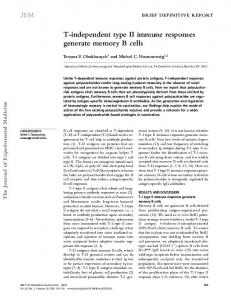
percentage of mononucleated cells
100
80
60
40
20
0 0
5
10
15
20
Days in culture
Fig. 1. MNGCs in culture. (A) Phase-contrast microscope observation of macrophages after 13 days in culture. Typical adherent macrophages and MNGCs surmounted by bright refractile structures are identified by arrowheads and arrows, respectively. Bar, 20 µm. (B) Scanning electron microscope examination. A typical MNGC consisting of a large flattened cell body surmounted by a cauliflower structure is shown in the middle of the field. Bar, 20 µm.
Fig. 2. Change in the proportion of mononucleated cells during a 13 day period of culture of chicken macrophages. Secondary macrophages were seeded at 1.6×10⁵ per 35 mm Petri dish. At regular intervals two dishes were fixed and stained with WrightGiemsa. An average of 400 cells were scored according to their nuclei content. The percentage of mononucleated is the ratio of mononucleated cells to the total number of cells. This represents the result of a typical experiment.
Generation of mono- from multi-nucleated cells 3235 represented less than 10% of the cell volume. Fluorescence images were taken with a SIT camera (Lhesa Electronique) and digitalized using an image processing system Crystal Sapphire (Quantel), coupled to a Bernoulli box image storage system. Images were printed using a color videoprinter from Sony. Chromatin labelling Viable cells were incubated in the presence of either acridine orange (10 µg/ml; Sigma) for 5 minutes at room temperature or Hoechst 33342 (1 µg/ml; Kodak) for 10 minutes at 37°C. Cells were then fixed with 4% paraformaldehyde in PBS, for 10 minutes. In some instances, cells were first fixed with 4% paraformaldehyde for 10 minutes, permeabilized in 0.5% Triton X-100 and incubated with either Hoechst 33258 (10 µg/ml; Kodak) or propidium iodide (50 µg/ml; Sigma) for 10 minutes. Confocal microscopy Confocal analysis was performed on propidium iodide-labelled cells that had been pretreated with RNase (170 µg/ml) for 20 minutes at room temperature, before propidium iodide labelling. Fluorescence images were obtained with a LSM10 laser scanning confocal microscope from Zeiss, using a 40× (NA 1.3) Zeiss Plan Neo Fluor objective. The illumination source was the 543 nm line from a helium-neon laser.
Fig. 3. Evolution of an MNGC studied after microinjection of Lucifer Yellow. A single MNGC was microinjected with Lucifer Yellow and observed over a 24 hour period. Phase-contrast micrographs (A,C,E) and the corresponding fluorescence images (B,D,F) were taken 15 minutes (A,B), 6 hours (C,D) and 22 hours (E,F) after microinjection. A single MNGC was microinjected (A, star), the Lucifer Yellow fluorescence was clearly restricted to that cell (B). Four hours later fluorescence appeared concentrated both in a large spot (D) corresponding to the former MNGC (C, star) and in a small spot (D, arrow) corresponding to an isolated cell (C, arrow); 22 hours after microinjection the fluorescent probe was found concentrated into 3 small spots (F, arrows) corresponding to 3 individual cells aggregated with other unlabelled cells (E, arrow). Bar, 20 µm.
RESULTS Chick peripheral blood monocytes plated at a density of 2.2×10⁴ cells/cm2 proliferated for 6-8 days and, upon reaching a density of about 12×104 cells/cm2, became quiescent, aggregated and then began to fuse. After 8-10 days in culture, large spread-out cells, multinucleated giant cells or MNGCs with a bright area in their center were observed (Fig. 1A; and Woods et al., 1995). A closer examination of MNGCs, by scanning electron microscopy, revealed that the bright area appears to be like a ‘cauliflower’ structure composed of highly convoluted membranes. It also revealed the abundance of membrane contacts between MNGCs and the smaller surrounding cells (Fig. 1B). At regular time intervals after seeding, cells were fixed and stained to evaluate the proportion of mononucleated cells versus the total number of cells (Fig. 2). Despite marked cell proliferation, the proportion of mononucleated macrophages rapidly dropped to about 25% of the total population after 6 days, indicating the active formation of multinucleated cells. Subsequently, the percentage of mononucleated cells rose again reaching 50% of the total cell number after 13 days. It was thus possible to distinguish two phases of culture: the first phase
3236 F. Solari and others appearing to correspond to the fusion of mononucleated macrophages to form MNGC; and the second phase that could either result from the proliferation of residual mononucleated cells or correspond to the generation of new mononucleated cells from MNGCs. Detailed microscopic observations favored the latter interpretation. To test this more rigourously we employed two complementary approaches, cell microinjection and cell cloning.
shown in Fig. 4, all Lucifer Yellow-fluorescing areas generated from a single microinjected MNGC (Fig. 4A and D) contained a single nucleus (Fig. 4B and E). It should be noted that about 10% of the small cells generated from the single large, microinjected MNGC contained two nuclei as illustrated on Fig. 4B (double arrow). These newly formed cells actively migrated; 36 hours after microinjection they were found several micrometers away from the microinjected MNGC (data not shown).
Formation of mononucleated cells from MNGCs: a study relying on microinjection of an ‘impermeable’ fluorescent probe MNGCs were microinjected with an impermeable fluorescent probe, Lucifer Yellow, which does not freely cross cell membranes. Immediately following microinjection, Lucifer Yellow fluorescence was observed only within the injected cell, not in the neighbouring cells (Fig. 3A-B). This indicated that no direct cytoplasmic contact existed between adjacent cells. To avoid ambiguity, only one MNGC was injected per Petri dish. Within 6 hours of microinjection, the fluorescence appeared to be concentrated in patches (Fig. 3D) and after 1824 hours, distinct and separate fluorescent areas were observed that appeared to correspond to individual single cells, as shown by phase-contrast microscopy (Fig. 3E-F). To confirm that these fluorescent areas indeed corresponded to individual cells, cells were stained with the chromatin dye, Hoechst 333342. As
Individually cloned MNGC continuously generate mononucleated cells To analyze further the behaviour of both single MNGC and generated mononucleated cells, we followed the fate of individual MNGC over a month. MNGCs detached from the dish and from any closely associated cells by trypsinization were seeded in separate culture wells. Within 1 hour after cloning, MNGCs began to flatten and attach to the surface of the dish. Within 4 hours, they acquired the typical morphology of adherent MNGCs. After 2 days in culture the single cloned MNGC was seen to contain large clustered oval nuclei, as seen by phase-contrast microscopy (Fig. 5A). After 5 days, formation of a small ‘cauliflower’ structure was clearly observed in the central area of the MNGC (Fig. 5B). This structure became very dense after 7 days in culture (Fig. 5C). At this time, new individual mononucleated cells could be seen in the vicinity of the MNGC (Fig. 5C, arrows). Three weeks
Fig. 4. Fluorescent structures generated from MNGCs represent nucleated cells. MNGC cultures were incubated with viable Hoechst 33342 to label chromatin. A single MNGC per Petri dish was microinjected with Lucifer Yellow and observed 48 hours later. The photographs presented here represent the results of 2 separate experiments (A,B,C, and D,E,F, respectively). Cultures were observed for Lucifer Yellow (A,D) or Hoechst (B,E) fluorescence as well as under a phase-contrast microscope (C,F). At 48 hours after Lucifer Yellow microinjection of a single MNGC, the fluorescent probe was found as individual spots (A,D). Each spot corresponded to a Hoechst-labelled nucleus (B,E, arrows), with sometimes 2 nuclei present in the spot (B, double arrow). The nucleated, Lucifer Yellow fluorescent spots could be identified as individual cells under phasecontrast observation (C, stars and F, arrowheads). Bar, 20 µm.
Generation of mono- from multi-nucleated cells 3237
Fig. 5. Generation of mononucleated cells from isolated MNGCs cultured individually. Single MNGCs were cultured separately in microwells and then regularly monitored under a phase-contrast microscope; the same cells was observed for 3 weeks. (A) Two days after cloning the MNGC exhibited a dark area (lower part of the cell in the picture) representing the nuclei of the cell. (B) At day 5 after cloning the MNGC presented vacuoles, as well as an expanding centrally located dark area identified as the cauliflower structure. (C) The cauliflower structure has expanded, but small individual cells indicated by arrows can be seen around the MNGC. (D) Three weeks after cloning the MNGC was still alive, with a compact cauliflower structure and the mononucleated cell compartment has covered the microwell. Some mononucleated cells were aggregated together or with the MNGC. Note the changes in morphology of the MNGC over time. Bar, 20 µm.
after cloning, the original MNGC was still alive and continued to generate mononucleated cells that progressively populated the dish (Fig. 5D). Subsequently, mononucleated cells began to aggregate and some of them began to fuse, forming a new generation of MNGCs, whereas others seemed to be fusing to the original MNGC (Fig. 5D). These results show that a single MNGC can generate many mononucleated cells that are themselves progenitors for new multinucleated cells. On the basis of morphological criteria, these cells resembled macrophages and so will be hereafter referred to as macrophage-like cells. Generation of mononucleated cell from MNGC is an Ara-C-insensitive event When MNGCs were labelled with any one of the following chromatin markers: acridine orange, Hoeschst 33342 or BrdU, no mitotic figures could be seen, suggesting that the generation of mononucleated cells from MNGC might be independent of mitosis (data not shown). To evaluate the role of mitosis in the budding process we analyzed the influence of a mitosis inhibitor on macrophage-like cell generation from
MNGCs. MNGCs cultured for 3 to 5 days in the presence of the antimitotic drug Ara-C were microinjected with Lucifer Yellow (Fig. 6A). The drug was maintained in the medium during the 48 hours post-injection period before staining the nuclei with Hoechst 33342. Under these conditions, formation of small distinct Lucifer Yellow-fluorescent areas from the initially diffusely labelled MNGC was still observed (Fig. 6B). These spots were again identified as cells by phase-contrast microscopy (Fig. 6C) and the presence of nuclei (Fig. 6D). These results confirm that mitotic events are not required for mononucleated cell generation from MNGC. Mononucleated cells are generated from MNGCs by a budding-like mechanism Having shown that MNGCs can generate mononucleated cells in the absence of mitosis, we then examined MNGCs that were active in the process of mononucleated macrophage-like cell generation by scanning electon microscopy (SEM). SEM observations readily showed the central raised structure, defined as a cauliflower structure by phase-contrast
3238 F. Solari and others
Fig. 6. Inhibition of mitosis by Ara-C did not prevent cell generation from MNGC. MNGCs cultures were maintained for 3 days in the presence of Ara-C to prevent mitosis before microinjection of Lucifer Yellow. A single MNGC per Petri dish was selected and microinjected with Lucifer Yellow (A). Cultures were maintained for another 2 days in the presence of Ara-C, then stained with Hoechst 33342 and checked for Lucifer Yellow fluorescence. Four positive fluorescent spots were found in the Petri dish, (B). They could be identified as individual cells (arrows in C) among other unlabelled cells by phase-contrast microscopy; and with bright fluorescent nuclei when observed under UV light with specific filter for Hoechst fluorescence (D, arrows). Bars, 20 µm.
microscopy, seen in Fig. 1. Fig. 7 shows a representative SEM view of MNGCs. From detailed observations such as these, it appeared that the mononucleated cells were arising from the apical face of the MNGC by some type of budding mechanism. To confirm this hypothesis, we looked at the nuclei distribution within MNGC. Chromatin staining with acridine orange revealed an unexpected arrangement of nuclei within MNGC. The bottom plane of the cell contained up to 20 large oval nuclei all clustered together in an orderly array in the central part of the cell (Fig. 8B, arrow). Some of them, with dark nucleoli, could be seen by phase-contrast microscopy (Fig. 8A, arrow) on the border of the cauliflower structure. Other nuclei were found in the upper planes (Fig. 8D). These nuclei were smaller, more irregular in shape, stained more brightly with the chromatin reagents (Fig. 8D, arrow) and corresponded exactly to the cauliflower structure (Fig. 8C). The structural organization of nuclei in MNGC was further analyzed in more detail by confocal microscopy following propidium iodide labelling. Serial optical sections (500 nm apart) through the central zone of a typical MNGC, corresponding to the cauliflower structure (Fig. 9A), where most of the nuclei were concentrated, are shown in Fig. 9B-E. The large oval nuclei were found 3-8 µm above the basal surface of the MNGC (in contact with the Petri dish), on the edge of the cauliflower structure (Fig. 9B). From 10 µm and up to 25 µm above the basal cell surface (Fig. 9C-E), smaller nuclei were observed. The computer-reconstituted image of the serial optical sections revealed that there was a continuum of labelled nuclei from the bottom (Fig. 9F, blue color) to the top of the cell (Fig. 9F, yellow and red colors) over a total thickness of about 25 µm.
Mononucleated cells could thus be generated by a buddinglike process from nuclei located near the top of the central cauliflower structure.
Fig. 7. Scanning electron microscope examination of an individual small MNGC. Cells were observed on a tilted screen to create a better image of the three-dimensional structure. Bar, 10 µm.
Generation of mono- from multi-nucleated cells 3239
Fig. 8. Organization of nuclei within MNGC. Cells labelled with acridine orange were observed. An MNGC was observed at two different focal depths under phase-contrast optics (A and C) or fluorescence (B and D). (A and B) The basal level of a large cell exhibiting the typical circular distribution of nuclei with a regular oval shape (arrows). (C and D) The apical region of the same cell. Note the cluster of small twisted nuclei (arrow). Bar, 20 µm.
DISCUSSION We report here the first evidence that in higher eucaryotes multinucleated cells are capable of generating small mononucleated cells in the absence of mitosis. These newly generated cells can be selectively harvested and are competent by themselves to give rise to a second generation of MNGCs (data not shown). Over three decades ago, it was shown by microcinematography as well as thymidine incorporation studies that nuclei and cytoplasm may shuttle between different individual osteoclasts (for review see Hancox, 1972). Several recent reports support this hypothesis. For example, during normal bone turnover, there is first a phase of resorption by osteoclasts followed by a phase of bone formation by osteoblasts. These two phases are separated by the reversal phase during which uncharacterized macrophage-like mononuclear cells are observed in the resorption bay (for review see Baron, 1993). It has been postulated that the cells colonizing the bone resorption lacunae during the later part of the resorptive sequence are derived directly from osteoclasts (Dr E. Eriksen, personal communication). Moreover, Merry et al. (1993) have shown that within completed bone resorption sites there are mononuclear cells sharing osteopontin expression in common with osteoclasts, suggesting to the authors that these mononuclear macrophage-like cells could be linked to the osteoclast lineage. Merry et al. (1993) interpreted these cells to be osteoclast pre-
cursors but in the light of our results reported here they could equally well be viewed as osteoclast progeny. We, and others, have previously shown that chicken hematopoietic macrophages in culture will give rise to multinucleated giant cells by fusion. These MNGCs share most of the characteristic features of postmitotic bona fide osteoclasts (Alvarez et al., 1991; Woods et al., 1995). Data obtained by microinjection of Lucifer Yellow into single MNGCs and by culturing individual MNGCs have unambiguously shown that they are able to generate mononucleated, or occasionally binucleated, macrophage-like cells without the disappearance of the MNGCs. It remains to be determined whether these mononucleated cells correspond to authentic macrophages or to cells already engaged in osteoclast differentiation. Normally, progeny cells are generated by a mitotic event. However, there are some instances where mitosis is not required; for example, the fragmentation of megakaryocytes, or the development of the syncitial Drosophila egg into blastoderm. The model we describe here is rather unusual in the way that mononucleated cells are continuously generated without disruption of the parental multinucleated cell. Morphological (after chromatin staining) and pharmacological (Ara-C treatment) analyses led us to conclude that the cell generation process, described here, is mitosis-independent. The unusual nuclei distribution within the cauliflower structure of the MNGC suggests a continuous flow of nuclei from the bottom to the top of the MNGC. Consequently, this suggests
3240 F. Solari and others
Fig. 9. Laser scan confocal microscope analysis of the distribution of nuclei within the caulifower structure of an MNGC. Cultures of MNGC on glass coverslips were fixed and chromatin was stained with propidium iodide. (A) Phase-contrast image of the same MNGC at higher magnification, focused on the central cauliflower structure. Bar, 20 µm. (B to E) Serial confocal images taken 5 µm (B), 12 µm (C), 19 µm (D), or 26 µm (E) above the bottom level. (F) Computer reconstituted image of the same area obtained after confocal microscopy. A laser beam was applied in order to scan the sample along the vertical axis every 500 nm. Basal nuclei are shown in blue (arrows), whereas apical nuclei are in yellow and in red at the top. Note that nuclei are present at all levels from the bottom of the MNGC up to its top, along a 25 µm thickness. Bar, 20 µm.
that cell generation from MNGCs could occur through a budding-like mechanism reminiscent of the budding process of the viral envelope packing of the viral genome at the surface of the host cell. Formation of mononucleated cells, while maintaining an active MNGC, implicates a constant flux of nuclei within the MNGC; constant sized MNGCs are maintained by import by cell fusion (Moudjou et al., 1989; Woods et al.,
1995; Zambonin-Zallone et al., 1984) and export by budding (our data; Hancox, 1972). The existence of about 10% of binucleated cells among newly formed cells also favours such a mechanism. We have consistently observed that many cells remain aggregated on the surface of the MNGCs at the level of the cauliflower structure. These cells, probably derived by budding, have been carefully collected and plated into new
Generation of mono- from multi-nucleated cells 3241 culture dishes; they re-formed a new generation of MNGCs capable of themselves actively budding after a few days in culture. The budding process would thus provide a mechanism for numerous rounds of multinucleated cell proliferation. It has recently been demonstrated that in urodele limb bud regeneration, prelabelled myotubes implanted into the regenerating stump can give rise to labelled mononucleated cells within a short period of time (Lo et al., 1993). In their report, however, the authors did not provide any information on the ability of the mononucleated cells to participate in the neoformation of multinucleated myotubes. In higher eucaryotes, there are few lineages consisting of mature syncitial cells, it remains to be determined if all multinucleated cell types have retained the capacity for budding off mononuclear cells with the characteristics of their own precursor cell type. We thank Jean Edens, who performed electron microscope observations; and Catherine Souchier, for her contribution in the confocal microscope analyses. This work was supported by research grants from CNRS and Association pour la Recherche sur le Cancer (ARC).
REFERENCES Alvarez, J. I., Teitelbaum, S. L., Blair, H. C., Greenfield, E. M., Athanasou, N. A. and Ross, F. P. (1991). Generation of avian cells resembling osteoclasts from mononuclear phagocytes. Endocrinology 128, 2324-2335. Baron, R. (1993). Anatomy and ultrastructure of bone. In Primer on the Metabolic Bone Disease and Disorders of Mineral Metabolism, 2nd edn, pp. 3-7. Publication of the ASBMR. Raven Press, NY. Gay, C. V. (1991). Avian osteoclasts. Calc. Tiss. Int. 49, 153-154.
Hancox, N. M. (1972). The osteoclast. In The Biochemistry and Physiology of Bone (ed. G. H. Bourne) Academic Press, San Francisco, London. Jotereau, F. V. and Le Douarin, N. (1978). The developmental relationship between osteocytes and osteoclasts: a study using the quail-chick nuclear marker in endochondral ossification. Dev. Biol. 63, 253-265. Lo, D. C., Allen, F. and Brockes, J. P. (1993). Reversal of muscle differentiation during urodele limb regeneration. Proc. Nat. Acad. Sci. USA 90, 7230-7234. Merry, K., Dodds, R., Littlewood, A. and Gowen, M. (1993). Expression of osteopontin mRNA by osteoclasts and osteoblasts in modelling adult human bone. J. Cell Sci. 104, 1013-1020. Moudjou, M., Lanotte, M. and Bornens, M. (1989). The fate of the centrosome-microtubule network in monocyte-derived giant cells. J. Cell Sci. 94, 237-244. Munari-Silem, Y., Audebert, C. and Rousset, B. (1991). Hormonal control of cell to cell communication: regulation by thyrotropin of the gap junctionmediated dye transfer between thyroid cells. Endocrinology 128, 3299-3309. Mundy, G. R. (1995). Bone Remodeling and its Disorders. Martin Dunitz. Nijweide, P. J. and de Grooth, R. (1992). Ontogeny of the osteoclast. In Biology and Physiology of the Osteoclast (ed. B. R. Rifkin and C. V. Gay). CRC Press, Boca Raton, FL. Sims, S. M., Kelly, M.E.M., Arkett, S. A. and Dixon, S. J. (1992). Electrophysiology of osteoclasts. In Biology and Physiology of the Osteoclast (ed. B. R. Rifkin and C. V. Gay). CRC Press, Boca Raton, FL. Takahashi, N., Udagawa, N., Tanaka, S., Murakami, H., Owan, I., Tamura, T. and Suda, T. (1994). Postmitotic osteoclast precursors are mononuclear cells which express macrophage-associated phenotypes. Dev. Biol. 163, 212-221. Woods, C., Domenget, C., Solari, F., Gandrillon, O., Lazarides E. and Jurdic, P. (1995). Antagonistic role of Vitamin D3 and retinoic acid on the differentiation of chicken hematopoietic macrophages into osteoclast precursor cells. Endocrinology 136, 85-95. Zambonin Zallone, A., Teti, A. and Primavera, M. V. (1984) Monocytes from circulating blood fuse in vitro with purified osteoclasts in primary culture. J. Cell Sci. 66, 335-342. (Received 9 May 1995 - Accepted 19 July 1995)