Multiple sclerosis impairs ability to detect abrupt appearance of a subliminal stimulus Simona Carrubba*, Alireza Minagar{, Eduardo Gonzalez-Toledo{, Andrew L. Chesson, Jr{, Clifton Frilot II1 and Andrew A. Marino*
Published by Maney Publishing (c) W. S. Maney & Son Limited
*Department of Orthopaedic Surgery, {Department of Neurology, {Department of Radiology and 1School of Allied Health Professions, LSU Health Sciences Center, Shreveport, LA, USA Objectives: The study was designed to find evidence that brain electrical activity associated with processing the abrupt appearance or disappearance of a sensory stimulus differed in the presence and absence of the neuropathological changes that are characteristic of multiple sclerosis (MS). Methods: A subliminal stimulus (electrical field) was applied, and the onset and offset responses from patients with MS were compared with the responses of study participants in two age- and gender-matched control groups, using a novel type of non-linear dynamical analysis that had been developed in earlier studies. Results: An onset response occurred in 27% of the patients with MS, compared with 85% in the control groups. Among the three patients who exhibited onset-induced changes in brain electrical activity, the average latency of the effect was less and the magnitude of the change was greater than the corresponding values in the control group. Discussion: Non-linear analysis of electroencephalograms recorded during the sudden presentation of a subliminal stimulus potentially could serve as the basis of a functional test to help diagnose MS. A larger cohort of patients with MS needs to be assessed to validate the results of this study. Keywords: Complexity conjecture, functional test, onset response, whole-brain states
Introduction Multiple sclerosis (MS) has typically been regarded as an immune-mediated disease of the human central nervous system that affects genetically predisposed young adults following exposure to yet unidentified environmental agents1. Recent neuropathological and imaging studies revealed significant gray-matter involvement in MS2, even during early-stage disease3. Although knowledge is scarce regarding the basic pathological processes in gray matter and their relation to those in white matter, the documented involvement of gray matter indicates that MS should be conceptualized as a disease that affects the whole brain4. Improved methods for assessing disease-related functional changes would also be helpful in diagnosing and treating MS. Cognitive activity is mediated by temporal–spatial electrical interactions between localized brain networks5. The instantaneous dynamical synchronization is reflected in and constitutes part of the electroencephalogram (EEG). In principle, therefore, analysis of scalp electrical measurements could provide the basis of a method for monitoring general brain function.
Correspondence and reprint requests to: Andrew A. Marino, PhD, Department of Orthopaedic Surgery, Louisiana State University Health Sciences Center, PO Box 33932, Shreveport, LA 71130-3932, USA. [
[email protected]] Accepted for publication September 2009. ß W. S. Maney & Son Ltd 2010 DOI 10.1179/016164109X12536042424135
Traditional electrophysiological measurements of stimulus-induced responses, visual evoked potentials (EPs) for example, are designed to evaluate specific neural pathways by means of linear techniques (time averaging or spectral analysis). Although they can demonstrate an EP in the fewest trials6,7, use of these techniques entails an assumption that the latency, duration and waveform of all EPs in a series of trials are identical, which is not true for successive wholebrain electrical states that occur during cognitive processing; instead, these states change profoundly from moment to moment because they are governed by non-linear dynamical laws8. The human brain contains specialized cortical and subcortical networks for detecting the abrupt appearance or disappearance of sensory stimuli9. Many studies indicated that the responses to the onset and offset of stimuli were mediated by independent pathways10–12. We generalized the notion of evoked potentials by developing a method to characterize electrical states of the whole brain in relationship to an abrupt change in a stimulus, without the necessity of assuming a linear response13,14. That method was used here to ascertain whether whole-brain electrical states in individual subjects were sensitive to the presence of MS. Specifically, we determined whether the onset and offset responses to a subliminal sensory stimulus differed between the presence and absence of MS. Neurological Research
2010
VOL
32
NO
3
297
Published by Maney Publishing (c) W. S. Maney & Son Limited
S. Carrubba et al.
298
MS impairs ability to detect subliminal stimulus
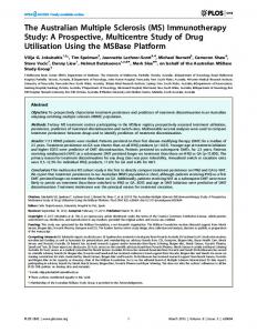
Figure 1 Detection of changes in the EEG induced by the presence of a subliminal stimulus (electrical field). (A) Complexity conjecture for effect of the stimulus on whole-brain electrical activity. The instantaneous strength of the connectivity between local neuronal networks is represented by the thickness of the line that joins them. (B) EEG trial showing the locations of the epochs used to detect the effect of the stimulus
Materials and methods Patients
Patients with MS were recruited from the outpatient neurology clinic from August 2007 to March 2008. Inclusion criteria were: definite MS15 with a relapsing–remitting course16, but in remission; expanded disability status scale (EDSS) score (3.017, assessed by the treating neurologist; absence of acute relapses and intravenous corticosteroid treatment for at least 60 days before inclusion in the study; no changes of EDSS score for at least 3 months before inclusion in the study. The criteria resulted in the identification of ten patients who volunteered to participate in the study, all of whom were females; an additional MS patient was recruited in April 2009 because the magnetic resonance imaging (MRI) scan of one of the enrolled patients was unavailable for quantitative analysis (see below). The average age of the patients was 33 years (range: 18–52 years); all patients were being treated with b-interferons or glatiramer acetate. Two gender- and age-matched control groups were used: (1) females assessed and examined in the neurology clinic who complained of headaches but who were otherwise healthy (average age: 33 years, range: 19–52 years); (2) females recruited from the general population who had no medical complaints (average age: 34 years, range: 24–53 years). All participants were informed of the goals, methods and general design of the investigation, but were not told exactly when during the experimental session, the stimulus would be applied or for how long. Written informed consent was obtained from Neurological Research
2010
VOL
32
NO
3
each participant. The institutional review board at the Louisiana State University Health Sciences Center approved all experimental procedures. Approach
Our approach was based on the complexity conjecture13,14 (Figure 1A). Stimuli are transduced by specialized cells resulting in afferent and efferent signals; cognition is mediated by electrical activity in localized neuronal networks and by internetwork electrical synchronization18–20. The overall process generates a time-dependent, spatially-extended threedimensional distribution of electrical potential that can be sampled on the scalp. Our hypothesis was that cognitive processing would be altered in the presence of MS. Stimulus
We chose an electrical field (350 V/m, 60 Hz) as the stimulus because the response it produces is subliminal21, thereby avoiding the possibility that the putative signature of the disease would be masked by the typical robust linear response from cortical generators that occurs during auditory or visual EP determinations. The electrical field was generated by applying a voltage to two parallel metal plates located on each side of the head. The stimulus was applied for 2 seconds, with a 5 second interstimulus period (7 second trials) to facilitate assessment of the onset and offset responses, which occur with latencies of 100–500 ms (Figure 1B)21. The study participants were exposed (eyes closed) in an isolation chamber to reduce the effect of
S. Carrubba et al.
random ambient stimuli. All electrical equipment was located outside the chamber; the absence of both uncontrolled sensory cues and direct perception of the field was verified by interviewing each subject at the end of the experimental session.
Published by Maney Publishing (c) W. S. Maney & Son Limited
Electroencephalograms
Electroencephalograms were recorded from O1, O2, C3, C4, P3 and P4 (International 10-20 System) referenced to linked ears, using gold-plated electrodes attached to the scalp with conductive paste; the signals were filtered (0.3–35 Hz), digitized and analysed offline. Application of the stimulus produced a spike artifact (30 ms) that was deleted before analysis. Trials containing artifacts (as assessed by visual inspection) were discarded (,5% of the trials). All results were based on data from at least 50 artifact-free trials. Each participant underwent 80 stimulus trials and 80 sham-stimulus trials; the former were used to determine the effect of the stimulus and the latter served as a negative control. Imaging
Brain MRI was performed using a 1.5 T scanner with a standard quadrature head coil; contiguous T2-weighted axial images (5 mm thick sections) were analysed using a conventional spin echo sequence with a 250 mm field of view and a 256 6 256 image matrix. A neuroradiologist (E. GonzalezToledo) identified the lesions based on predetermined guidelines22 and measured lesion volume using morphometric analysis (MIPAV, National Institutes of Health, Bethesda, MD, USA) while blinded to the electrophysiological data. Separate measurements were made for periventricular23 and non-periventricular lesions. Analysis and statistics
The EEG from each derivation was analysed to detect the effects of stimulus onset and offset; a portion of the interstimulus period (t55.1–5.5 seconds) served as the control (Figure 1B). Our method13 involved embedding the EEG in a mathematical phase space, calculating recurrence plots, and quantifying them using two distinct but related quantifiers called percent determinism and percent recurrence24. The measured characteristics of the
MS impairs ability to detect subliminal stimulus
response were the magnitude of quantifiers (expressed as a percent of the corresponding value of the control) and the latency of the response (in millisecond), assessed at the mid-point of the response13. A statistically reliable change in the quantifiers that occurred in association with application of the stimulus was a direct indication of a change in brain electrical activity. For each statistical test involving an onset or offset response, a comparable test was carried out on the sham data and the results were used to calculate the pairwise error rate (number of false-positive effects in the sham data divided by the total number of tests performed). The error rate thus determined was used to estimate the family-wise error rate (PFW) for the decision that a subject had exhibited a stimulusinduced change in brain electrical activity14. To examine for the occurrence of linear changes, the EEG was also evaluated directly (no unfolding in phase space) by time averaging7; family-wise error was determined as described above. We regarded a change as non-linear if it was detected by recurrence analysis but not by time averaging.
Results Onset responses occurred in only 27% of the patients with MS (Table 1), compared with 90% of those in the migraine group (Table 2) and 80% of the normal subjects (Table 3); the onset detection rate in the patients with MS was significantly less than either control group (p,0.05, chi-square test).The rate of detection of stimulus offset in the patients with MS (70%) was comparable to that in the control groups (50 and 70%, respectively, in the migraine and normal groups) (Tables 2 and 3). Among the three patients who exhibited onsetinduced changes in brain electrical activity (MS-4, MS-6 and MS-9, in Table 1), the average latency of the effect was less and the magnitude of the change was greater than the corresponding values in the control group (p,0.05, t-test) (Table 4). Stimulus-related changes were not seen in any participant when the EEGs were evaluated by timeaveraging (data not shown). Lesion load was quantified in eight of the patients with MS (Table 1); in two other cases (MS-10 and
Table 1 Stimulus-induced changes in brain electrical activity in study participants with multiple sclerosis Lesion load (cm3) MS participant
Total
Peri.
Onset stimulus
PFW
Offset stimulus
PFW
MS-1 (40) MS-2 (34) MS-3 (52) MS-4 (32) MS-5 (19) MS-6 (30) MS-7 (18) MS-8 (27) MS-9 (50) MS-10 (31) MS-11 (38)
1.77 1.59 7.67 0.47 0.14 NA 2.44 1.82 3.40 Diffuse Diffuse
0.64 0.39 5.63 0.47 0 NA 0.92 0.96 2.56 Diffuse Diffuse
NE* NE NE O1 O2 C3 C3 C4 NE O2 O2 C3 NE C3 C4 P4 NE NE NE
… … … 0.003 … 0.029 … 0.029 … … …
NE O1 O2 O2 P4 O1 C3 C3* C3 C4 C4 O2 C3 P3 O2 C3 P3 C3 C4 C4 P3 P4 O2 C4 P4 NE O1 O1 P3 O2 O2 C4
… 0.000 0.084 0.014 0.001 0.001 0.000 0.024 … 0.005 0.005
Results found using the EEG quantifier percent recurrence and percent determinism are shown in non-bold and bold, respectively. Age (years) in parentheses. NE: no effect; NA: not available; Peri.: periventricular; PFW: family-wise error. *False-positive detection. Neurological Research
2010
VOL
32
NO
3
299
S. Carrubba et al.
MS impairs ability to detect subliminal stimulus
MS-11), the brain structural changes were too diffuse for measurement. In the onset detectors for which an MRI was available (MS-4 and MS-8), the average total load was 1.14 ml, compared with 2.84 ml in the onset non-detectors; the respective average periventricular loads were 0.72 and 1.63 ml, respectively.
putative stimulus-induced change in brain activity could be anticipated to vary from trial to trial; if so, real effects would be averaged away and hence would appear non-existent. We therefore generalized the notion of the evoked potential by quantifying the stimulus-induced change before the averaging step in the analysis, using a non-linear mathematical algorithm. We report here the results obtained using a subliminal stimulus that was not consciously perceived. The rates at which clinically normal subjects exhibited changes in brain electrical activity in response to the onset and offset of the stimulus (Table 3) were as expected based on previous studies13,14,21; the detection rates in the migraine group (Table 2) were similar to those in the normal group. In the MS group, in contrast, the rate of occurrence of onset responses was significantly lower (Table 1). If the absence of a change due to stimulus onset was taken as indicating the presence of MS, then the sensitivity of the test was 73% (eight of 11). A rough measure of test reliability can be obtained from a consideration of the sham data. A total of 62 tests were performed (onset and offset in 31 patients),
Published by Maney Publishing (c) W. S. Maney & Son Limited
Discussion Cortical and subcortical networks sensitive to the abrupt appearance or disappearance of sensory stimuli facilitate unconscious shifting of attention to environmental events, for example, an immediate awareness of the sudden cessation of a sound, like birds singing9. Different networks are involved in attention to stimuli onset and offset11,25–29. Because structural abnormalities in white matter and gray matter are pathognomic for MS, we hypothesized that a general functional test for the disease might be based on measurements of the electrophysiological correlates associated with detection of the sudden appearance and/or disappearance of stimuli. Analysing responses by directly averaging the EEG, studies of visual evoked potentials for example, offered little hope of success because the characteristics of the
Table 2 Stimulus-induced changes in brain electrical activity in study participants who complained of headache Headache participant
Onset stimulus
PFW
Offset stimulus
PFW
H-1 (53) H-2 (29) H-3 (35) H-4 (35) H-5 (28) H-6 (40) H-7 (34) H-8 (32) H-9 (28) H-10 (24)
O1 C3 C4 P4 O1 C3 C3 C3 C3 P3 O2 C3 C4 P3 P4 O1 O1 P4* C4 C4 P3 P4 P4 O1 C4 C4 NE O2 C3 P3 O2 O2 P4
0.000 0.013 0.005 0.000 0.040 0.029 0.025 … 0.001 0.040
O1 O1 C3 NE C3 C3 C4 O2 C3 C4 P3 P4 O1 O1 O2 O2 NE NE O1 C3 C4 O1 O2 C3 P3 NE
0.013 … 0.035 0.001 0.001 … … 0.052 0.000 …
Results found using the EEG quantifier percent recurrence and percent determinism are shown in non-bold and bold, respectively. Age (years) in parentheses. NE, no effect; PFW, family-wise error. *False-positive detection. Table 3 Stimulus-induced changes in brain electrical activity in study participants who had no medical complaints Normal participant
Onset stimulus
PFW
Offset stimulus
PFW
N-1 (51) N-2 (66) N-3 (22) N-4 (26) N-5 (23) N-6 (23) N-7 (23) N-8 (46) N-9 (23) N-10 (25)
O2 O2 C3 O2 C3 C3 P4 NE C3 C4 C4 P3 C3 C4 P4 C3 C3 C4 C4 O1 C3 C3 P3 O1 O1 C3 O1 O2 C4 C4 P3 P4 P3 P3 P4*
0.031 0.001 … 0.001 0.001 0.001 0.004 0.005 0.000 0.084
C3 C4 P3 C4 C4 P4 O2 O2 P3 NE O2 C4 P3 C4 P4 P4 O1 O2 C3 P3 P4 O1 C3 P3 C3 C3 P3 P3 C3 P3 P4
0.077 0.040 0.059 … 0.011 0.005 0.000 0.001 0.000 0.001
Results found using the EEG quantifiers percent recurrence and percent determinism are shown in non-bold and bold, respectively. NE: no effect; PFW: family-wise error. Age (years) in parentheses. *False-positive detection. Table 4 Stimulus-induced changes in brain electrical activity (mean ¡ SD) Group
Onset latency (ms)
Magnitude (%)
Offset latency (ms)
Magnitude (%)
MS Migraine Normal
288.0 ¡ 56.3 (3, 11)* 291.3 ¡ 50.4 (9, 32)* 333.3 ¡ 44.3 (9, 34)
53.2 ¡ 20.9 (3, 11)* 32.7 ¡ 11.6 (9, 32) 30.8 ¡ 12.2 (9, 34)
295.9 ¡ 57.5 (9, 30) 263.1 ¡ 40.5(6, 23)* 307.6 ¡ 55.3 (9, 31)
35.6 ¡ 14.5 (9, 30) 30.3 ¡ 11.2 (6, 23) 31.0 ¡ 12.6 (9, 31)
Numbers in parentheses indicate the number of subjects who exhibited a response and the number of electrode derivations at which a change in brain activity was found, respectively (Tables 1 and 3). The magnitude of stimulus-induced changes is expressed as a percent difference from the control value of the recurrence quantifier. *p,0.05 (t-test) with respect to the normal group. 300
Neurological Research
2010
VOL
32
NO
3
Published by Maney Publishing (c) W. S. Maney & Son Limited
S. Carrubba et al.
and only four cases of a false-positive result EP were found (Tables 1–3). Thus, the method was unlikely to report a stimulus-induced change where none existed. In the three cases where an onset response was found, its characteristics (latency and magnitude) differed significantly from the controls, on average. Although the number of patients was small, the results raised the possibility that the functional test was sensitive to the presence of MS even in patients who detected the stimulus. The lesion load appeared to be smaller in the detectors (Table 1), but too few patients were studied to permit a realistic assessment of the correlation between load and ability to detect the stimulus. Overall, the results suggested that nonlinear dynamical analysis of changes in brain electrical activity induced by the abrupt onset of the stimulus might be useful for characterizing brain function in patients with MS. We previously showed that electrical fields were perceived subliminally21. We used a field as the stimulus in this study to lay emphasis on the role of subcortical networks. Moreover, the stimulus receptor cell, believed to be a force-transducing ion channel similar to that present in lower life forms21, has been located in the head30, possibly the cerebellum31. Thus, we had good reason to suspect that the subcortical networks mediated even the early post-transduction steps in the cognitive processing triggered by the stimulus. The relative advantages and disadvantages of using ordinary stimuli remain to be explored. Presently, there are no adequate functional tests to assist in diagnosing MS or to characterize end points in longitudinal studies and clinical trials. Evoked potentials have frequently been assessed in patients with MS, but several factors limit their clinical usefulness. Most studies reported increased average latency, but decreased latencies also occurred in particular patients in the MS group32,33. Thus, the complex time- and spatially-dependent interplay of degenerative and regenerative processes that occur in the central nervous system of patients with MS prevents both interpretation of latency changes in terms of specific neurological function and determination of the extent of latency changes that can reliably be regarded as clinically meaningful34–36. Another restriction on the use of EPs for assessing patients with MS stems from the common use of time-averaging to evaluate the data. Although timeaveraging facilitates detection of lesions in specific sensory pathways, it cannot characterize whole-brain electrical activity because only the aspects of the responses that are identical in the trials are captured; the variable parts are averaged away. Functional magnetic resonance imaging (fMRI) can help reveal the neural basis of motor and cognitive impairment in patients with MS, usually by indicating relative increases in the degree of activation within given brain regions37,38. However, changes in activation may be due to differences in task performance between the groups tested, rather than direct effects of the underlying disease process39. Additionally, fMRI is susceptible to artifacts related to head motion.
MS impairs ability to detect subliminal stimulus
The method described in our study permits an assessment of the extent of synchronization between brain networks, which is precisely the kind of highlevel brain function that we would expect would be impaired, given our present perspective that MS is a whole-brain disease. Complicated, expensive equipment is not needed to implement the method, and the algorithms necessary to evaluate the data are available as freeware40. The potential advantages of non-linear analysis of whole-brain electrical states are opposed by some notable limitations and uncertainties: (1) the fundamental results of the analysis are not expressed as a brain image, or a familiar scalar quantity such as time or voltage, but rather in terms of unfamiliar quantifiers that have no direct physiological interpretation or meaning; (2) scalp electrical signals can be affected by vascular pathology, brain tumor or stroke; the extent to which altered tissue perfusion affects the interpretation of the dynamical electrical changes has not been evaluated; (3) the disease specificity of the response is an unresolved issue. We found that patients in the headache and MS groups could easily be distinguished, but the specificity issue must be evaluated by considering other diseases like Alzheimer’s and Parkinson’s diseases; (4) the potential influence of the treatment (b-interferons and glatiramer acetate) on the observed response should be assessed; (5) it remains unclear whether or to what extent the difference in onset response between those who do and do not have MS can be explained by a difference in baseline brain electrical activity. In summary, non-linear analysis of EEGs recorded during the sudden presentation of a subliminal stimulus could potentially serve as the basis of a functional test to help diagnose MS. A larger cohort of patients with MS needs to be assessed to validate the results of this study.
Acknowledgement We thank Erin L. Eaton for help in identifying study participants and verifying that they met the entry criteria.
References 1 2 3
4 5 6 7
8
9
Noseworthy JH, Lucchinetti C, Rodriguez M, et al. Multiple sclerosis. N Engl J Med 2000; 343: 938–952 Minagar A. Gray matter involvement in multiple sclerosis: A new window into pathogenesis. J Neuroimaging 2003; 13: 291–292 de Stefano N, Matthews PM, Filippi M, et al. Evidence of early cortical atrophy in MS: Relevance to white matter changes and disability. Neurology 2003; 60: 1157–1162 Minagar A. Multiple sclerosis: Current knowledge and future directions. Neurol Res 2006; 28: 227–229 Gazzaniga MS. The New Cognitive Neurosciences, Cambridge, MA: MIT Press, 2000 Leocani L, Comi G. Neurophysiological markers. Neurol Sci 2008; 29 (Suppl. 2): S218–S221 Regan D. Human Brain Electrophysiology: Evoked Potentials and Evoked Magnetic Fields in Science and Medicine, Amsterdam: Elsevier Science, 1989 Rubinov M, Sporns O, van Leeuwen C, et al. Symbiotic relationship between brain structure and dynamics. BMC Neurosci 2009; 10: 55 Downar J, Crawley AP, Mikulis DJ, et al. A multimodal cortical network for the detection of changes in the sensory environment. Nat Neurosci 2000; 3: 277–283 Neurological Research
2010
VOL
32
NO
3
301
Published by Maney Publishing (c) W. S. Maney & Son Limited
S. Carrubba et al.
302
MS impairs ability to detect subliminal stimulus
10 Bair W. No doubt about offset latency. Vis Neurosci 2004; 21: 671– 674 11 Pratt H, Starr A, Michalewski HJ, et al. The auditory P50 component to onset and offset of sound. Clin Neurophysiol 2008; 119: 376–387 12 Yamashiro K, Inui K, Otsuru N, et al. Somatosensory off-response in humans: An MEG study. Neuroimage 2009; 44: 1363–1368 13 Carrubba S, Frilot C, Chesson A, et al. Detection of nonlinear event-related potentials. J Neurosci Meth 2006; 157: 39–47 14 Carrubba S, Frilot C, Chesson AL, Jr, et al. Evidence of a nonlinear human magnetic sense. Neuroscience 2007; 144: 356–367 15 McDonald WI, Compston A, Edan G, et al. Recommended diagnostic criteria for multiple sclerosis: Guidelines from the International Panel on the diagnosis of multiple sclerosis. Ann Neurol 2001; 50: 121–127 16 Lublin FD, Reingold SC. Defining the clinical course of multiple sclerosis: Results of an international survey. National Multiple Sclerosis Society (USA) Advisory Committee on Clinical Trials of New Agents in Multiple Sclerosis. Neurology 1996; 46: 907–911 17 Kurtzke JF. Rating neurologic impairment in multiple sclerosis: An expanded disability status scale (EDSS). Neurology 1983; 33: 1444–1452 18 Freeman WJ, Skarda CA. Spatial EEG patterns, non-linear dynamics and perception: The neo-Sherringtonian view. Brain Res 1985; 357: 147–175 19 Go´mez-Herrero G, Atienza M, Egiazarian K, et al. Measuring directional coupling between EEG sources. Neuroimage 2008; 43: 497–508 20 Moeller S, Freiwald WA, Tsao DY. Patches with links: A unified system for processing faces in the macaque temporal lobe. Science 2008; 320: 1355–1359 21 Carrubba S, Frilot II C, Hart FX, et al. The electric field is a sufficient physical determinant of the human magnetic sense. Int J Radiat Biol 2009; 85: 622–632 22 Gawne-Cain ML, O’Riordan JI, Coles A, et al. MRI lesion volume measurement in multiple sclerosis and its correlation with disability: A comparison of fast fluid attenuated inversion recovery (fFLAIR) and spin echo sequences. J Neurol Neurosurg Psychiat 1998; 64: 197–203 23 Nakashima I, Fujihara K, Miyazawa H, et al. Relevance of callosal and periventricular MRI lesions to oligoclonal bands in multiople sclerosis. Acta Neurol Scand 2006; 113: 125–131 24 Zbilut JP, Webber CL, Jr. Recurrence quantification analysis. In: Akay M, ed. Wiley Encyclopedia of Biomedical Engineering, Hoboken, NJ: John Wiley & Sons, 2006: pp. 2979–2986 25 Bandini F, Pierantozzi M, Bodis-Wollner I. Parkinson’s disease changes the balance of onset and offset visual responses: An evoked potential study. Clin Neurophysiol 2001; 112: 976–983
Neurological Research
2010
VOL
32
NO
3
26 Clementz BA, Keil A, Kissler J. Aberrant brain dynamics in schizophrenia: Delayed buildup and prolonged decay of the visual steady-state response. Cogn Brain Res 2004; 18: 121–129 27 Kreegipuu K, Allik J. Detection of motion onset and offset: Reaction time and visual evoked potential analysis. Psychol Res 2007; 71: 703–708 28 Okada T, Honda M, Okamoto J, et al. Activation of the primary and association auditory cortex by the transition of sound intensity: A new method for functional examination of the auditory cortex in humans. Neurosci Lett 2004; 359: 119–123 29 Tanaka E, Inui K, Kida T, et al. Common cortical responses evoked by appearance, disappearance and change of the human face. BMC Neurosci 2009; 10: 38–46 30 Marino AA, Nilsen E, Frilot C. Localization of electroreceptive function in rabbits. Physiol Behav 2003; 79: 803–810 31 Frilot C 2nd, Carrubba S, Marino AA. Magnetosensory function in rats: Localization using positron emission tomography. Synapse 2009; 63: 421–428 32 Gundogan FC, Demirkaya S, Sobaci G. Is optical coherence tomography really a new biomarker candidate in multiple sclerosis? – A structural and functional evaluation. Invest Ophthalmol Vis Sci 2007; 48: 5773–5781 33 van Dijk JG, Jennekens-Schinkel A, Caekebeke JF, et al. What is the validity of an ‘abnormal’ evoked or event-related potential in MS? Auditory and visual evoked and event-related potentials in multiple sclerosis patients and normal subjects. J Neurol Sci 1992; 109: 11–17 34 Aminoff MJ, Davis SL, Panitch HS. Serial evoked potentials studies in patients with definite multiple sclerosis. Clinical relevance. Arch Neurol 1984; 41: 1197–1202 35 Davis SL, Aminoff MJ, Panitch HS. Clinical correlations of serial somatosensory evoked potentials in multiple sclerosis. Neurology 1985; 35: 359–365 36 Leocani L, Comi G. Neurophysiological investigations in multiple sclerosis. Curr Opin Neurol 2000; 13: 255–261 37 Filippi M, Rovaris M, Rocca MA. Imaging primary progressive multiple sclerosis: The contribution of structural, metabolic, and functional MRI techniques. Mult Scler 2004; 10 (Suppl. 1): S36– S44 38 Sweet LH, Rao SM, Primeau M, et al. Functional magnetic resonance imaging response to increased verbal working memory demands among patients with multiple sclerosis. Human Brain Mapp 2006; 27: 28–36 39 Phillips MD. Functional faults: fMRI in MS. Neurology 2008; 70: 248–249 40 Webber CL, Jr. Recurrence quantification analysis. Available at: http://homepages.luc.edu/ycwebber[June 2009]