wortmannin. Activation of three processes is involved in the stimulation of fatty acid synthesis from glucose by insulin, namely glucose uptake, acetyl-CoA ...
595
Biochem. J. (1995) 311, 595-601 (Printed in Great Britain)
Multiple signalling pathways involved in the stimulation of fatty acid and glycogen synthesis by insulin in rat epididymal fat cells S. Kelly MOULE,* Nigel J. EDGELL, Gavin 1. WELSH, Tricia A. DIGGLE, Emily J. FOULSTONE, Kate J. HEESOM, Christopher G. PROUD and Richard M. DENTON Department of Biochemistry, School of Medical Sciences, University Walk, Bristol BS8 1TD, U.K.
We have investigated the signalling pathways involved in the stimulation of glycogen and fatty acid synthesis by insulin in rat fat cells using wortmannin, an inhibitor of phosphatidylinositol 3-kinase, and rapamycin, which blocks activation of p70 ribosomal S6 protein kinase (p7OS6K). Insulin produced a decrease in the activity of glycogen synthase kinase-3 which is likely to be important in the observed stimulation of glycogen synthase. Both of these actions were found to be sensitive to inhibition by wortmannin. Activation of three processes is involved in the stimulation of fatty acid synthesis from glucose by insulin, namely glucose uptake, acetyl-CoA carboxylase and pyruvate dehydrogenase. Whereas wortmannin largely abolished the effects of insulin on glucose utilization and acetyl-CoA carboxylase activity, it was without effect on the stimulation of pyruvate dehydrogenase. Although epidermal growth factor
stimulated mitogen-activated protein kinase to a greater extent than insulin, it was unable to mimic the effect of insulin on glycogen synthase, glycogen synthase kinase-3, glucose utilization, acetyl-CoA carboxylase or pyruvate dehydrogenase. Rapamycin also failed to have any appreciable effect on stimulation of these parameters by insulin, although it did block the effect of insulin on p70s6K. We conclude that the activity of phosphatidylinositol 3-kinase is required for the effects of insulin on glycogen synthesis, glucose uptake and acetyl-CoA carboxylase, but is not involved in signalling to pyruvate dehydrogenase. Activation of mitogen-activated protein kinase or p7OS6K, however, does not appear to be sufficient to bring about the stimulation of fatty acid or glycogen synthesis. Altogether it seems likely that at least four distinct signalling pathways are involved in the effects of insulin on rat fat cells.
INTRODUCTION
pathways can be investigated using the fungal metabolite wortmannin, which is considered to be a specific inhibitor of the kinase at nanomolar concentrations [12-14]. PI 3-kinase may be involved in regulation of the MAPK cascade, as wortmannin has recently been shown to block almost completely the activation of MAPK by insulin in muscle L6 cells [15] and in Chinese hamster ovary cells stably overexpressing the human insulin receptor (CHO.T cells) [16]. Increases in the activity of PI 3-kinase have also been implicated in the activation of p70 ribosomal S6 protein kinase (p7os6K) in response to insulin [17,18]. Although increases in the activity of MAPK may account for the mitogenic effects of insulin and may also be involved in the selective increase in the transcription of certain genes (see ref. [5]), the mechanism by which insulin brings about its major effects on metabolism, such as the stimulation of fatty acid and glycogen synthesis in adipose tissue, has yet to be elucidated. Insulin is thought to increase the rate of fatty acid synthesis from glucose in this tissue by acting on three main control points. First, it stimulates the rate of glucose transport into the cell, primarily via increased translocation of the GLUT4 glucosetransporter isoform to the plasma membrane, although increases in GLUTI translocation may be responsible for some of the increase in glucose uptake (see ref. [19] for a review). At present there is no clear evidence for the involvement of increases in MAPK activity in the effect of insulin on GLUT4 translocation [20-22]. The effect of insulin on fatty acid synthesis also involves the activation ofboth pyruvate dehydrogenase (PDH) and acetylCoA carboxylase (ACC). Increases in PDH activity arise from increased dephosphorylation of the enzyme by a specific PDH
Despite substantial recent advances in our understanding of intracellular signalling, the mechanisms by which insulin regulates metabolic processes are still unclear. Interaction of insulin with its receptor at the cell surface stimulates the intrinsic tyrosine kinase activity of the receptor and increases the tyrosine phosphorylation of a number of intracellular substrates such as insulin receptor substrate 1 (IRS-1) [1]. Increased phosphorylation of tyrosine residues on IRS-1 creates a number of recognition sites for proteins containing SH2 domains, including GRB-2 and the p85 regulatory subunit of phosphatidylinositol 3kinase (PI 3-kinase) (for a review see ref. [2]). The binding of GRB-2, through its association with the guanine nucleotide exchange factor, son-of-sevenless (mSOS), leads to increases in the level of active GTP-bound p2Ira8. GTP-p2Ira8 recruits the serine/threonine protein kinase Raf-l to the plasma membrane, increasing the activity of this protein kinase by some, as yet undefined, mechanism, hence stimulating the mitogen-activated protein kinase (MAPK) cascade (see refs. [3] and [4] for reviews). Increased MAPK activity in response to insulin has been shown in many insulin-sensitive tissues and cell lines (see ref. [5]). Association of the p85 subunit with IRS-I increases the activity of the pl 10 catalytic subunit of PI 3-kinase [6,7]. At present it is not clear how the products of PI 3-kinase are involved in insulin action. The plO subunit has been shown to exhibit serine protein kinase activity, although to date the only known substrates for this serine kinase activity are the p85 subunit [8] and IRS-1 [9-11]. The involvement of PI 3-kinase in insulin signalling
Abbreviations used: ACC, acetyl-CoA carboxylase; GSK-3, glycogen synthase kinase 3; IRS-1, insulin receptor substrate 1; MAPK, mitogen-activated protein kinase; PDH, pyruvate dehydrogenase; PI 3-kinase, phosphatidylinositol 3-kinase; PP-1G, protein phosphatase-lG; SH2, Src-homology-2; pgorSk, p90 ribosomal S6 protein kinase; p7Qs6K, p70 ribosomal S6 protein kinase; EGF, epidermal growth factor. To whom correspondence should be addressed. *
596
S. K. Moule and others
phosphatase [23], whereas changes in ACC activity are associated with increased phosphorylation by a specific insulin-stimulated ACC kinase [24,25]. Insulin increases the rate of glycogen synthesis in muscle, liver and adipose tissue primarily via dephosphorylation of a number of sites on the C-terminus of glycogen synthase (for recent reviews see refs. [3] and [26]). The phosphorylation of these Cterminal serine residues is controlled by the relative activities of glycogen synthase kinase-3 (GSK-3) and the glycogen-associated form of protein phosphatase-I (PP-1G) [27-30]. GSK-3 activity is decreased in response to insulin in a number of cell types [15,31,32], and a potential explanation for this phenomenon is suggested by the ability of both p90 ribosomal S6 protein kinase (p9Orsk) and p70S6K to phosphorylate and inactivate GSK-3 in vitro [33,34]. The activity of p9078k is increased by insulin on phosphorylation by MAPK [35,36]. The p70S6K is phosphorylated and activated by insulin via a signalling pathway distinct from the MAPK cascade [17]. A number of recent studies using cultured cells and rapamycin, an immunosuppressant that blocks activation of p7OS6K without affecting activation of MAPK [37], suggest that stimulation of p70S6K may not be involved in the regulation of GSK-3 activity by insulin in vivo [15,16]. Activation of muscle PP-i G in response to insulin has been proposed to be a consequence of phosphorylation of the subunit that binds the phosphatase to glycogen particles by p9Oruk [38,39]. The aim of the present study was to characterize further the signalling pathways involved in the stimulation of fatty acid and glycogen synthesis by insulin. Because established cell lines are dividing and are not necessarily fully differentiated, insulin signalling pathways in such cells may be different from those present in the cells of insulin target tissues in vivo. In the present study we have used fresh rat epididymal fat pads and isolated fat cells. The inhibitors wortmannin and rapamycin, along with epidermal growth factor (EGF), a potent activator of MAPK in fat cells [40,22], have been used to extend the work of others mainly in cultured cells [41,42] and to explore the relative importance of PI 3-kinase, MAPK and p705S6K in a wide range of effects of insulin in rat fat cells.
EXPERIMENTAL Materials Male Wistar rats (180-200 g) were fed ad libitum up to the time of killing on a stock laboratory diet (CRM; Bioshore, Lavender Hill, Manea, Cambs., U.K.). [y-32P]ATP and the ECL Westernblotting detection kit were obtained from Amersham International (Amersham, Bucks., U.K.). Collagenase was purchased from Worthington Diagnostic Systems (Freehold, NJ, U.S.A.) and pepstatin, leupeptin and antipain were from Cambridge Research Biochemicals (Harston, Cambridge, U.K.). The synthetic peptides used to assay MAPK, GSK-3 and p7OS6K activities were synthesized by Dr. G. Bloomberg of this department. EGF was obtained from Collaborative Biomedical Products (Bedford, MA, U.S.A.). Rapamycin was purchased from Affiniti Research Products Limited (Nottingham, U.K.). Microcystin and dithiothreitol were obtained from Calbiochem (Nottingham, U.K.) and GSH was from Boehringer-Mannheim (Lewes, E. Sussex, U.K.). The anti-(C-terminal p7oS6K) antibody used for immunoblotting was purchased from UBI (Lake Placid, NY, U.S.A.), and the anti-p70S6K antibody used for immunoprecipitation was raised by E. J. F. in rabbits immunized with a synthetic peptide based on residues 502-525 from the human p7OS6K cDNA [43]. The anti-GSK-3 serum was a gift from Dr. J. Vandenheeden (Katholieke Universiteit, Leuven, Belgium). All other chemicals and biochemicals, including wortmannin, were from Sigina
Chemical Co. or BDH (both of Poole, Dorset, U.K.). Wortmannin and rapamycin (5 mM and 100 mM stock solutions respectively in DMSO) were stored in aliquots at -20 'C.
Incubatfon of epididymal fat pads and preparation of Isolated adipocytes Epididymal fat pads were preincubated at 37 'C for 15 min in gassed Krebs-Henseleit buffer [44] containing 10 mM Hepes and 5.6 mM glucose before incubation in fresh buffer containing further additions as described in the Figure legends. After incubation, pads were extracted by homogenization in the appropriate ice-cold extraction buffer (2 ml/g of tissue) using a polytron homogenizer PT1O. Pad extracts were centrifuged (10000 g; 4 'C; 10 min) and the infranatant removed for enzyme activity measurements. Isolated adipocytes were prepared from epididymal fat pads as described previously [45]. Cells (routinely 150-200 mg cell dry weight/ml) were preincubated in gassed Krebs-Henseleit buffer containing 10 mM Hepes, 2 mM glucose and 1 % BSA for 15 min and then incubated in the presence of further additions as described in the Figure legends. Adipocytes were then extracted by vortexing in the appropriate ice-cold extraction buffer in a glass tube [46] and centrifuged as described above for epididymal fat-pad extracts. All incubations of fat-pad and adipocyte extracts contained 0.1 % (v/v) DMSO as this was the vehicle used to prepare stock solutions of both wortmannin and rapamycin. Control studies showed that this concentration of DMSO had no appreciable effect on any of the parameters measured. Preliminary investigations showed that the full effect of wortmannin on the stimulation of glucose utilization by insulin was obtained if the adipocytes were incubated with the inhibitor for 30 min before the addition of insulin. The doseresponse curve for wortmannin on glucose utilization was similar to that reported previously [14], with an IC50 of approx. 30 nM. Under the conditions used here, wortmannin does not alter adipocyte ATP levels (T. A. Diggle, unpublished work).
Assay of MAPK, GSK-3 and p70uk MAPK activity in isolated adipocytes was measured in anti(p42/p44 MAPK) immunoprecipitates as described by Young et al. [47], except that a synthetic peptide substrate based on the sequence around Thr-669 of the EGF receptor (KRELVEPLTPSGEAPNQALLR) was used at 0.2 mM instead of the more commonly used myelin basic protein. This peptide has been shown to be a more specific substrate for MAPK than myelin basic protein [48]. Cells (150-200 mg dry weight) were extracted in 1 ml of kinase extraction buffer (50 mM ,-glycerophosphate, 1.5 mM EDTA, 1 mM benzamidine, 0.5 mM Na3VO4, 1 mM dithiothreitol, 1 ,M microcystin, 0.1 mM PMSF and 1 ,g/ml pepstatin, leupeptin and antipain, pH 7.4). MAPK was immunoprecipitated from 500 p1 of cell extract by incubation with 10 mg of Protein A-Sepharose and 5 Iu1 of anti-(p42/p44 MAPK) serum [47] for 2 h at 4 'C. Immunoprecipitates were washed three times in extraction buffer before resuspension to a final volume of 80 ,d. The activity of GSK-3 in isolated adipocyte extracts was determined as described previously [16] using the synthetic peptide substrate YRRAAVPPSPSLSRHSSPHQS(P)EDEEE (60 ,uM) and the negative control peptide YRRAAVPPSPSLSRHSSPHQAEDEEE in which the 'priming' serine phosphate residue is replaced with alanine. The same peptides were also used to measure GSK-3 activity in anti-GSK-3 immunoprecipitates as described by Van Lint et al. [49]. The activity of p7OS6K in adipocytes was determined in antip7OS6K immunoprecipitates as described above for MAPK, except
Multiple signalling pathways involved in the metabolic effects of insulin that the S6 peptide KEAKEKRQEQIAKKRRLSSLRASTSKSESSQK was used as the peptide substrate. Western blotting for p70S6K, MAPK and GSK-3 was carried out after separation of proteins by SDS/PAGE (10 % gel). The relevant antisera were used at a 1: 1000 dilution and immunoreactant proteins were visualized using a chemiluminescence detection system as described previously [50].
597
0 0
300 o30
> 200
Measurement of glucose utilization and enzyme activities The utilization of glucose by the epididymal fat pads was a2sessed by measuring disappearance of glucose from the incubation medium [51]. For the measurement of ACC activity, fat pads were extracted in ACC extraction buffer (0.25 M sucrose, 10 mM Tris, 20 mM Mops, 2 mM EGTA, 10 mM GSH, 3 % defatted BSA, 2 mM benzamidine and 1 ,ug/ml each pepstatin, leupeptin and antipain, pH 7.4) and ACC activity was measured both before and after incubation with its allosteric activator, citrate (20 mM), as described previously [25]. For the measurement of PDH activity, fat pads previously frozen in liquid N2, were extracted in PDH extraction buffer (100 mM KH2PO4, 2 mM EDTA, 1 mM dithiothreitol and 50 #Il/ml rat serum, pH 7.3). The activity of PDH was measured as described by Rutter et al. [52] before and after incubation with PDH phosphatase. Glycogen synthase activity was measured in isolated adipocyte extracts by the method of Thomas et al. [53]. Briefly, cells were extracted with 300 ml/ 150-200 mg dry weight glycogen synthase extraction buffer (0.25 M sucrose, 50 mM Mops, 5 mM EDTA, 25 mM NaF, 5 mM dithiothreitol and 50 ,l/ml rat serum, pH 7.0) and the activity ratio was determined by following the incorporation of UDP-[U-'4C]glucose into glycogen in the presence or absence of the allosteric activator, glucose 6-phosphate (15 mM).
ATP cltrate-lyase phosphorylation Adipocytes were incubated in low-phosphate (0.4 mM) KrebsHenseleit buffer containing 10 mM Hepes, 5.6 mM glucose, 1 % BSA and [32P]P1 (500c.p.m./pmol) for 2h. Cells were then preincubated for 30min with wortmannin or rapamycin as indicated before addition of insulin. After a further 10 min, cells were extracted in 1 ml/200-250 mg dry weight ACC extraction buffer. Extracts were then spun at 10000 g for 10 min at 4 °C to remove the fat and then solubilized in SDS/PAGE sample buffer. Phosphoproteins were separated on an SDS/7.5 % polyacrylamide gel [54], and subsequently visualized by overnight radioautography using preflashed Amersham Hyperfilm.
RESULTS AND DISCUSSION Effects of wortmannin and rapamycin on MAPK activity In adipocytes Initial experiments were performed to determine the time course of MAPK activation seen in adipocytes on treatment with insulin or EGF. Both insulin and EGF rapidly activated MAPK in isolated rat adipocytes (Figure 1), with maximal stimulation of kinase activity seen after 2-5 min. EGF increased MAPK activity approximately 4-fold after 5 min, whereas the maximum effect seen with insulin was more modest (2.5-fold). These increases are similar to those reported by Sevetson and co-workers [40] and by Lin and Lawrence [22]. The time course for activation of MAPK in freshly isolated adipocytes was broadly similar to that reported for the effects of insulin and EGF on MAPK activity in 3T3-Ll adipocytes [20]. The identity of the kinases measured was confirmed by Mono Q chromatogaphy and immunoblotting (results not shown).
U to
le
4-
a
0.3
tB a CuQ
Rapamycin
-
-
(a
S Q~ .c co
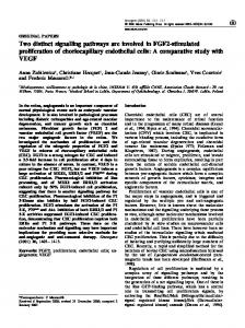
Figure 2 Effect of wortmannin and rapamycin on the stimulation of MAPK by insulin and EGF
300
F
0.1
F
00 C>:
0)2 0
Adipocytes were incubated with wortmannin (100 nM) or rapamycin (100 nM) for 30 min before the addition of 83 nM insulin or 100 nM EGF for 10 min. Results are expressed as a percentage of the control value (no additions) for MAPK activity (2.27+0.65 pmol of 32p incorporated into substrate peptide/min per g dry cell weight; n = 10) and are means+S.E.M. for the number of separate cell preparations shown in parentheses. Significance as assessed by Student's t test is indicated by a single letter code as follows: a, P < 0.05 compared with control; b, P < 0.05 compared with insulin-stimulated result; c, P < 0.05 compared with EGFstimulated result; d, P < 0.05 control + wortmannin compared with insulin or EGF + wortmannin; e, P < 0.05 control + rapamycin compared with insulin or EGF + rapamycin. The insert shows the effect of rapamycin on the phosphorylation of p7QS6K by insulin. Cells were incubated with or without insulin and rapamycin as above. The phosphorylation state of p70s6K in the cell extracts was determined by Western blotting using an anti-p70S6K antibody as described in the Experimental section. The tracks shown are: C, control; I, insulin; C+R, control plus rapamycin (20 nM); + R, insulin plus rapamycin. Similar results were obtained from -four separate cell preparations.
0.2
O0
> C 0)
m _+
0'I
P.",
(6) (6) (4) Insulin EGF Wortmannin Rapamycin
-
+
-
(5) (4)
(4) (4)
-
-
+
-
+
+
-
-
-
-
-
-
-
+
+
-
-
-
-
-
-
-
+
+
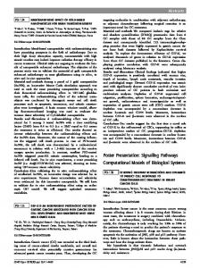
Figure 4 Effect of wortmannin and rapamycin on changes synthase activities in response to Insulin and EGF
in
GSK-3 and
glycogen
GSK-3 (a) and glycogen synthase (b) activities were determined in cell extracts prepared from adipocytes incubated as described in the legend for Figure 2. The activity of glycogen synthase is expressed as a ratio of its activity in the absence of glucose 6-phosphate to that in the presence of 15 mM glucose 6-phosphate. Extracts prepared from untreated control cells showed glycogen synthase activities of 34.02 + 2.60 and 3.53 + 0.37 nmol/min per g cell dry weight (n= 6) in the presence and absence of glucose 6-phosphate respectively. All results are expressed as means + S.E.M. of the number of separate cell preparations shown in parentheses. Significance is indicated where appropriate as given in the legend for Figure 2.
._ >
¢fr 0u, 0
Cm
C
0)
200 200-
however, had
~,0.
.-
no
detectable effect
on
GSK-3 activity in adi-
pocytes, although it has been reported to decrease its activity in
(oo-
E
0
0
Figure 3 Time
course
5
10
15 20 Time (min)
25
of the effect of insulin and EGF
30
on
GSK-3 activity
GSK-3 activity was determined in cell extracts prepared from adipocytes incubated as described in the legend for Figure 1 with 83 nM insulin (-) or 100 nM EGF (*). Results are expressed as means+S.E.M. for observations on four separate cell preparations.
kinase'. More recently it has been shown that this kinase is the a-isoform of GSK-3 [56]. Insulin has also been shown to decrease GSK-3 activity in a number of other cell types [15,16]. EGF,
A431 and NIH/3T3 cells [57,58]. Figure 4(a) shows that the effect of insulin was abolished by preincubation with wortmannin. Rapamycin was unable to block the inhibition of GSK-3 seen in response to insulin (Figure 4a). In agreement with the observed changes in GSK-3 activity, insulin increased the activity ratio of glycogen synthase approximately 3.4-fold over basal levels (Figure 4b). Although rapamycin had no effect on the stimulation of glycogen synthase by insulin, the increase in glycogen synthase activity was abolished by wortmannin. As expected from its lack of effect on GSK-3 activity, EGF did not stimulate glycogen synthase in adipocytes (see also ref. [22]).
Effects of wortmannin and rapamycin on the control of lipogenesis by insulin Wortmannin has recently been shown to block the increase in 2deoxyglucose uptake by isolated adipocytes in response to insulin
Multiple signalling pathways involved in the metabolic effects of insulin
increase in the disappearance of glucose from the medium. In contrast, Okada et al. [14], and Lin and Lawrence [22] were unable to detect any effects of EGF on either the translocation of the GLUT4 isoform of the glucose transporter to the adipocyte plasma membrane or the uptake of 2-deoxyglucose into adipo-
5-
(a) ' 4
3
.
cytes.
4)a@ 2
' 0
E1 0
50
(b) 40 0
0
30
>
* 20 10