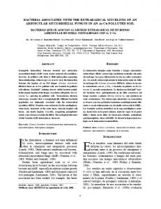
JOURNAL OF CLINICAL MICROBIOLOGY, Dec. 2009, p. 4067–4077 0095-1137/09/$12.00 doi:10.1128/JCM.00112-09 Copyright © 2009, American Society for Microbiology. All Rights Reserved.
Vol. 47, No. 12
Multiplex Detection of Bacteria Associated with Normal Microbiota and with Bacterial Vaginosis in Vaginal Swabs by Use of Oligonucleotide-Coupled Fluorescent Microspheres䌤† Tim J. Dumonceaux,1* John Schellenberg,2 Vanessa Goleski,1 Janet E. Hill,3,5 Walter Jaoko,7 Joshua Kimani,1,7 Deborah Money,4,5 T. Blake Ball,1,2,6 Francis A. Plummer,1,2 and Alberto Severini1,2 National Microbiology Laboratory, Canadian Science Centre for Human and Animal Health, 1015 Arlington St., Winnipeg, MB, Canada R3K 0R21; Departments of Medical Microbiology and Immunology, University of Manitoba, 730 William Avenue, Winnipeg, MB, Canada R3E 0W32; Department of Veterinary Microbiology, University of Saskatchewan, 52 Campus Drive, Saskatoon, SK, Canada S7N 5B43; Department of Obstetrics and Gynecology, University of British Columbia, 2H30, 4500 Oak Street, Vancouver, BC, Canada4; Women’s Health Research Institute, Provincial Health Services Authority, B325B, 4500 Oak Street, Vancouver, BC, Canada5; National HIV and Retrovirology Laboratories, Public Health Agency of Canada, 1015 Arlington St., Winnipeg MB, Canada R3K 0R26; and Department of Medical Microbiology, University of Nairobi, P.O. 19676, Nairobi, Kenya7 Received 19 January 2009/Returned for modification 2 April 2009/Accepted 10 September 2009
Bacterial vaginosis (BV) is a recurrent condition that is associated with a range of negative outcomes, including the acquisition of human immunodeficiency virus and other sexually transmitted diseases, preterm births, and pelvic inflammatory disease. In contrast to the Lactobacillus-dominated normal vaginal microbiota, BV is characterized by a lack of lactobacilli and an abundance of anaerobic and gram-negative organisms, including Gardnerella vaginalis and Atopobium vaginae. To date, the laboratory diagnosis of BV has relied upon the fulfillment of criteria determined by microscopic observation of Gram-stained vaginal swabs. We describe a molecular-based method for the easy determination of the species profile within the vaginal microbiota based on the amplification of the chaperonin-60 genes of all bacteria present in the swab and hybridization of the amplicon to species-specific oligonucleotide-coupled fluorescent beads that are identified by flow cytometry with a Luminex instrument. We designed a nineplex Luminex array for characterization of the vaginal microbiota and applied it to the analysis of vaginal swabs from individuals from Africa and North America. Using the presence of A. vaginae or G. vaginalis, or both, as the defining criterion for BV, we found that the method was highly specific and sensitive for the diagnosis of BV using microscopy as a gold standard. of Nugent et al. (29) or Ison and Hay (20). These scoring systems are based on the relative abundance of Lactobacillus morphotypes (large gram-positive rods) compared to the abundance of the bacterial morphotypes suggestive of BV (gramnegative or gram-variable coccobacilli and curved rods and gram-positive cocci). Although this method provides reliable information and is particularly well suited for use in resourcepoor settings, it requires well-trained, highly experienced individuals to interpret the results and is simplistic in its reflection of the variety of organisms present in the microbial communities present in healthy individuals with a normal vaginal flora and patients with BV. Potential problems with the subjective interpretation of the Gram stain result, along with an increasing appreciation of the fact that the complexity of the vaginal microbiota can be missed by these classical methods, have resulted in a search for other methods for the diagnosis of BV that can also identify the different species present (5, 25). In-depth studies of the makeup of vaginal microbial communities are quite common, including studies that use the older culture-based approaches (31) and more recent molecular studies that involve the construction of a clone library on the basis of the 16S rRNA gene (30) or the chaperonin-60 (cpn60) (16) target. While the information generated by these studies is critical to obtaining a greater understanding of the
Bacterial vaginosis (BV), which is defined by a reduction in the vaginal Lactobacillus populations and an increase in the number of microbial species present, including Gardnerella spp., Atopobium spp., and others, is increasingly recognized as an important risk factor for adverse reproductive health outcomes, such as miscarriage and premature birth, and sexually transmitted diseases, including human immunodeficiency virus infection (25). Despite intense investigation, the etiology and clinical course of BV have not been well defined, probably because the normal variation of the vaginal microbial communities between individuals and their dynamics over time are complex and poorly understood. Consequently, the accurate diagnosis of BV, as well as the elaboration of effective prevention and treatment strategies, remains a major challenge (26, 44). The current “gold standard” for the diagnosis of BV in the laboratory setting relies on microscopic profiling of microbial types in Gram-stained vaginal swab smears by using the criteria * Corresponding author. Present address: Agriculture and AgriFood Canada Saskatoon Research Centre, 107 Science Place, Saskatoon, SK S7N 0X2, Canada. Phone: (306) 956-7653. Fax: (306) 9567247. E-mail:
[email protected]. † Supplemental material for this article may be found at http://jcm .asm.org/. 䌤 Published ahead of print on 30 September 2009. 4067
4068
DUMONCEAUX ET AL.
J. CLIN. MICROBIOL.
TABLE 1. PCR primers and oligonucleotide capture probes used in this study Reference or source
Primer/probe name
Target
Sequence (5⬘–3⬘)a
Luminex capture probeb
Gvg56P Avg127P Lin365P Lcp155P Lgs154P Pan164P Lvg496P Pbv344P Ljn223P
G. vaginalis A. vaginae L. iners L. crispatus L. gasseri P. anaerobius L. vaginalis P. bivia L. jensenii
CCAGCAACAACATTCTTTAAGCCTTCGTG CACTCTTCATTTCTTCTACCGCAGCATTAACA TCGGCTTCCATCTTGTCATTGTCAGTAAC TGGTCTCTTGATTCAACCTTGTGGCTAATC CATCCTTAGTGCTTACCTTGTGGCTGATC TGGGCTATCTTCTCTTCTGTGTTTATCTCCTTA CTTCACCAGTAATGTCATCGGCAATAATCAGG ACGAAATAGCCTGAAAGGTAACCACGATC CCATAGCGTCAGCAATCAAGTTACCTACT
This This This This This This This This This
qPCR
Lcp1 Lcp2 Lgs1 Lgs2 Gvg1 Gvg2 Lin169 Lin239 Avg333 Avg550
L. crispatus
TCCTTACATTTTGATCACTGA GAGCTTCACCAGTAACGTC ACAAGGTAAGCACTAAGGATG CCCTTTGATTCCTCAATTGTG CGCATCTGCTAAGGATGTTG CAGCAATCTTTTCGCCAACT GCTTCTGTTTCTTCAGCATCA TCGTGTCCTACTCGTTCCATT CTTTGCAACCAACAATGACACC CAGCGCATACGGTAAGCGTAC
8
Primer/probe use
Generation of amplicon for Luminex analysis
a b
H279BP H280 H1612BP H1613
L. gasseri G. vaginalis L. iners A. vaginae Universal
Biotin-OEFOGAIIIIGCIGGIGAYGGIACIACIAC YKIYKITCICCRAAICCIGGIGCYTT Biotin-OEFOGAIIIIGCIGGYGACGGYACSACSAC CGRCGRTCRCCGAAGCCSGGIGCCTT
study study study study study study study study study
8 24 This study This study This study 17 This study 18
O, phosphorothioate-C; E, phosphorothioate-G; F, phosphorothioate-A; I, inosine; Y, C or T; R, A or G; K, T or G; S, C or G. The Luminex capture probes had a 5-amino-C12 linker at the 5⬘ end.
variability and ecological dynamics of vaginal microbial communities, they remain far too labor-intensive at present for the screening of large numbers of samples or for the routine laboratory diagnosis of BV. Most recently described molecular approaches for the diagnosis of BV are based on quantitative PCR (qPCR) of the 16S rRNA gene or other targets in bacteria extracted from vaginal samples. For example, one recent study found that the simultaneous quantification of the Gardnerella cpn60 target and the Atopobium 16S rRNA gene target resulted in the sensitive and specific diagnosis of BV in comparison to the sensitivity and specificity of the diagnosis by use of the scoring system of Nugent et al. (29). The advantages of the cpn60 universal target (UT) as an alternative to the 16S rRNA gene for the identification of bacterial species are increasingly recognized and include the increased resolution available at the species level and its presence at a single copy per genome, improving the reliability of quantitation (7). Multiplexed, bead-based flow cytometric detection and quantification of bacterial targets by use of the Luminex platform have been described previously (3, 9, 14, 38). The main advantage of this approach is its ability to detect a large number of molecular targets simultaneously after a single PCR, making high-throughput analysis of multiple samples from large study groups or longitudinal studies technically feasible. In the context of BV, the use of this multiplex approach, coupled with highly discriminatory cpn60-specific probes, facilitates screening for multiple species of Lactobacillus and a number of BV-associated bacteria simultaneously, providing an unprecedented level of information about the variability of vaginal bacterial communities, defined as those
associated with a normal vaginal flora and those associated with BV, in a single, rapid assay. The goal of the present study was the design of a flow cytometric bead array for the rapid profiling of microbial communities extracted from vaginal samples and its evaluation as a potential technique for the rapid diagnosis of BV in the laboratory setting. MATERIALS AND METHODS Study population. Samples were collected from African women as part of a larger study, ongoing since the 1980s, examining the factors associated with resistance to human immunodeficiency virus infection in a cohort of commercial sex workers in Nairobi, Kenya (22). Other samples were collected from young women from North America within the scope of a research program aimed at developing methodologies for studying the vaginal microbiota (36). The study procedures were reviewed and approved by the Health Research Ethics Boards of the University of Manitoba and the University of Nairobi. Vaginal swabs and microscopy. For the samples from Africa, swabs of the midvaginal canal were collected at the time of speculum examination and were rolled onto a glass slide, air dried, stained with Gram stain reagents, and scored by using the criteria of Nugent et al. (29) for the laboratory diagnosis of BV by two trained and experienced evaluators. Samples were collected from young women in Canada, and Gram-stained slides were evaluated by using the criteria of Ison and Hay (20), as described previously (36). For both study groups, a duplicate vaginal swab was collected simultaneously. The swab was immersed in 1 ml freezing buffer consisting of potassium phosphate buffer, NaCl, (NH4)2SO4, MgSO4, EDTA, Na2CO3, dithiothreitol, and 10% glycerol (21) within 3 h of sample collection and then stored at ⫺80°C for batch processing later. After the sample was thawed, it was eluted from the swab by vortexing, the swab head was discarded, and the eluted sample was washed twice in 1 ml phosphate-buffered saline (0.02 M sodium phosphate, 0.9% NaCl, pH 7.2). DNA was extracted by using the Instagene matrix (Bio-Rad Laboratories, Hercules, CA), according to the manufacturer’s recommendations, and was used in subsequent experiments. Species-specific cpn60 UT-directed PCR primer and oligonucleotide capture probe design. PCR primers (Table 1) that amplify the cpn60 UT from Lactobacillus iners and Atopobium vaginae to the exclusion of other species were designed
VOL. 47, 2009
FLUORESCENT MICROSPHERE DETECTION OF VAGINAL BACTERIA
by using a combination of primer design software, as described previously (7). The Beacon Designer program (version 7.0; Premier Biosoft International, Palo Alto, CA), which is written to design primer and probe sets for quantitative PCR assays, was used to design oligonucleotide capture probes targeting each of nine species chosen to represent the vaginal microbiota (Table 1). For probe design, the program was set to design a TaqMan nuclease assay with an amplicon length of 100 to 500 bp and PCR primers with a melting temperature of 60 ⫾ 1°C. The primers suggested for use by the software were not used; rather, amplicons for all bacterial target species were generated by using the cpn60 universal PCR primers, as described below. The corresponding probes were set to hybridize within 250 bp of either primer, with the optimum length being 29 to 33 bp, and to have a melting temperature of 5 ⫾ 1°C higher than that of the primers. Only antisense probes were selected. To select probes that were likely to be specific for the bacterial target of interest (Table 1), unique signatures were identified in the target cpn60 UT sequence by using SigOligo software, as described previously (7, 8). Probes that were suggested for use by the Beacon Designer program were selected on the basis of their locations within the identified signatures, and then the probes were screened against the chaperonin-60 sequence database (cpnDB; http://cpndb.cbr.nrc.ca) by using a modified BLAST algorithm (32) designed for short, nearly exact matches. Coupling of oligonucleotide capture probe to fluorescent beads. Oligonucleotide capture probes were modified by the addition of a 5-amino-C12 linker at the 5⬘ end to facilitate their covalent coupling to the carboxylated fluorescent microspheres. An aliquot of 400 l (5 ⫻ 106 beads) of a fluorescent bead suspension (Luminex Corporation, Austin, TX) was centrifuged at 14,000 ⫻ g for 1 min and resuspended in 50 l of 0.1 M 2-(N-morpholino)ethanesulfonic acid (pH 4.5; Sigma, St. Louis, MO). To this suspension was added 1 nmol of capture oligonucleotide and 2.5 l of freshly prepared aqueous 10-mg/ml 1-ethyl-3(3-dimethylaminopropyl) carbodiimide-HCl (EDC; Pierce Chemical Co., Rockford, IL), and the suspension was incubated at room temperature in the dark for 30 min. Another 2.5-l aliquot of EDC was then added, and the suspension was incubated as described above. The bead suspension was washed with 1 ml of 0.02% Tween 20, centrifuged at 14,000 ⫻ g for 1 min, and resuspended in 1 ml 0.1% sodium dodecyl sulfate. The beads were centrifuged again and resuspended in 100 l of TE buffer (10 mM Tris-HCl, pH 8.0, 1 mM EDTA). The concentration of beads was determined with a Coulter counter (Beckman Coulter, Fullerton, CA). The beads were stored at 4°C. For the preparation of the bead master mix, each of the different bead suspensions was mixed at a concentration of 100 beads/l in TE buffer. Generation of amplicon for analysis with the Luminex instrument. Plasmid templates or vaginal swab DNA extracts were used as the templates for the PCR with modified universal cpn60-targeted primers (17). To maximize the community representation, a cocktail of universal primers was used, as described previously (18). For analysis with the Luminex instrument, the two universal PCR primers that amplify the 5⬘ end of the cpn60 UT were modified by the addition of biotin and four phosphorothioated bases at the 5⬘ end of the primer (Table 1). The phosphorothioate modification protects the sense strand of the cpn60 amplicon from degradation by the double-strand-specific T7 exonuclease during processing for analysis with the Luminex instrument (27). The PCR proceeded in 1⫻ PCR buffer (Invitrogen) containing 2.5 mM MgCl2, 200 M each deoxynucleoside triphosphate, 125 nM each of primers H279BP and H280, 375 nM each of primers H1612BP and H1613, and 2.5 U of Taq DNA polymerase (Invitrogen). The PCR conditions were 95°C for 2 min, followed by 40 cycles of 95°C for 30 s, 42°C for 30 s, and 72°C for 30 s, followed by a final extension at 72°C for 2 min. Analysis of cpn60 UT amplicons with the Luminex instrument. The parameters of the Luminex hybridization assay, including the optimal T7 exonuclease exposure time, the optimal hybridization time, and the optimal hybridization temperature, were optimized prior to use with clinical samples (data not shown). The PCR product was treated with 20 U of T7 exonuclease (New England Biolabs, Pickering, Ontario, Canada) at room temperature for 40 min. Seventeen microliters of the single-stranded PCR product was denatured at 95°C for 10 min, after which 33 l of the fluorescent bead master mix was added. Hybridization of the oligonucleotide-coupled fluorescent beads to the single-stranded PCR product proceeded at 60°C for 10 min, followed by the addition of 25 l of streptavidin–R-phycoerythrin (Invitrogen) diluted to 20 g/ml in 3 M tetramethyl ammonium chloride (Sigma)–0.1% Sarkosyl–50 mM Tris-HCl, pH 8.0–4 mM EDTA. This mixture was incubated at 60°C for ⬃5 min prior to analysis on a Luminex instrument (Qiagen). The sample volume was 50 l, and the heater temperature was 60°C. The Luminex instrument reported the mean fluorescence intensity (MFI) of the phycoerythrin reporter dye for each of the coupled bead types. For statistical determination of sample positivity, samples were tested in triplicate or quadruplicate and the MFI was compared to that for the no-
4069
TABLE 2. Composition of the mixed vaginal panela Organism
Source
Lactobacillus iners ....................................................Library clone Lactobacillus crispatus .............................................Library clone Lactobacillus jensenii................................................Library clone Lactobacillus vaginalis..............................................Vaginal swab isolate Lactobacillus gasseri .................................................Library clone Gardnerella vaginalis ................................................Vaginal swab isolate Peptostreptococcus anaerobius.................................Vaginal swab isolate Prevotella bivia ..........................................................Library clone Bifidobacterium bifidum...........................................Library clone Atopobium vaginae ...................................................Library clone Streptococcus lutetiensis ...........................................Vaginal swab isolate Lactobacillus sp. strain L6 ......................................Vaginal swab isolate Bifidobacterium infantis ...........................................Vaginal swab isolate Lactobacillus salavarius............................................Vaginal swab isolate Lactobacillus sp. strain N27....................................Vaginal swab isolate Lactobacillus sp. strain N19....................................Vaginal swab isolate Lactobacillus sp. strain N20....................................Vaginal swab isolate Lactobacillus johnsonii.............................................Vaginal swab isolate Lactobacillus plantarum...........................................Vaginal swab isolate Streptococcus gallolyticus .........................................Library clone a Each organism was represented by its cloned cpn60 UT PCR product and was mixed in equal copy numbers.
template control by a one-tailed Student’s t test, with significance being set at a P value of 0.001. Diagnostic sensitivity and specificity of the assay with the Luminex instrument compared with those of microscopy. To compare the method with the Luminex instrument to the gold standard of microscopy by using the methods of Nugent et al. (29) or Ison and Hay (20), we considered samples that were positive by the criterion presented above, that is, for the sample to contain Gardnerella vaginalis or A. vaginae, or both, to be from women with BV and samples in which neither organism was detected to be from individuals with a normal vaginal flora. Analysis with the Luminex instrument was performed in a blinded manner, such that the microscopy results were unknown prior to the analysis with the Luminex instrument and correlations were done afterwards. Sensitivity and specificity, along with 95% confidence intervals, were calculated by standard procedures (2). Determination of oligonucleotide capture probe target specificity. In order to evaluate whether the oligonucleotide capture probes specifically hybridized to the target of interest, we constructed an artificial vaginal microbiota sample (referred to as the “mixed vaginal panel”), which consisted of equal numbers of copies of cloned cpn60 UT PCR products from 20 species relevant to the vaginal tract microbiota (Table 2). Each probe was evaluated with the mixed vaginal panel and the cpn60 UT amplicon generated as described above (test sample) or with the mixed vaginal panel without the target cpn60 UT (control sample) as the template DNA. The signals generated by the test and control samples were compared to the signal generated from the no-template control. Probes that generated the strongest statistically significant positive signal from the test sample and a statistically significant negative control sample were chosen for each of the target species. qPCR. The bacteria in vaginal swab DNA extracts were quantified by qPCR with species-specific cpn60 UT-targeted PCR primers (Table 1) and a standard curve consisting of serial dilutions of cloned cpn60 UT plasmids, as described previously (7). Briefly, 2 l of template DNA was added to a solution of 1⫻ SYBR green master mix (Applied Biosystems, Foster City, CA) and 500 nM of each primer in a final volume of 25 l. The reactions were quantified with an ABI 7500 Fast system (Applied Biosystems). Quantification of L. iners used 1⫻ LC480 SYBR green master mix (Roche) in a final volume of 20 l and a LightCycler 480 quantitative PCR apparatus (Roche). The results were expressed as the number of genomes detected per qPCR (7).
RESULTS Target template specificity of each oligonucleotide capture probe. In order to verify that the hybridization signal measured for each of the nine capture probes reflected the detection of the target of interest, an amplicon was generated by using the
4070
DUMONCEAUX ET AL.
artificial mixed vaginal panel as the template. Three to five probes were evaluated for each of the target organisms. As shown in Fig. 1, the probes that were designed had various performance characteristics in this artificial context, even though they all showed little sequence similarity to anything other than the target template in cpnDB. Nevertheless, it was possible to choose at least one acceptable probe for each of the target organisms. The probes that were chosen for each of the target bacteria on the basis of this analysis are shown in Table 1. Nineplex assay with plasmid templates. To determine whether the simultaneous detection of all targets postamplification was feasible, equivalent volumes of the amplicon generated from each target template were mixed and analyzed by use of a nineplex Luminex bead master mix. While the signal amplitude varied for each of the probes, from approximately 300 MFI for the L. jensenii probe to about 1,200 MFI for the G. vaginalis probe, it was possible to detect all of the targets simultaneously in this artificial context (Fig. 2). Quantification of target template amount by the Luminex assay. Since the amount of template DNA should correlate with the amount of product generated, at least over a limited range of concentrations, we determined the effect of the amount of input template DNA on the MFIs for four targets: L. iners, Peptostreptococcus anaerobius, G. vaginalis, and A. vaginae. Typical standard curves generated with the L. iners probe are shown in Fig. 3. With a complex template (mixed vaginal panel), the correlation between the input template amount and the signal amplitude was excellent over the range of ⬃5 ⫻ 105 to 50 copies of each template (r2 values, 0.97 for L. iners, 0.97 for G. vaginalis, 0.98 for A. vaginae, and 0.99 for P. anaerobius). Single-target templates typically had poorer correlations over the same range of template amounts (r2 values, 0.97 for L. iners, 0.89 for G. vaginalis, 0.82 for A. vaginae, and 0.95 for P. anaerobius). For both single-target and complex templates, the MFI did not increase linearly with the target template amount beyond ⬃106 copies of template DNA (not shown). qPCR and Luminex assay analysis of vaginal swabs from individuals from Africa. Vaginal swabs from a cohort of African sex workers were analyzed by microscopy and were scored for BV by using the criteria of Nugent et al. (29). DNA was extracted from the same samples and was used to detect and quantify individual species by qPCR targeting G. vaginalis, A. vaginae, L. iners, L. crispatus, and L. gasseri. Primer sets targeting the cpn60 UT of G. vaginalis, L. crispatus, and L. gasseri were published previously (Table 1); and similar primer sets targeting A. vaginae and L. iners were designed for this study. Validation of the applicability of these primer sets is presented in Fig. S6 in the supplemental material. The efficacy of the DNA extraction technique with the Instagene matrix was investigated by spiking vaginal swabs with serial 10-fold dilutions of Bifidobacterium animalis cells and determining the recovery of B. animalis DNA in the extract by qPCR with cpn60-directed primers specific for B. animalis. This experiment confirmed that B. animalis DNA was recovered in the expected proportions by using Instagene matrix (data not shown). A fiveplex Luminex assay was employed with the same samples and probes for detection of the same organisms. Nine samples considered to be from individuals with a normal vaginal flora
J. CLIN. MICROBIOL.
(score, 1) and nine samples considered to be from individuals with a diagnosis of BV (score, 3) were included in the analysis. There was, in general, good agreement between the results of the analysis by use of the Luminex instrument (defined as positive or negative, where a positive result is an MFI that is significantly greater than that for the negative control at P ⬍ 0.001; see Materials and Methods) and the results of qPCR analysis (Fig. 4). The few exceptions served to illustrate the detection limits of the method with the Luminex instrument with complex templates: samples with less than ⬃104 genomes in the qPCR were typically negative by analysis by the Luminex assay, although there were some exceptions in which samples with higher levels were negative (e.g., the sample from individual 31 for L. iners). None of the samples that were scored as being from individuals with a normal vaginal flora by microscopy were positive by use of the Luminex instrument for either of the BV-associated organisms, G. vaginalis and A. vaginae, but all were positive for one or more of the Lactobacillus species by use of the Luminex instrument and qPCR (Fig. 4). Among the nine samples that were scored as being from individuals with BV by microscopy, all were positive for G. vaginalis and/or A. vaginae by use of the Luminex instrument. Seven of the samples from individuals with BV and seven of the samples from individuals with a normal vaginal flora contained some level of L. iners that was detectable by use of the Luminex instrument and/or qPCR, but all of the samples from individuals with BV were negative for L. crispatus by use of the Luminex instrument, and only two samples (samples 77 and 104) had low levels of this organism, as measured by qPCR. In contrast, samples from five of the nine healthy individuals contained relatively high levels of L. crispatus. Longitudinal analysis of vaginal swabs from individuals from North America. Self-sampled vaginal swabs were collected from 16 female adolescents from North America at multiple time points (36) and were analyzed by the use of microscopy, qPCR, and the Luminex instrument. Detailed results comparing the results obtained by microscopy and with the Luminex instrument for selected individuals are shown in Fig. 5. As was observed in the samples from Africa, there was generally good agreement between the results of qPCR and those obtained by use of the Luminex instrument for the five targets sought by qPCR (G. vaginalis, A. vaginae, L. iners, L. crispatus, and L. gasseri); samples that were positive by qPCR but negative by use of the Luminex instrument tended to have low levels of the target organism (data not shown). By considering only the results for samples that were positive by both qPCR and with the Luminex instrument, there was a statistically significant positive correlation at the 0.05 significance level between the MFIs and the quantities determined by qPCR for all targets analyzed except L. crispatus (Table 3). This is consistent with the positive correlations between the number of target template input copies and the MFIs at lower copy numbers observed by using plasmid templates (Fig. 3). Comparison of the results obtained with the Luminex instrument and by microscopy for individuals over multiple time points yielded some insight into the composition and dynamics of the vaginal microbiota. For example, many of the samples that scored normal by microscopy showed that one individual (individual 1) or more individuals (individuals 13 and 14) were infected with Lactobacillus spp. by analysis with the Luminex
VOL. 47, 2009
FLUORESCENT MICROSPHERE DETECTION OF VAGINAL BACTERIA
4071
FIG. 1. Template specificity for the Luminex capture probes evaluated in this study. For each candidate probe targeting each organism, templates consisted of water (white bars), the mixed vaginal panel including the target organism (gray bars), or the mixed vaginal panel excluding the target organism (black bars). The MFI of each probe for each of the target organisms was measured in triplicate, and the results are expressed as means ⫾ standard deviations. The probes that were ultimately chosen for inclusion in the Luminex array are indicated by an asterisk. In addition to the eight targets shown here, three probes that targeted Lactobacillus vaginalis (data not shown) were evaluated, leading to the selection of probe Lvg496p (Table 1).
4072
DUMONCEAUX ET AL.
J. CLIN. MICROBIOL.
FIG. 2. Nineplex detection of cloned cpn60 UT plasmid mixtures by use of the Luminex instrument. An amplicon was generated from each target organism separately and was then mixed postamplification and hybridized to the nineplex bead mix (gray bars). White bars, no-template control. Values are shown as the means of duplicate measurements ⫾ standard deviations.
instrument. In contrast, samples from individuals with BV tended to have a more diverse vaginal microbiota with few or no lactobacilli but with A. vaginae, G. vaginalis, Prevotella bivia, and, in some cases, L. iners being detected (individuals 5, 11, 16, and 25). The composition of the vaginal microbiota of certain individuals changed over the time course of this analysis, illustrating the shifts in the composition of the microbiota that are detectable by the use of this method. For example, individual 11 changed from having BV to having a normal vaginal flora over the course of the analysis, and this was reflected in the profile of the vaginal microbiota that was revealed by analysis with the Luminex instrument. For this individual, the MFI observed for G. vaginalis came to a peak and then waned, while the signal obtained for L. iners increased steadily, consistent with the observed transition from BV to a normal microbiota over this time course. The opposite transition was observed for individual 16; at the first three time points that individual scored as having a normal vaginal flora and displayed a steadily decreasing MFI for L. iners (Fig. 5), while at the final time point the individual was scored as having BV and displayed a mixed microbiota that included A. vaginae, G. vaginalis, P. bivia, and L. iners.
FIG. 3. Standard curves correlating input target template amount with the postamplification MFI. Amplicons were generated by using the mixed vaginal panel (MVP; 䡺) or equivalent copy numbers of L. iners cpn60 UT plasmid DNA (f) and were analyzed in duplicate with a fourplex bead mixture. The results are shown as means ⫾ standard deviations.
Sensitivity and specificity of use of Luminex instrument for diagnosis of BV. The results of the analysis with the Luminex instrument with samples from women from Africa and North America (Fig. 4; Fig. 5) showed that there was reasonable agreement between the diagnosis of BV by traditional microscopy and the detection of G. vaginalis and/or A. vaginae by the method with the Luminex instrument described here. By using a definition of BV as the detection of one or both of these organisms, the specificity and the sensitivity of the method with the Luminex instrument compared to those of microscopy were calculated. The sensitivity of the method with the Luminex instrument for the diagnosis of BV was lower for the samples from North America (0.86) than for the samples from Africa (1.00), and the overall sensitivity of the method was 0.90 (Table 4). The method was also highly specific; samples in which neither of the two organisms was detected were likely to have been considered to be from individuals with a normal vaginal flora by microscopy (Table 4).
DISCUSSION To our knowledge, this is the first time that a Luminex instrument-based analysis has been applied to the evaluation of vaginal microbial communities. We describe a novel method for characterizing the vaginal microbiota based on the amplification of the cpn60 UT of all bacteria present in a vaginal swab using universal PCR primers (18), followed by the detection of individual species using oligonucleotides coupled to fluorescent beads and flow cytometry. This method is conceptually similar to previously described methods for the detection and typing of cultured isolates or environmental DNA (6, 10, 39), but it offers the additional advantage of the species specificity that is achievable by the use of cpn60-based probes (7). This method also compares favorably to whole-genome hybridization, as described by Nikolaitchouk et al. (28), which provides a similar snapshot of the vaginal microbiota but which is necessarily limited to the analysis of cultured isolates. We have targeted nine bacterial species for oligonucleotide capture probe development and have shown that it is feasible to design probes specific for all of the targeted species. Our choice of bacterial targets was based on a literature survey of isolates that have been cultured from the vaginal microbiota. Given the increasing amount of information regarding the
VOL. 47, 2009
FLUORESCENT MICROSPHERE DETECTION OF VAGINAL BACTERIA
4073
FIG. 4. Paired Luminex and qPCR analysis of samples from individuals from Africa who were classified as having a normal vaginal flora (A) or as having BV (B) by the criteria of Nugent et al. (29). Samples were analyzed by a fiveplex Luminex method for the detection of G. vaginalis, A. vaginae, L. iners, and L. crispatus, as well as L. gasseri (data not shown). ⴱ, statistically significantly positive Luminex assay results (P ⬍ 0.001).
4074
DUMONCEAUX ET AL.
J. CLIN. MICROBIOL.
VOL. 47, 2009
FLUORESCENT MICROSPHERE DETECTION OF VAGINAL BACTERIA
TABLE 3. Spearman rank correlation between qPCR and Luminex-positive MFI values for different target organismsa Target organism
P
No. of degrees of freedom
G. vaginalis L. iners L. crispatus A. vaginae All pooled
0.6522 0.4756 0.4235 0.6892 0.4686
0.0028 0.017 0.099 0.0098 0.00001
20 24 14 13 81
a
The correlations and P values were calculated as described by Wessa (43).
composition of the vaginal microbiota accruing from cultureindependent approaches (16, 19, 40, 42, 46), the first-generation vaginal Luminex array described here could easily be improved upon to yield more information regarding the composition of a vaginal sample. We have successfully developed a 46-plex Luminex instrument-based test for the detection of human papillomaviruses (V. Goleski and A. Severini, unpublished data), and it is theoretically possible to include up to 99 different probes in each well of a Luminex array. Therefore, there is considerable room to expand upon the current Luminex array for vaginal bacteria to include other relevant targets. In addition to offering a means of characterizing the organisms in a complex sample quickly and easily, we found that this approach is semiquantitative, in that the MFI reading correlates to the number of copies of target DNA used as the template. This correlation is strongly linear at lower copy numbers (⬃50 to 500,000 input copies), particularly in the case of mixed templates, but the MFI becomes saturated at higher input copy numbers. The lower dynamic range of the Luminex assay compared with that of qPCR is an expected result, since the Luminex assay is a postamplification method. Furthermore, even though the Luminex assay MFI data are linear and the qPCR data are logarithmic, we observed positive correlations between these two types of quantitative data (Table 3), indicating that changes in the amplitude of a Luminex assay result for a given organism over time may be indicative of trends in the abundance of that organism. An elegant mathematical model that is able to compute the number of copies of a given target DNA present in a mixed amplicon by a fluorescent bead-based approach has been described (38), and this method has been used to characterize the dynamics of individual gastrointestinal communities (45). Such a method might also be useful in the current context; however, as a technique based on the analysis of the PCR endpoint, the results of the analysis with the Luminex instrument can best be viewed as being semiquantitative and only when lower template copy numbers are used. We used the Luminex method to characterize the vaginal microbiota in a cohort of African sex workers. We found that
4075
individuals with a normal vaginal microbiota, as defined by microscopy, typically contained one or more of the Lactobacillus species analyzed and undetectable levels of A. vaginae and G. vaginalis. In contrast, samples from individuals with BV were much more likely to contain A. vaginae and/or G. vaginalis DNA. African individuals with BV often had detectable levels of L. iners but not L. crispatus. These results are consistent with those presented in other reports describing the association of A. vaginae and G. vaginalis with BV (4, 11–13, 15, 24, 35, 41). We used an expanded nineplex analysis of a longitudinal series of samples from adolescents from North America and compared the results of the Luminex assay with those of microscopy and qPCR. In general, the agreement among the methods was excellent; the microbiota of individuals who scored normal by microscopy was usually dominated by one or more of the lactobacilli for which assays were conducted, and individuals with BV generally had more complex microbiota, with A. vaginae and G. vaginalis commonly being detected. We noted that some of the samples from individuals with BV contained both G. vaginalis and P. bivia (individuals 5 and 16) and that these samples had low levels of lactobacilli. Furthermore, samples containing L. crispatus or L. gasseri had undetectable levels of G. vaginalis and P. bivia, while L. iners could be found in either samples from individuals with a normal vaginal flora or samples from individuals with BV. These observations are consistent with the symbiotic relationship between G. vaginalis and P. bivia that has been reported (34) and with the ability of certain vaginal lactobacilli to inhibit the growth of G. vaginalis and P. bivia (1). However, despite the symbiotic relationship between P. bivia and P. anaerobius (33), the latter organism was not found in any of the samples. Considering the samples from women from both Africa and North America, we determined the sensitivity and the specificity of this multiplex bead-based method for the diagnosis of BV using microscopy as a gold standard. By using only the presence of A. vaginae or G. vaginalis, or both, as the defining criterion for BV, the method described here performed very well compared to the performance of microscopy and provided additional information regarding the species composition of each sample. The difference in sensitivity between the two sample groups that was measured may relate to the different scoring systems that were used, since the samples from Africa were evaluated by using the criteria of Nugent et al. (29), while the samples from North America were evaluated by using the criteria of Ison and Hay (20). While the overall sensitivity of the Luminex method was not as high as that of the qPCRbased method described by Menard et al. (24), we expect that this method could be improved upon by adding other BVassociated targets to the Luminex array and/or by increasing the number of replicates analyzed for each individual. Since BV is a polymicrobial syndrome with unique individual profiles
FIG. 5. Longitudinal nineplex analysis with the Luminex instrument of vaginal swabs from selected individuals in North America. Each swab was analyzed by microscopy and was scored by using the criteria of Ison and Hay (20); a diagnosis of BV is indicated above each time point. DNA extracts were taken from the same swabs and analyzed with the Luminex instrument for the presence of each of the bacterial species targeted in this study. The MFI of each probe was measured in duplicate assays, and the results were compared to those for the no-template control. Error bars indicate standard deviations. ⴱ, statistically significant positive results (P ⬍ 0.001). The results obtained with the Luminex instrument for L. vaginae and P. anaerobius were negative for all samples and are not shown.
4076
DUMONCEAUX ET AL.
J. CLIN. MICROBIOL.
TABLE 4. Sensitivity and specificity of the cpn60 UT-targeted oligonucleotide probe-based method for diagnosis of BV by microscopya Samples from individuals with a normal vaginal flora (BV negative)
Samples from patients with BV (BV positive) Vaginal swab source
Africa North America Total
No. of samples
No. of samples
Luminex assay positive
Total no. of samples tested
Sensitivity (%)
Luminex assay negative
Total no. of samples tested
Specificity (%)
9 18
9 21
1.00 (0.63–1.00)b 0.86 (0.63–0.96)
9 23
9 27
1.00 (0.63–1.00) 0.85 (0.65–0.95)
27
31
0.90 (0.72–0.97)
32
35
0.89 (0.73–0.96)
a
The samples were scored for BV by using as the gold standard the criteria of Nugent et al. (29) for samples from Africa and the criteria of Ison and Hay (20) for the samples from North America. By using the criteria of Nugent et al., (29), a score of 1 to 3 was considered normal, while a score of 7 to 10 was considered diagnostic of BV. By using the criteria of Ison and Hay (20), a score of 1 or 2 was considered normal, while a score of 3 was considered diagnostic of BV. Samples were scored BV positive by the Luminex assay if the MFI was significantly greater (P ⬍ 0.001, one-tailed Student’s t test) than the MFI for the controls for either G. vaginalis or A. vaginae, or both. Samples were scored BV negative if neither of these two organisms were detected. b Values in parentheses are 95% confidence intervals.
(15), it is important to inform the choice of probes by sequence analysis of the DNA from a range of individuals with BV. Some of the samples showed the limitations of the current Luminex array. For example, individual 31, who was from North America, was scored as having BV at time zero, yet the only organism detected was L. crispatus; it is likely that organisms not represented in the current array were responsible for the diagnosis of BV. Even though two of the probes targeted organisms that are known to be associated with the vaginal microbiota (L. vaginalis and P. anaerobius), use of those probes did not yield positive results for any of the samples, suggesting that these organisms were not present in quantities in the sample sufficient to be detected by this method. In addition, all samples were negative by use of another probe targeting Bifidobacterium bifidum (data not shown), an organism associated with the vaginal microbiota (23). Consistent with this finding, we did not find these organisms in any appreciable abundance by ultradeep-pyrosequencing analysis of the vaginal microbiota of the cohort from Africa and the samples from Winnipeg, Manitoba, Canada (37; J. Schellenberg et al., unpublished data). Future Luminex arrays that target the vaginal microbiota will benefit from culture-independent analysis in order to guide the choice of probes. For example, there is an abundance of uncultivated Megasphaera-like organisms that are unique to the vaginal microbiota and that would be good targets for probe development (46); we have observed similar Megasphaera-like species in cpn60-based culture-independent studies of the vaginal microbiota in individuals from Africa (Schellenberg et al., unpublished). In summary, we describe a multiplex method for the detection and generation of semiquantitative data for organisms within the vaginal microbiota based on the amplification of the cpn60 UT and hybridization to species-specific oligonucleotidecoupled fluorescent beads. This method is rapid and simple, and it provides a means of identifying and quantifying the key species present in a vaginal swab at a given point in time. Since BV is often a transient, recurring condition that is difficult to treat (44), diagnostic methods that facilitate the longitudinal profiling of the vaginal microbiota are desirable. By the method that we describe, it was possible to detect the presence of up to nine species in a single PCR; and further multiplexing, which would take advantage of the full capabilities of the
Luminex instrument, is certainly possible. The ease of specific oligonucleotide capture probe design and validation, along with the ability to multiplex the detection and quantification of bacterial species, makes this approach very attractive for characterization of the vaginal microbiota. Future expansion and improvement of the Luminex array with information obtained from in-depth molecular studies will allow the detailed profiling of vaginal microbial communities, as well as the rapid and reproducible diagnosis of BV in the clinical setting. ACKNOWLEDGMENTS We thank all research participants, as well as Charles Wachihi, Jane Njeri Mungai, and the clinical/laboratory staff at the University of Nairobi, Nairobi, Kenya, and Margo Lane, Heather McIntosh, Charisse Maliwanag, and the staff at Children’s Hospital, Health Sciences Centre, Winnipeg, Manitoba, Canada. REFERENCES 1. Atassi, F., D. Brassart, P. Grob, F. Graf, and A. L. Servin. 2006. Lactobacillus strains isolated from the vaginal microbiota of healthy women inhibit Prevotella bivia and Gardnerella vaginalis in coculture and cell culture. FEMS Immunol. Med. Microbiol. 48:424–432. 2. Banoo, S., D. Bell, P. Bossuyt, A. Herring, D. Mabey, F. Poole, P. G. Smith, N. Sriram, C. Wongsrichanalai, R. Linke, R. O’Brien, M. Perkins, J. Cunningham, P. Matsoso, C. M. Nathanson, P. Olliaro, R. W. Peeling, and A. Ramsay. 2006. Evaluation of diagnostic tests for infectious diseases: general principles. Nat. Rev. Microbiol. 4:S21–S31. 3. Baums, I. B., K. D. Goodwin, T. Kiesling, D. Wanless, M. R. Diaz, and J. W. Fell. 2007. Luminex detection of fecal indicators in river samples, marine recreational water, and beach sand. Mar. Pollut. Bull. 54:521–536. 4. Bradshaw, C. S., S. N. Tabrizi, C. K. Fairley, A. N. Morton, E. Rudland, and S. M. Garland. 2006. The association of Atopobium vaginae and Gardnerella vaginalis with bacterial vaginosis and recurrence after oral metronidazole therapy. J. Infect. Dis. 194:828–836. 5. Brotman, R. M., and J. Ravel. 2008. Ready or not: the molecular diagnosis of bacterial vaginosis. Clin. Infect. Dis. 47:44–46. 6. Diaz, M. R., and J. W. Fell. 2004. High-throughput detection of pathogenic yeasts of the genus Trichosporon. J. Clin. Microbiol. 42:3696–3706. 7. Dumonceaux, T. J., J. E. Hill, S. A. Briggs, K. K. Amoako, S. M. Hemmingsen, and A. G. Van Kessel. 2006. Enumeration of specific bacterial populations in complex intestinal communities using quantitative PCR based on the chaperonin-60 target. J. Microbiol. Methods 64:46–62. 8. Dumonceaux, T. J., J. E. Hill, S. M. Hemmingsen, and A. G. Van Kessel. 2006. Characterization of intestinal microbiota and response to dietary virginiamycin supplementation in the broiler chicken. Appl. Environ. Microbiol. 72:2815–2823. 9. Dunbar, S. A., and J. W. Jacobson. 2007. Quantitative, multiplexed detection of Salmonella and other pathogens by Luminex xMAP suspension array. Methods Mol. Biol. 394:1–19. 10. Dunbar, S. A., C. A. Vander Zee, K. G. Oliver, K. L. Karem, and J. W. Jacobson. 2003. Quantitative, multiplexed detection of bacterial pathogens:
VOL. 47, 2009
11.
12.
13.
14.
15. 16.
17.
18.
19.
20.
21.
22.
23.
24.
25. 26. 27.
28.
FLUORESCENT MICROSPHERE DETECTION OF VAGINAL BACTERIA
DNA and protein applications of the Luminex LabMAP system. J. Microbiol. Methods 53:245–252. Ferris, M. J., A. Masztal, K. E. Aldridge, J. D. Fortenberry, P. L. Fidel, Jr., and D. H. Martin. 2004. Association of Atopobium vaginae, a recently described metronidazole resistant anaerobe, with bacterial vaginosis. BMC Infect. Dis. 4:5. Fredricks, D. N., T. L. Fiedler, and J. M. Marrazzo. 2005. Molecular identification of bacteria associated with bacterial vaginosis. N. Engl. J. Med. 353:1899–1911. Fredricks, D. N., T. L. Fiedler, K. K. Thomas, B. B. Oakley, and J. M. Marrazzo. 2007. Targeted PCR for detection of vaginal bacteria associated with bacterial vaginosis. J. Clin. Microbiol. 45:3270–3276. Gilmour, M. W., D. M. Tracz, A. K. Andrysiak, C. G. Clark, S. Tyson, A. Severini, and L. K. Ng. 2006. Use of the espZ gene encoded in the locus of enterocyte effacement for molecular typing of Shiga toxin-producing Escherichia coli. J. Clin. Microbiol. 44:449–458. Hay, P. 2005. Life in the littoral zone: lactobacilli losing the plot. Sex. Transm. Infect. 81:100–102. Hill, J. E., S. H. Goh, D. M. Money, M. Doyle, A. Li, W. L. Crosby, M. Links, A. Leung, D. Chan, and S. M. Hemmingsen. 2005. Characterization of vaginal microflora of healthy, nonpregnant women by chaperonin-60 sequence-based methods. Am. J. Obstet. Gynecol. 193:682–692. Hill, J. E., R. P. Seipp, M. Betts, L. Hawkins, A. G. Van Kessel, W. L. Crosby, and S. M. Hemmingsen. 2002. Extensive profiling of a complex microbial community by high-throughput sequencing. Appl. Environ. Microbiol. 68: 3055–3066. Hill, J. E., J. R. Town, and S. M. Hemmingsen. 2005. Improved template representation in cpn60 polymerase chain reaction (PCR) product libraries generated from complex templates by application of a specific mixture of PCR primers. Environ. Microbiol. 8:741–746. Hyman, R. W., M. Fukushima, L. Diamond, J. Kumm, L. C. Giudice, and R. W. Davis. 2005. Microbes on the human vaginal epithelium. Proc. Natl. Acad. Sci. USA 102:7952–7957. Ison, C. A., and P. E. Hay. 2002. Validation of a simplified grading of Gram stained vaginal smears for use in genitourinary medicine clinics. Sex. Transm. Infect. 78:413–415. Kilic, A. O., S. I. Pavlova, S. Alpay, S. S. Kilic, and L. Tao. 2001. Comparative study of vaginal Lactobacillus phages isolated from women in the United States and Turkey: prevalence, morphology, host range, and DNA homology. Clin. Diagn. Lab. Immunol. 8:31–39. Kimani, J., R. Kaul, N. J. Nagelkerke, M. Luo, K. S. MacDonald, E. Ngugi, K. R. Fowke, B. T. Ball, A. Kariri, J. Ndinya-Achola, and F. A. Plummer. 2008. Reduced rates of HIV acquisition during unprotected sex by Kenyan female sex workers predating population declines in HIV prevalence. AIDS 22:131–137. Korshunov, V. M., Z. A. Gudieva, B. A. Efimov, A. P. Pikina, V. V. Smeianov, G. Reid, O. V. Korshunova, V. L. Tiutiunnik, and I. I. Stepin. 1999. The vaginal Bifidobacterium flora in women of reproductive age. Zh. Mikrobiol. Epidemiol. Immunobiol. 4:74–78. Menard, J. P., F. Fenollar, M. Henry, F. Bretelle, and D. Raoult. 2008. Molecular quantification of Gardnerella vaginalis and Atopobium vaginae loads to predict bacterial vaginosis. Clin. Infect. Dis. 47:33–43. Money, D. 2005. The laboratory diagnosis of bacterial vaginosis. Can. J. Infect. Dis. Med. Microbiol. 16:77–79. Morris, M., A. Nicoll, I. Simms, J. Wilson, and M. Catchpole. 2001. Bacterial vaginosis: a public health review. Br. J. Obstet. Gynecol. 108:439–450. Nikiforov, T. T., R. B. Rendle, M. L. Kotewicz, and Y. H. Rogers. 1994. The use of phosphorothioate primers and exonuclease hydrolysis for the preparation of single-stranded PCR products and their detection by solid-phase hybridization. PCR Methods Appl. 3:285–291. Nikolaitchouk, N., B. Andersch, E. Falsen, L. Strombeck, and I. MattsbyBaltzer. 2008. The lower genital tract microbiota in relation to cytokine-,
29.
30.
31.
32. 33.
34.
35.
36.
37.
38.
39.
40.
41.
42.
43. 44. 45.
46.
4077
SLPI- and endotoxin levels: application of checkerboard DNA-DNA hybridization (CDH). APMIS 116:263–277. Nugent, R. P., M. A. Krohn, and S. L. Hillier. 1991. Reliability of diagnosing bacterial vaginosis is improved by a standardized method of gram stain interpretation. J. Clin. Microbiol. 29:297–301. Oakley, B. B., T. L. Fiedler, J. M. Marrazzo, and D. N. Fredricks. 2008. Diversity of human vaginal bacterial communities and associations with clinically defined bacterial vaginosis. Appl. Environ. Microbiol. 74:4898– 4909. Onderdonk, A. B., G. R. Zamarchi, J. A. Walsh, R. D. Mellor, A. Munoz, and E. H. Kass. 1986. Methods for quantitative and qualitative evaluation of vaginal microflora during menstruation. Appl. Environ. Microbiol. 51:333– 339. Pearson, W. R., and D. J. Lipman. 1988. Improved tools for biological sequence comparison. Proc. Natl. Acad. Sci. USA 85:2444–2448. Pybus, V., and A. B. Onderdonk. 1998. A commensal symbiosis between Prevotella bivia and Peptostreptococcus anaerobius involves amino acids: potential significance to the pathogenesis of bacterial vaginosis. FEMS Immunol. Med. Microbiol. 22:317–327. Pybus, V., and A. B. Onderdonk. 1997. Evidence for a commensal, symbiotic relationship between Gardnerella vaginalis and Prevotella bivia involving ammonia: potential significance for bacterial vaginosis. J. Infect. Dis. 175:406– 413. Rodriguez Jovita, M., M. D. Collins, B. Sjoden, and E. Falsen. 1999. Characterization of a novel Atopobium isolate from the human vagina: description of Atopobium vaginae sp. nov. Int. J. Syst. Bacteriol. 49(Pt 4):1573–1576. Schellenberg, J., T. Blake Ball, M. Lane, M. Cheang, and F. Plummer. 2008. Flow cytometric quantification of bacteria in vaginal swab samples selfcollected by adolescents attending a gynecology clinic. J. Microbiol. Methods 73:216–226. Schellenberg, J., M. G. Links, J. E. Hill, T. J. Dumonceaux, G. A. Peters, S. D. Tyler, T. B. Ball, A. Severini, and F. A. Plummer. 2009. Pyrosequencing of the chaperonin-60 universal target as a tool for determining microbial community composition. Appl. Environ. Microbiol. 75:2889–2898. Spiro, A., and M. Lowe. 2002. Quantitation of DNA sequences in environmental PCR products by a multiplexed, bead-based method. Appl. Environ. Microbiol. 68:1010–1013. Spiro, A., M. Lowe, and D. Brown. 2000. A bead-based method for multiplexed identification and quantitation of DNA sequences using flow cytometry. Appl. Environ. Microbiol. 66:4258–4265. Sundquist, A., S. Bigdeli, R. Jalili, M. L. Druzin, S. Waller, K. M. Pullen, Y. Y. El-Sayed, M. M. Taslimi, S. Batzoglou, and M. Ronaghi. 2007. Bacterial flora-typing with targeted, chip-based pyrosequencing. BMC Microbiol. 7:108. Tabrizi, S. N., C. K. Fairley, C. S. Bradshaw, and S. M. Garland. 2006. Prevalence of Gardnerella vaginalis and Atopobium vaginae in virginal women. Sex. Transm. Dis. 33:663–665. Verstraelen, H., R. Verhelst, G. Claeys, M. Temmerman, and M. Vaneechoutte. 2004. Culture-independent analysis of vaginal microflora: the unrecognized association of Atopobium vaginae with bacterial vaginosis. Am. J. Obstet. Gynecol. 191:1130–1132. Wessa, P. 22 October 2009, accession date. Free statistics software, version 1.1.23-r3. http://www.wessa.net. Wilson, J. 2004. Managing recurrent bacterial vaginosis. Sex. Transm. Infect. 80:8–11. Wireman, J., M. Lowe, A. Spiro, Y. Z. Zhang, A. Sornborger, and A. O. Summers. 2006. Quantitative, longitudinal profiling of the primate fecal microbiota reveals idiosyncratic, dynamic communities. Environ. Microbiol. 8:490–503. Zozaya-Hinchliffe, M., D. H. Martin, and M. J. Ferris. 2008. Prevalence and abundance of uncultivated Megasphaera-like bacteria in the human vaginal environment. Appl. Environ. Microbiol. 74:1656–1659.