52.2. 52.2. GGG-FAM. 65.2. 53.7. 54.2. 58.7. 59.7. 61.7. 58.2. 55.7. 67.2. 55.2. 55.7. 61.2. 61.7. 63.7. 61.2. 59.7. GAA-HEX. 45.7. 61.2. 41.7. 50.2. 35.2. 45.2. 41.7.
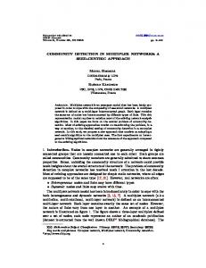
2 Multiplex Detection of Mutations David S. Perlin, Sergey Balashov, and Steven Park Abstract Rapid and reliable detection of mutations at the genetic level is an integral part of modern molecular diagnostics. These mutations can range from dominant single nucleotide polymorphisms within specific loci to codominant heterozygotic insertions and they present considerable challenges to investigators in developing rapid nucleic acid-based amplification assays that can distinguish wild-type from mutant alleles. The recent improvements of real-time polymerase chain reaction (PCR) using self-reporting fluorescence probes have given researchers a powerful tool in developing assays for mutation detection that can be multiplexed for high-throughput screening of multiple mutations and cost effectiveness. Here we describe an application of a multiplexed real-time PCR assay using Molecular Beacon probes for the detection of mutations in codon 54 of the CYP51A gene in Aspergillus fumigatus conferring triazole resistance. Key Words: Real-time PCR; molecular beacons; Aspergillus fumigatus; triazole resistance; multiplex.
1. Introduction The ability to detect multiple targets in a single assay represents one of the most powerful applications of real-time self-reporting probes. Multiplex assays increase throughput and reduce assay costs, which provides important benefits for diagnostic laboratories and research applications (1). Most current instrumentation is limited to the simultaneous discrimination of four separate fluorophores per assay reaction, although newer instruments can handle six-color detection. Thus, in most assays only four defined targets can be distinguished if each target is labeled with a separate probe. Other limitations can include interference from the inclusion of multiple primer sets for amplification of separate targets within the assay reaction. However, depending on the requirement of the output data many more targets can be distinguished. For example, if the assay From: Methods in Molecular Biology, Vol. 429: Molecular Beacons: Signalling Nucleic Acid Probes, Methods and Protocols Edited by: A. Marx and O. Seitz © Humana Press Inc., Totowa, NJ
23
24
Perlin et al.
is intended to distinguish between a wild-type sequence and multiple mutant sequences within the probe domain, then labeling the wild-type probe with one fluorophore and all others with a different fluorophore allows simultaneous detection of numerous targets with two color discrimination that can be detected with most real-time polymerase chain reaction (PCR) instruments. Such an approach is particularly amenable to the detection of drug resistance alleles, which can be confined to a single residue or to short sequence stretches (2–4). In such cases, allele discrimination within a common target region is critical. Molecular beacons are ideally suited for both allele discrimination and multiplex applications (5–8). The development of a robust multiplex assay for distinguishing multiple targets is shown for codon 54 of AfCYP51A, which encodes the sterol biosynthetic enzyme lanosterol demethylase (4). Mutations at this locus confer resistance to the triazole drug itraconazole in the invasive mould Aspergillus fumigatus (9,10), which is a major cause of death in severely immunosuppressed patients. Depending on the data output requirement, a multiplex assay can be established with either four (or six depending on instrument) outputs (four color) corresponding to each specific allele or with either two outputs (two color) representing the wild-type target sequence and any mutant allele. 2. Materials 2.1. Probe Development and Validation 1. Beacon Designer 2.12 software (PREMIER Biosoft Int., Palo Alto, CA). 2. Oligo, version 4.04, software (Molecular Biology Insights, Inc., Cascade, CO). 3. Molecular beacons were purchased from Biosearch Technologies, Inc. (Novato, CA). The probes were labeled at the 5�-end with fluorophores 5-carboxyfluorescein (FAM) and 6-carboxy-2�,4,4�,5�,7,7�-hexachlorofluorescein (HEX), carboxy-X-rhodamine (ROX), or Quasar 670 (Q670) and dabcyl (4-((-4-(dimethylamino)-phenyl)azo)-benzoic acid) or BHQ-1 at the 3�-end stored at �20°C until used. 4. Beckman Coulter System Gold high-pressure liquid chromatography (HPLC) (Beckman Coulter, Fullerton, CA). 5. C-18 reverse-phase column (Waters, Milford, MA). 6. 20% to 70% acetonitrile in 0.1 M triethylammonium acetate (pH 6.5) filtered and degassed. 7. Ethanol, 200 proof (Sigma Aldrich, St Louis, MO) stored at �20°C until used. 8. 0.3 M sodium acetate (Sigma Aldrich) stored at 4°C until used. 9. Eppendorf Vacufuge concentrator (Brinkmann, Westbury, NY). 10. DNA grade water (Fisher Scientific, Fairlawn, NJ), stored at room temperature until used. 11. Hybridization reaction mixture contained Stratagene Core PCR buffer (Stratagene, La Jolla, CA), 4 mM MgCl2, 100 pmol individual target oligonucleotide and 5 pmol molecular beacon. Each reagent was stored at �20°C until used. 12. Stratagene Mx4000 Multiplex Quantitative PCR System (Stratagene).
Multiplex Detection of Mutations
25
2.2. Real-Time PCR 1. Stratagene Core PCR buffer stored at �20°C until used.
2.3. DNA Extraction and PCR Amplification of Template 1. Sabouraud’s dextrose medium (SAB): 4% dextrose, 1% bactopeptone (pH 5.7) stored at room temperature until used. 2. FastDNA® kit (Qbiogene, Inc., Carlsbad, CA). 3. FastPrep® instrument (Qbiogene). 4. PCR amplification mix contained 25 pmol of each of Af210F (GTCTCTCATTCGTCCTTGTCCT) and Af709R (CGTTGAGAATAAACTCGTTCCC) primer, 2.5 U iTaq DNA polymerase (Bio-Rad Laboratories, Hercules, CA), 0.5 mM dNTPs, 50 mM KCl, 4 mM MgCl2, 20 mM Tris–HCl (pH 8.4), and about 100 ng A. fumigatus chromosomal DNA. 5. iCycler thermal cycler (Bio-Rad Laboratories). 6. The cycling conditions were 1 cycle of 3 min at 95°C, 35 cycles of 30 s at 95°C, 30 s at 55°C, 1 min at 72°C 1 cycle of 3 min at 72°C. 7. Montage PCR purification kit (Millipore, Bedford, MA).
3. Methods 3.1. DNA Extraction and PCR Amplification of Template 1. The sequence of A. fumigatus gene CYP51A (accession number AF338659) was used for primer design for PCR amplification of a 500 nt product containing codon 54 to be used as a template for real-time PCR assay development. 2. A. fumigatus chromosomal DNA is isolated using the FastDNA® kit (Qbiogene, Inc., Carlsbad, CA). Cells grown 24 h in SAB are first homogenized for a 30-s interval at 6000 m/s using a FastPrep® instrument (Qbiogene). 3. Amplification of a 500 bp fragment of cyp51A is performed on an iCycler thermal cycler (Bio-Rad Laboratories). Each 100 �L PCR contain 25 pmol of each of Af210F (GTCTCTCATTCGTCCTTGTCCT) and Af709R (CGTTGAGAATAAACTCGTTCCC) primer, 2.5 U iTaq DNA polymerase (Bio-Rad Laboratories), 0.5 mM dNTPs, 50 mM KCl, 4 mM MgCl2, 20 mM Tris–HCl (pH 8.4), and about 100 ng A. fumigatus chromosomal DNA. The cycling conditions were 1 cycle of 3 min at 95°C, 35 cycles of 30 s at 95°C, 30 s at 55°C, 1 min at 72°C 1 cycle of 3 min at 72°C. PCR products were purified using the Montage PCR purification kit (Millipore).
3.2. Design Considerations for Multiplexing Allele Discriminating Molecular Beacons 1. Molecular beacons covering the locus of diversity corresponding to codon 54 of CYP51A were designed using Beacon Designer 2.12 software utilizing default software parameters (see Note 1). The following sequence was designed for the wild-type CYP51A codon 54 region including the six nucleotide complementary molecular beacon stems; CGCGATCATCAGTTACGGGATTGATCCATCGCG (codon 54 in bold).
26
Perlin et al.
2. Analysis of the CYP51A region surrounding the Gly54 codon for secondary structures was performed using Zuker DNA—RNA folding server (http://www. bioinfo.rpi.edu/~zukerm/) (see Note 2). 3. PCR primers were designed with Beacon Designer, version 2.12, software and Oligo, version 4.04, software and were purchased from Sigma-Genosys (Woodlands, TX). 4. A molecular beacon complementary to the test locus (wild-type allele) was synthesized with a 21-nucleotide probe target sequence (hairpin loop) domain (CATCAGTTAC- GGGATTGATCC; codon 54 in bold) and a six-nucleotide stem domains (CGCGAT) with the 5�-end labeled with FAM as the fluorophore and the sixnucleotide 3�-end (ATCGCG) modified with a dabcyl quencher (see Note 3). 5. Molecular beacons designed with probe sequences recognizing mutant alleles were otherwise identical to the wild-type beacon, and they were labeled with either HEX alone (two color multiplex) or HEX, ROX, FAM, and Quasar 670 (four color multiplex) at the 5�-end and with dabcyl at the 3�-end as described: http://www. molecular-beacons.org/PA_protocol.html#cap2 (see Note 4). 6. Molecular beacons were purchased from Biosearch Technologies, Inc. as either fully- or semi-purified products (see Steps 7 and 8), which were subjected to HPLC purification for the latter. 7. Purification of fluorophore- and/or quencher-coupled oligonucleotides was achieved by HPLC on a C-18 reverse-phase column (Waters), utilizing a linear elution gradient of 20% to 70% acetonitrile in 0.1 M triethylammonium acetate (pH 6.5), which was filtered and degassed and run for 25 min at a flow rate of 1 ml/min. The elution of nucleic acid peaks were monitored at 260 and 495 nm (FAM), 535 nm (HEX), 575 nm (ROX), or 649 nm (Quasar 670), as described: http://www.molecular-beacons.org/PA_protocol.html#cap2. The final product was precipitated with ethanol at 2.5:1 (v/v) and 0.3 M sodium acetate, washed with 70% ethanol, dried with a Vacufuge (Eppendorf) concentrator, and final resuspension in DNA grade nuclease-free water.
3.3. Validating Molecular Beacons 1. Molecular beacon—target hybridization was investigated using the Stratagene Mx4000 Multiplex Quantitative PCR System (Stratagene). The “Molecular Beacon Melting Curve” option was chosen in the Mx4000 software for data monitoring and analysis. 2. Each hybridization reaction mixture contained 1x Stratagene Core PCR buffer, 3 or 5 mM MgCl2, 100 pmol individual oligonucleotides, and 5 pmol molecular beacons. 3. The test reactions were subjected to heating at 95°C for 3 min and cooling to 80°C with subsequent cooling down to 25°C using 112 30-s steps with a temperature gradient �0.5°C. Fluorescence output was measured at the end of each step. 4. The final data of the “Molecular Beacon Melting Curve” experiment were converted to a “SYBR Green (with Dissociation Curve)” output. The values for melting temperatures (Tm) were calculated by Mx4000 software as a temperature points corresponding to maximal values of the first derivative of the fluorescence output (–Rn�(T)).
Multiplex Detection of Mutations
27
Fig. 1. Molecular beacon CYP51A codon 54 melting curves. This summarizes the results of nine separate experiments on hybridization of GGG-FAM (wild-type) molecular beacon with complement target GGG-T (�), seven mutant allele-specific targets GTG-T (*), AGG-T (▼), GAG-T (▲), TGG-T (�), CGG-T (�), AAG-T (�), GAA-T (●), and no-template control (×).
5. Molecular beacon must show appropriate thermal behavior (Fig. 1). In the absence of target, molecular beacons should show self-annealing of the stem sequence as temperature decreases resulting in fluorescence quenching (see Note 5). 6. Hybridization with a perfect complementary target should show maximum fluorescence response at temperatures below predicted Tm (Fig. 1). 7. The efficiency for annealing to different targets varies depending on target nucleotide content with experimentally derived Tm values as illustrated for each of the eight beacon-target hybrids as shown in Table 1. In this case, the stability of intermolecular hybrids of GGG-FAM molecular beacons and artificial oligonucleotide targets decreased in the order of GGG > GTG > AGG > GAG > TGG > CGG > AAG > GAA. Hybrids with double mismatches are expected to possess the lowest stability (see Note 6). 8. The temperature interval between Tm of the most stable mismatched beacon—target hybrid and Tm of the complement beacon—target hybrid represented the condition allowing specific allele-discriminative binding of the molecular beacon or window of discrimination. Accordingly, the window of discrimination for GGG-FAM molecular beacon was within the temperature ranges of 61.7–65.2°C under conditions of 3 mM Mg2+ and 63.7–67.2°C at 5 mM Mg2+ (Table 1) (see Note 7). Under these
28
aT s m
GGGT 48.7 65.2 45.7 46.2 55.7 54.7 54.7 56.2 54.7 52.7 52.7 42.2 44.7
GAAT 44.7 53.7 61.2 39.7 55.7 42.2 47.7 47.2 40.7 39.2 40.7 58.2 46.2
AAGT 62.2 54.2 41.7 61.2 56.7 55.2 48.7 53.7 47.7 49.7 47.7 42.7 47.7
GAGT 52.7 58.7 50.2 52.2 63.2 49.7 54.7 52.7 48.2 46.2 46.7 48.7 55.2
AGGT 53.2 59.7 35.2 51.7 50.2 61.7 49.7 58.7 54.2 56.2 53.7 39.7 42.7
GTGT 48.2 61.7 45.2 49.7 57.7 49.7 63.7 53.7 50.2 48.2 49.7 42.7 61.2
TGGT 50.7 58.2 41.7 48.2 50.7 56.2 51.7 63.7 54.7 62.7 53.2 39.2 49.7
CGGT 49.7 55.7 36.2 47.2 50.7 58.2 49.7 60.2 65.2 58.2 64.2 41.2 48.2
of complement molecular beacon–target hybrids are in boldface.
Beacon AAG-FAM GGG-FAM GAA-HEX AAG-HEX GAG-HEX AGG-HEX GTG-HEX TGG-HEX CGG-HEX TGG-ROX CGG-ROX GAA-Q670 GTG-Q670
3 mM for target GGGT 49.7 67.2 46.7 48.7 57.2 56.7 56.2 57.7 55.2 54.2 54.2 46.7 49.7
Table 1 Melting Temperatures (Tm) for CYP51A Codon 54 Molecular Beacons Mg concentrationa
GAAT 45.2 55.2 63.7 44.7 56.2 44.7 48.7 48.2 42.7 39.7 42.2 61.7 48.2
AAGT 63.7 55.7 47.7 63.2 57.2 57.2 51.2 54.7 49.2 51.2 48.7 42.7 50.2
GAGT 54.2 61.2 52.7 53.2 64.2 51.7 57.2 53.2 50.2 49.2 49.7 51.2 56.2
AGGT 54.7 61.7 41.2 53.2 51.7 63.7 50.7 59.7 55.2 57.7 54.7 40.2 49.2
5 mM for target GTGT 54.2 63.7 46.7 51.2 58.7 52.2 65.2 55.2 51.7 50.2 51.2 48.2 64.2
TGGT 52.2 61.2 42.2 50.7 52.7 58.2 53.7 65.2 57.2 63.7 55.2 44.7 51.7
CGGT 52.2 59.7 41.7 49.7 52.2 59.7 52.2 61.7 66.2 59.7 65.7 43.2 50.2
Multiplex Detection of Mutations
29
conditions, all molecular beacons (mutant and wild type) could be run in a real-time PCR experiment at the same annealing temperature of 61°C and showed excellent discrimination against both wild-type and mutant alleles.
3.4. Multiplex Panels and Real-Time PCR 1. Once target-specific hybridization with complement DNA target was validated for each beacon, the probes can be combined in a simple multiplex real-time PCR format. The advantage of this format is that, in a single assay, mutant (resistant) and wild-type (susceptible) strains can be easily distinguished. However, this format cannot be used to identify specific mutations of Gly54 because each mutant beacon was labeled with the same fluorophore (see Section 3.4.2.). 2. A single-reaction real-time PCR assay with eight different molecular beacons (one wild-type beacon labeled with FAM and seven mutant beacons labeled with HEX) and a 0.5-kb template corresponding to each cyp51A allele can be performed. In this format, the fluorescence signal indicated the presence of either wild-type or mutant sequences corresponding to itraconazole susceptibility or resistance, respectively. 3. To expand the applicability of the multiplex assay to distinguish separate alleles, the molecular beacons were labeled with different fluorophores: FAM, HEX, ROX, or the CY5 analog Q670 (Table 1). This configuration required two PCRs to assess all seven mutations, since only four colors could be distinguished in a single reaction. The first PCR mixture contained molecular beacons with high Tms against complement DNA targets: GGG-FAM, GAG-HEX, GTG-Q670, and CGG-ROX. The second PCR mixture contained molecular beacons with somewhat lower Tms: AAG-FAM, AGG-HEX, GAA-Q670, and TGG-ROX. The system specificity was further optimized by adjusting the Mg2+ concentration up to 3 mM for the first reaction and to 5 mM for the second reaction. A uniform annealing temperature of 57°C was used for both reactions. 4. The assay system was tested by adding individual 0.5-kb cyp51A templates to each of two reaction mixtures. Under these conditions, specific hybridization with complement cyp51A alleles was observed for all molecular beacons except the GGG-FAM wild-type beacon, which exhibited some level of nonspecific hybridization with the GTG allele. To avoid any possible false-positive results coming from nonspecific hybridization of the wild-type GGG-FAM beacon, the threshold fluorescence level was adjusted to values close to those obtained for complement beacon DNA polymerase, 0.4 mM dNTPs, 3 or 5 mM MgCl2, and 100 ng chromosomal DNA. 5. PCRs were performed using the parameters, as follows: 1 cycle of 10 min at 95°C, 45 cycles of 30 s at 95°C, 30 s at 61°C and 30 s at 72°C. Annealing temperatures of 55°C and 57°C are used when PCR experiments are performed in multiplex format. The filter gain set of the Mx4000 System is changed to FAM-940 HEX-720 CY5700 ROX- with the aim of equalization of the fluorescence signals from different molecular beacons. 6. The fluorescence is measured during the annealing step. This multiplex format allowed all seven specific cyp51A alleles bearing mutations in the Gly54 codon, along with the wild-type allele, to be distinguished in a real-time assay.
30
Perlin et al.
7. Real-time PCR experiments were performed on a Stratagene Mx4000 Multiplex Quantitative PCR System. The “Quantitative PCR (Multiple Standards)” setting was used for all real-time PCR experiments. Reagents from Brilliant® QPCR Core Reagent Kit were used for all reactions. Each 50-�l PCR contained 1x Stratagene Core PCR buffer, 20 pmol molecular beacon, 25 pmol of each of the CYP51AS and CYP51AA primers, 2.5 U Stratagene SureStart® Taq.
3.5. Data Processing Fluorescence signals coming from Mx4000 System during PCR amplification were monitored using Mx4000 software in real time. At the end of each run, the amplification plots data were converted to graphic format and stored as image files or exported into Microsoft Office Excel and stored as spreadsheet files. In the case of multiplex beacons PCRs, the final results of PCR amplifications were converted from “Quantitative PCR (Multiple Standards)” type of experiment to the “Quantitative Plate Read” type of experiment. 4. Notes 1. Amplicons