Anal Bioanal Chem DOI 10.1007/s00216-014-7894-5
RESEARCH PAPER
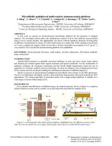
Multiplexed microfluidic enzyme assays for simultaneous detection of lipolysis products from adipocytes Colleen E. Dugan & William P. Cawthorn & Ormond A. MacDougald & Robert T. Kennedy
Received: 26 March 2014 / Revised: 7 May 2014 / Accepted: 13 May 2014 # Springer-Verlag Berlin Heidelberg 2014
Abstract Microfluidics has enabled new cell biology experiments. Incorporating chemical monitoring of cellular secretion into chips offers the potential to increase information content and utility of such systems. In this work, an integrated, multilayer polydimethylsiloxane microfluidic chip was developed to simultaneously measure fatty acids and glycerol secreted from cultured adipocytes on chip in near real time. Approximately 48,000 adipocytes were loaded into a cell chamber in a reversibly sealed chip. Cells were perfused at 0.75 μL/min. Cell perfusate was split and directed to separate, continuously operating fluorescent enzyme assay channel networks. The fluorescent assay products were detected simultaneously near the outlet of the chip. The fatty acid and glycerol assays had linear dynamic ranges of 150 and 110 μM and limit of detection (LOD) of 6 and 5 μM, respectively. Surface modifications including pretreatment with sodium dodecyl sulfate were utilized to prevent adsorption of fatty acids to the chip surface. Using the chip, basal fatty acid and glycerol concentrations ranged from 0.18 to 0.7 nmol × 10 6 Electronic supplementary material The online version of this article (doi:10.1007/s00216-014-7894-5) contains supplementary material, which is available to authorized users. C. E. Dugan : R. T. Kennedy Department of Chemistry, University of Michigan, Ann Arbor, MI 48109, USA W. P. Cawthorn : O. A. MacDougald Department of Molecular and Integrative Physiology, University of Michigan, Ann Arbor, MI 48109, USA O. A. MacDougald Department of Internal Medicine, University of Michigan, Ann Arbor, MI 48109, USA R. T. Kennedy (*) Department of Pharmacology, University of Michigan, Ann Arbor, MI 48109, USA e-mail:
[email protected]
cell−1 min−1 and from 0.23 to 0.85 nmol×106 cell−1 min−1, respectively. Using valves built into the chip, the perfusion solution was switched to add 20 μM isoproterenol, a βadrenergic agonist, which stimulates the release of glycerol and fatty acids in adipocytes. This manipulation resulted in a rapid and stable 1.5- to 6.0-fold increase of non-esterified fatty acid (NEFA) and glycerol. The ratio of NEFA:glycerol released increased with adipocyte age. These experiments illustrate the potential for performing multiple real-time assays on cells in culture using microfluidic devices. Keywords Adipocytes . Enzyme assay . Integration . Microfluidics . Multiplex
Introduction Microfluidic and lab-on-chip devices offer many benefits for analytical measurements over traditional techniques including reduced reagent and sample volumes, improved temporal resolution for monitoring, and increased automation. Such devices have also become powerful tools for cell biology [1, 2]. Specific cell lines or individual cells can be isolated in the reduced dimensions of a microfluidic device [3, 4]. Cells can be perfused in two or three dimensions with continual replenishment of media to more closely mimic the in vivo environment compared to traditional static incubation methods [5, 6]. Microfluidics also provides a way to miniaturize and provide a higher throughput of cell culture-based experiments. Although growing cells in chips can be useful, in many cases, it is also necessary to assess cell function. Visual inspection and fluorescence measurements of cells are straightforward on chips that are optically clear; however, chemical analysis of the cellular environment often requires other assays. Although chemical measurements can be performed off-chip [7], integrating analytical measurements with cells on microfluidic
C.E. Dugan et al.
systems eliminates the need for sample collection and off-line analyte detection and can provide real-time recording of cellular dynamics [8, 9]. Several examples of this approach have been described including on-line immunoassays [10], sensors [11], and enzyme assays [12]. Most such systems perform a single assay on cells. In this report, we describe an advance on this capability in a system fabricated in polydimethylsiloxane (PDMS) that uses on-line dual-enzyme assays to monitor metabolic activity in near real time. We also demonstrate a method to avoid chemical loss to absorption by PDMS that improves the sensitivity of measurements of hydrophobic compounds such as fatty acids. Adipocytes were used as a model system for these experiments. Adipocytes are fat-storing cells that secrete glycerol and non-esterified fatty acids (NEFAs) as a result of the catabolism of triglycerides through lipolysis. Flux of lipolytic products is regulated by a variety of signals. The adipocyte reesterifies (recycles) a percentage of NEFAs back into triglycerides, as a method of regulation for systemic NEFA supply [13]. Depending on the physiological energy state, different enzymes and glycerol-3-phosphate precursors dictate the pathway and amount of recycling [14, 15]. Increased adiposity, as in obese individuals, is often associated with a number of disorders including type 2 diabetes [16–19]. Thus, understanding the mechanisms of fatty acid recycling and the factors that lead to its dysfunction could be fundamental to providing improved treatment for obesity-related disorders. Adipocytes have been previously mounted in microfluidic devices, mainly to monitor differentiation and culture of the on-chip cells [20, 21]. The influence of adipocyte secretions on other cell lines in a multichamber chip has also been monitored [22]. Despite these advances in using microfluidics to better understand adipocyte biology, automated and realtime quantification of adipocyte secretion is lacking. Previous reports from our group have shown the ability to monitor either NEFA or glycerol secretion from 3T3-L1 adipocytes on glass microfluidic devices [23, 24]. These chips showed the potential to detect adipocyte secretion on-line, but individual measurements of NEFA or glycerol cannot provide information on fatty acid re-esterification. The concentration of glycerol secreted from adipocytes is a direct indication of the rate of lipolysis; glycerol is not directly recycled by the adipocyte because of the lack of sufficient glycerol kinase in the cell [25–27]. If NEFA secretion can be monitored at the same time as glycerol, from the same group of cells, the amount of fatty acid re-esterification can be inferred based on the rate of lipolysis and concentration of secreted NEFAs. To accomplish dual-analyte measurement, a microfluidic chip with two enzyme assay reaction networks was designed. Cultured murine 3T3-L1 adipocytes were perfused in a cell chamber, and the resulting perfusate was split into the two enzyme assay reaction channels. The chip design and reagent flow rates were chosen to provide good on-line assay
sensitivity and response time. PDMS was chosen as the microfluidic substrate because of many convenient properties. PDMS is biocompatible and gas permeable, making it well suited for cell studies [28]. The inherently elastomeric PDMS permitted easier integration of a valve system, which was constructed upstream of the cell chamber to allow automated stream selection, to reduce dead volume, and to prevent flow disruption when changing solutions. Despite the advantages of PDMS, it can absorb hydrophobic molecules. Since NEFAs are being detected on chip, a surface modification was performed to prevent loss of the analyte. These improvements allow a rapid, automated measurement method for monitoring adipocyte fatty acid re-esterification and can be extended to other adipocyte secretion studies.
Experimental Reagents The fatty acid assay reagents and standard solution (oleic acid) were purchased in a kit, HR Series NEFA-HR(2), through Wako Chemicals USA, Inc. (Richmond, VA). Amplex UltraRed Reagent, Hank’s buffered salt solution (HBSS) (Cat. No. 14175), and cell culture reagents were received from Life Technologies (Carlsbad, CA). Triton X-100 was purchased from Bio-Rad Laboratories (Hercules, CA). Free glycerol reagent, standard glycerol solution, fatty acid free bovine serum albumin (BSA), isoproterenol hydrochloride, sodium dodecyl sulfate (SDS), dimethyl sulfoxide (DMSO), and hexamethyldisilazane (HMDS) were obtained from SigmaAldrich (St. Louis, MO). Microfluidic chip fabrication The multilayer PDMS chips were fabricated with soft lithography techniques as previously described [28–30]. All of the PDMS used in the chip fabrication was RTV-615 (Curbell Plastics, Livonia, MI) and had a base to curing agent ratio of 10:1. The four PDMS layers are denoted as follows: “control layer,” “mixing channel layer,” “lower cell chamber layer,” and “blank layer.” For the fabrication of the control layer, HMDS was spun on a 3-in. silicon wafer (University Wafer, Boston, MA) to facilitate photoresist adhesion. SU-8 2075 photoresist (MicroChem, Newton, MA) was spun onto the wafer to create a 60-μm layer and was patterned using a darkfield photomask with the design shown as the black lines in Fig. 1a. A thick layer (~1 cm) of PDMS was poured over the mold and heated to 80 °C for at least 30 min. The mold for the mixing channel layer was made the same as the control layer, but with the pattern shown as the black lines in Fig. 1b. After the SU-8 photoresist was developed, a 12-μm thick layer of AZ-9260 photoresist (Capitol Scientific, Austin, TX) was
Multiplexed microfluidic enzyme assays
peeled from the mixing channel layer mold, bonded to the blank layer PDMS to enclose the channels, and heated to 80 °C for 15 min. The three bonded layers were peeled from the wafer, and the blank layer PDMS was removed from inside the upper portion of the cell chamber using a scalpel blade. A 4.8 mm×4.8 mm piece of cover glass (No. 1, Fisher Scientific, Waltham, MA) was bonded to the inside of the upper cell chamber. The lower cell chamber layer was peeled from the wafer mold and bonded to a glass slide (0.55 mm thick; Telic, Valencia, CA), feature side exposed, and heated to 80 °C for 15 min. The side view of the assembled chip is shown in Fig. 2.
Adipocyte culture
Fig. 1 Design of the three different fabricated layers in the PDMS multilayer chip. The control layer (a), the reaction channel layer (b), and the base chamber layer (c) fabrication patterns are shown. The fluorogenic dye, Amplex UltraRed, is abbreviated as AUR. The blue circles in a represent the points where access holes were punched through the PDMS, and the red line in b indicates the portion of the design that was fabricated with AZ photoresist
spun onto the wafer. The final pattern of the AZ photoresist is shown in Fig. 1b as the red line. The wafer was heated at 125 °C for 5 min to round the AZ features. PDMS was spun on the wafer to create a 60-μm-thick layer and heated to 80 °C for at least 30 min. The fabrication of the lower cell chamber layer was done with SU-8 2075 as described above, but to a thickness of 250 μm, with the pattern shown in Fig. 1c. PDMS was poured over the mold and manually distributed across the wafer to create a ~1-mm-thick layer. The PDMS was allowed to settle for 10 min before it was heated to 80 °C for at least 30 min. Finally, PDMS was spun on a blank wafer to a thickness of 170 μm and heated to 80 °C for at least 30 min, to create the blank layer. The control layer PDMS was peeled away from the mold, and access holes were punched at the points indicated in blue in Fig. 1a using a blunt 20-gauge needle (Small Parts, Logansport, IN). The control layer was bonded to the top of the mixing channel layer using corona discharge and was heated to 80 °C for 15 min. The two bonded layers were
Glass coverslips (No. 1, Fisher Scientific) were cut to 4.8 mm×4.8 mm squares using a dicing saw. The coverslips were sterilized in 70 % ethanol for at least 1 h and were then dried in a sterile culture hood. Three to four coverslips were placed in each 35-mm petri dish using sterile tweezers prior to seeding preadipocytes. Murine 3T3-L1 preadipocytes were seeded into the 35-mm dishes (200,000 cells per dish), maintained in Dulbecco’s modified Eagle’s medium (Cat. No. 11965-092, Life Technologies) with 8 % v/v bovine calf serum (Denville Scientific, South Plainfield, NJ), 100 units/mL penicillin, 100 μg/mL streptomycin, 2 mM L-glutamine, and 1 mM sodium pyruvate and were stored in an incubator with 10 % CO2. Two days after the cells became confluent, differentiation (adipogenesis) was induced by adding 500 μM methylisobutylxanthine, 1 μM dexamethasone, and 5 μg/mL insulin. Two days post-differentiation, medium was replaced with adipogenic medium that contained 5 μg/mL insulin. Adipogenic medium was the same as the preadipocyte medium except that 10 % fetal bovine serum replaced the bovine calf serum. Every 2 days following, culture medium was refreshed with adipogenic medium that did not contain insulin. Adipocytes were matured to at least 14 days postinduction before on-chip experiments.
Fig. 2 Side view of chip slice showing fabrication layers and direction of flow. The dotted lines represent the interfaces between layers that were irreversibly bonded
C.E. Dugan et al.
Computational modeling The perfusion of the cell chamber and mixing channels was modeled using COMSOL Multiphysics 4.3b (COMSOL, Inc., Burlington, MA). The “laminar flow” and “transport of diluted species” physics models were used to monitor the flow split and theoretical temporal resolution of the system. All simulations assumed water perfusing through the chip, with a density of 998 kg m−3 and a viscosity of 1.002×10⁻³ Pa·s’ (20 °C). The cell chamber and mixing channel network were modeled separately in order to reduce computation times with finer meshes. The cell chamber was modeled in a 3D geometry and had dimensions of 5.0 mm×5.0 mm×0.33 mm with a 4.8 mm×4.8 mm×0.15 mm box removed from top to mimic the bonded coverslip in the actual chip. The inlet and outlet channels for the cell chamber were 60 μm tall. Based on initial cell experiments, the optimal flow rate to perfuse over the cells was 0.75 μL/min, so the cell chamber inlet was also set to that flow rate. The channels in the assay reaction channel network were modeled in 2D with the shallow channel approximation set to 60 μm. Reversible chip assembly The PDMS was cut so that the ends of the channels were exposed. Fluidic connections were made by inserting 50 μm ID/150 μm OD fused silica capillaries (Polymicro Technologies, Phoenix, AZ), ~13 cm long, with rounded tips into the exposed inlets (between the mixing channel layer and the blank layer). Sheath capillaries (180 μm ID/360 μm OD) were glued to the opposite ends of the 50/150 capillaries, to allow for easier connections to external pumps. An additional 17 cm of 100/360 capillary was connected to the valve waste capillaries during experiments to balance the back pressure. The enzyme assay mixing channels were modified by pumping 20 mM SDS solution through both of the mixing channel outlets at 1.5 μL/min for 45 min. The channels were then flushed with water for an additional 20 min at the same flow rate. After every use of the chip, this process was repeated to refresh the treatment. The control channels were operated by connection to an inhouse built pneumatically actuated system. A stainless steel tube (23 gauge, ~1 cm long) was inserted halfway into Tygon tubing (1/16 in. OD, 0.5 mm ID; IDEX, Lake Forest, IL). The other half was inserted into the top of the chip where the access holes were punched in the control layer. The Tygon tubing was filled with water, and 15 psi of nitrogen gas was applied to the desired channels with the aid of an in-house written LabView program. Before the chip was used for an experiment, it was degassed in a vacuum chamber for ~1 h to reduce the presence of bubbles in the channels during perfusion. The channels were primed with water after the chip was degassed. The
two parts of the PDMS chip were rinsed with 70 % ethanol, and dust was removed with Scotch tape. For cell experiments, a coverslip with ~48,000 attached 3T3-L1 adipocytes was removed from the culture dish with sterile tweezers and placed in the lower cell chamber. Cells per coverslip were roughly estimated by assuming an even distribution of cells on the culture plate so that the number of cells equaled the fraction of plate area occupied by the coverslip times the total number of cells. Total number of cells was estimated from the number of preadipocytes seeded onto the plate (measured by hemocytometer) and the replication rate during differentiation. About 10 μL of culture medium was added to prevent trapping bubbles during sealing. For calibration experiments, a blank coverslip was placed in the lower cell chamber. The upper cell chamber was aligned over the lower chamber, and the two parts of the chip were pressed to make a conformal contact. A compression frame to enclose the PDMS chip was built in-house from two sheets of acrylic plastic. Holes were drilled through both sheets along the outer edge to allow screws to pass through both sheets and tighten the compression frame. Additionally, four holes were drilled on the top for access to the control channel tubing, and a large chamfered hole was drilled on the bottom to allow the microscope objective to have closer access to the detection channels. The control tubing of the assembled chip was threaded through the compression frame, and screws tightened the compression frame over the chip. Figure 3 shows the assembled chip in the compression frame. Perfusion The fatty acid assay reagents were reconstituted as follows: Color Reagent A (CR-A) was reconstituted with 30 mL of solvent A (creating a solution 1.66 times more concentrated than manufacturer’s specifications), and Color Reagent B (CR-B) was reconstituted with Solvent B according to manufacturer’s instructions. CR-A was further modified by adding 200 μL 10 % (v/v) Triton X-100 to every 800 μL CR-A. The free glycerol reagent was reconstituted with 40 mL 18.0 MΩ/cm distilled water. Amplex UltraRed was reconstituted with DMSO to make a 10 mM stock solution. The Amplex UltraRed stock solution was further diluted to make a 1 mM solution in 35 % DMSO, which was used for the on-line assays. Prior to performing cell studies, calibrations were performed to characterize the actual delay and rise times of the two multiplexed assays. For these experiments, standards were diluted in HBSS containing 2 % BSA (w/v) and pumped through the two valve inlets at 0.75 μL/min. When an additional standard was required (i.e., when creating a calibration curve with multiple standard concentrations), the solution that was being directed to waste through the valve was replaced, which prevented disruption in the flow. The glycerol assay
Multiplexed microfluidic enzyme assays
Fig. 3 The assembled chip in compression frame. Blue food dye is being perfused through one of the valves for better visualization. The arrows highlight the capillary connections and the direction of fluid flow
was performed by adding the glycerol reagent and 1 mM Amplex UltraRed solution onto the chip at 0.29 μL/min (each) at the locations indicated in Fig. 1b. The modified CR-A, CR-B, and 1 mM Amplex UltraRed solutions were pumped at 0.30 μL/min (each) through the respective inlets shown in Fig. 1b. All of the solution pumping was controlled by external syringe pumps. During cell experiments, one valve inlet was perfused with 2 % BSA in HBSS (to monitor basal secretion), and the other valve inlet was perfused with 20 μM isoproterenol in HBSS with 2 % BSA (to create lipolysis-stimulated conditions).
glycerol phosphate oxidase [23]. Both assays create hydrogen peroxide from their respective reactions, which can then be reacted with peroxidase and Amplex UltraRed to create a fluorescent product. These chips demonstrated important principles such as loading predifferentiated adipocytes on glass coverslips into reversibly sealed chips, prevention of shear stress on the fragile cells during perfusion, and continuous monitoring which were used in this work. To achieve simultaneous analysis capability, we designed a chip (Fig. 1) that split the perfusate and directed the flows to two separate, continuously operating enzyme assay streams. The detection points for the two assay channels were placed near each other, so they could be imaged on a fluorescence microscope. Because the enzyme assays yielded the same fluorescent product, this arrangement allowed glycerol and NEFA to be monitored on-line simultaneously as they were secreted from the cells. Other improvements were also made including lowering the flow rate to cell ratio to reduce dilution of secreted chemicals by 11-fold. The new chip also integrates valves to automate physiologic experiments that require changing the perfusion conditions, e.g., adding drugs to the cells. Because valves are easily incorporated into PDMS by multilayer lithography, we used this material instead of glass. This change in material necessitated surface modification procedures that prevented adsorption of hydrophobic analytes to the chip. Simultaneous assay chip design and flow splitting
Detection An Olympus IX71 inverted microscope was used to collect the signal from the fluorescent assay product. The light path was directed by a 10×, 0.40 numerical aperture objective (Olympus Part No. 1-U2B823) and the appropriate mirrors/ filters for resorufin detection. A Hamamatsu ImagEM X2 CCD camera allowed simultaneous detection of the two outlet channels at the point shown in Fig. 1b. Data were collected at 2 Hz with an exposure time of 250 ms and were analyzed with Microsoft Excel 2007.
Results and discussion Overview of microfluidic chip design In previous work, we demonstrated different chips that could monitor NEFA and glycerol release from adipocytes [23, 24]. In these chips, cells were perfused, enzyme assay reagents were added on-line to mix with the perfusate, and reaction products were measured by fluorescence detection. The reagents for the NEFA assay contained coenzyme A (CoA), ATP, acyl-CoA synthetase, and acyl-CoA oxidase [24]. The glycerol assay reagent contained ATP, glycerol kinase, and
Stable and reliable split of the perfusate flow for each enzyme assay required careful consideration of the geometry and flow rates used. Although the assay reaction channels had approximately the same flow resistance, the different number and placement of reagent inlets required for each assay (see Electronic Supplementary Material for pilot studies on individual assays, incl. Fig. S1) created differences in back pressure. These differences made it difficult to predict how reagent flow would affect the perfusate flow into the two assay channels. To assist in determining the flow conditions, a COMSOL model was developed. For simplicity, we operated the chip with a 50:50 split of perfusate to each assay channel. We also fixed the NEFA assay flow rates and adjusted glycerol reagent flow rates because the glycerol assay was more sensitive and adaptable to changes in analyte to reagent ratios than that of the NEFA assay. Based on data from the single channel chip for NEFA detection, the highest sensitivity response was achieved when the analyte to reagent ratio was set to 1.25:1:1:1 NEFA/CR-A/ CR-B/Amplex UltraRed. Assuming an even flow split and a flow rate of 0.75 μL/min through the cell chamber, this ratio required that the NEFA reagents be set to 0.3 μL/min. To determine the glycerol reagent flow rates that were needed, we used the COMSOL model to vary flow rates from 0.1 to 1 μL/ min. The model showed that the two reagent inlets on the
C.E. Dugan et al.
glycerol assay side of the chip should be set to 0.29 μL/min to achieve even flow split. The velocity map of the flow split using these flow rates is shown in Fig. 4a. The map shows an even split under these conditions. It also shows a slight decrease in velocity before the split, which is due to slight widening of the channel before the split. Additionally, it was determined that if the glycerol reagent flow rates were higher than 0.7 μL/min, 100 % of the flow coming from the cell chamber would be directed to the NEFA assay channel showing the sensitivity of the system to changes in flow. Similarly, we found that abrupt changes in pressure, such as from rapidly inserting capillaries or making connections, could result in unstable flows in the system. To confirm that the flow was symmetrical through both assay channels, we measured the flow rate through each channel separately. To do this, water was perfused through the chip, and fractions were collected at the outlets every 2 min and weighed. The results were compared to theoretical values of 0.96 μL/min at the glycerol outlet and 1.28 μL/min at the NEFA outlet, assuming an even flow split of the cell chamber perfusate into the two reaction channel networks. (The NEFA channel should have a higher net flow out because of the extra reagent channel.) The flow rate exiting the chip was 1.0±0.02 μL/min on the glycerol side and 1.3±0.08 μL/ min on the NEFA side (n=4). The results show that the
Fig. 4 COMSOL modeling of the dual chip. a The velocity of laminar flow at the flow split allowed the ratio of cell perfusate that would be directed into each assay reaction channel to be determined. b The mixing efficiency could be measured at a reagent inlet by injecting a 5 μM chemical species at the flow split and a blank solution at the reagent inlet and finding the point in which complete diffusion has occurred across the width of the channel. c A concentration gradient moving through the cell chamber when a 5 μM chemical species injected was modeled to show the profile of flow across the width of the chamber
appropriate flow rates were exiting the chip and confirm an equal flow split. We also used the model to confirm that complete mixing of reagents and sample would occur at the flow rates and channels used. As shown in Fig. 4b, complete mixing was found after the fluid traveled 7 mm through the 100-μm-wide serpentine channels. Another important issue for design is determining whether the cells are evenly perfused and if the chamber is well swept. COMSOL modeling also assisted in establishing the appropriate flow profile through the cell chamber. Varying the number and angle of the channels leading to and from the chamber changed the profile and temporal response of the chip. It was desired to reduce the rise time of the system to detect cellular dynamics; therefore, a flat even flow across the chamber was ideal. Figure 4c illustrates a simulation of a chemical species injection into the chamber to allow visualization of the flow profile of the cell chamber used in these experiments. Chip characterization Figure 5a shows the detector response to a series step changes in analyte concentration performed on chip. Figure 5b illustrates the resulting calibration curves for both assays. The limit of detection (LOD) was calculated as the concentration corresponding to a signal that was three times the standard deviation of the blank. The glycerol assay had a LOD of 5 μM and was linear through 110 μM. The NEFA assay had a LOD of 6 μM and had a linear calibration through 150 μM. The calibration curves were reproducible over multiple experiments using the same chip with relative standard deviation (RSD) of slope and intercept below 6 % (n=4 chips, each used for at least three trials). These data also allow us to characterize the temporal response of the chip. The rise time was defined as the amount of time between a 10 and 90 % change in signal when standard concentrations were switched. The delay time was calculated as the time between switching a solution and the initial 10 % rise in signal. The rise times for the complete assembled chip were 2.3±0.6 min for both the glycerol and NEFA assays. The delay times were 10.2±0.3 min. The COMSOL model of the chip (including the same flow components such as valves, cell chamber, and mixing channels) predicts 4.6 and 15.4 min for the rise and delay times of the glycerol assay and 4.7 and 17.8 min for the rise and delay times of the NEFA assay, respectively. The longer times predicted than observed suggest a difference in geometry that is not accounted for by the model. A likely source of the discrepancy is reduction of channel height caused by the compression frame closing off the channels of the compliant PDMS. Because the flow rates were through each channel as predicted (see above), the compression must have an equal effect on both sides of the chip. Experimental values were consistent across multiple
Multiplexed microfluidic enzyme assays
Fig. 6 Step changes on glycerol assay independent chip from high to low concentration standards without (a) and with (b) fabricated valves
Fig. 5 Dual chip calibration. a Detector trace from step changes of glycerol and NEFA standards in the dual chip, which are simultaneously detected. The resulting calibration curve for the glycerol assay (b) for four replicate experiments shows a linear response through 112 μM, a LOD of 5 μM, and R2 of 0.998. The fatty acid assay (c) is linear through 150 μM and has a LOD of 6 μM and R2 of 0.999 for four replicate experiments
chips and were used for subsequent calculations, such as accounting for delay times, related to cell experiments. An important aspect of cell physiology experiments is manipulating the solutions flowing over cells. In prior work [23, 24], changing solutions was accomplished by switching connections to the chip. Making manual connections requires a stopping and starting flow and creates a change in pressure on chip, which affects the detected signal. This effect is especially problematic in a dual-channel chip with multiple flow paths. Figure 6a illustrates the effect on signal of such a switch. The use of fabricated valves on chip significantly improved the stability of the flow through the chip when solutions were switched, creating smooth step changes, as shown in Fig. 6b. This addition also reduced dead volumes of the perfused solutions and increased automation of the device. Assay and PDMS modifications Preliminary experiments revealed low sensitivity and signal drift in the NEFA assay. Because this had not been observed
with the glass devices and it is well-known that PDMS can adsorb hydrophobic molecules [31, 32], the problems with signal were attributed to adsorption of the hydrophobic fatty acids by PDMS. A variety of PDMS modifications that might prevent NEFA adsorption, including sol-gels, polyelectrolyte multilayers, and dynamic coating with SDS, polyethylene glycol, and Pluronic F-127, were tested for their effect on the NEFA signal [33–37]. The best results, as determined by NEFA assay sensitivity and stability, were obtained when the channels were dynamically coated with a 20 mM SDS solution [38, 39]. For the SDS treatment, it was important that chips were modified a few days after fabrication. This aging effect was attributed to corona discharge used for bonding. Corona discharge creates a hydrophilic surface, which slowly reverts back to its native hydrophobic state that would be better able to bind SDS [40, 41]. The NEFA assay sensitivity increased from 7.6±2.5 relative fluorescence units (RFU)μM−1 (n=3) to 11.1±2.3 RFU μM−1 (n=3) with SDS modification (see Electronic Supplementary Material Fig. S2 for more details on effectiveness of SDS). Day-to-day experiments on the same chip modified with SDS had reproducible calibration curve slopes with a RSD of 2.1 % (n=3). The coating was refreshed every day to maintain this performance. To reduce the exposure of fatty acids to the PDMS, a glass coverslip was bonded to the top of the cell chamber. This lessened the surface area that the fatty acids could contact the PDMS while in the cell chamber, resulting in a further 44 %
C.E. Dugan et al.
improvement in assay sensitivity. The addition of the coverslip also had the benefit of directing flow towards the cells, allowing improved sampling of cell secretion. Although these measures improved the sensitivity, we still observed drift in signal for the fatty acid assay over time. Addition of the non-ionic surfactant, Triton X-100, to CR-A reagent reduced the drift of the signal at high standard concentrations so that the RSD improved from >12 to 1–5 %. This effect may be due to both prevention of adsorption and improving enzyme solubility. Adipocyte perfusion Representative simultaneous traces of adipocyte secretion of glycerol and NEFAs are shown in Fig. 7a, b. Basal concentrations, i.e., secretion prior to drug treatment, were above the LOD of both assays for all cases. Upon addition with 20 μM isoproterenol, a drug that stimulates lipolysis, a simultaneous increase in concentration of both analytes was observed between two- and sixfold depending on the batch of cells used. Previous work with static culture systems have reported a two- to sevenfold increase in lipolysis products with similar stimulation [42, 43]. The simultaneous measurement of NEFA and glycerol allows an assessment of recycling. For example, for the case in Fig. 7a, the NEFA:glycerol ratio was 1:1, implying some recycling that did not change after stimulation. In contrast, for the cells used in Fig. 7b, we observed NEFA:glycerol ratio of 1:3 that increased to 2:1 with isoproterenol, suggesting high recycling that decreased with stimulation. Previous studies have reported a wide range of NEFA:glycerol ratios, e.g., from 1:2 [44] to 2:1 [42] for 3T3-L1 adipocytes. In general, values are less than the theoretical maximum of 3:1, similar to our results. Few Fig. 7 Representative traces of simultaneous detection of NEFA and glycerol secretion from adipocytes and trends in day-to-day experiments. Traces were collected during basal secretion and during lipolysis stimulation by the application of isoproterenol, represented by the black bar. a Cells day 14 post-induction (PID). b Cells day 35 post-induction. c The NEFA/glycerol ratio decreased for basal secretion the longer the adipocytes remained in culture. d As the cells matured, the ratio of average isoproterenol-stimulated cell response to the average basal response increased for the NEFA assay and decreased for the glycerol assay
results have been reported on NEFA:glycerol ratio for 3T3L1 cells with isoproterenol treatment; however, studies of primary adipocytes have reported that isoproterenol generally evokes higher NEFA:glycerol ratios (less recycling) than basal as we observed for most of our trials. For example, one report showed a 1.3-fold increase in NEFA:glycerol ratio [45]. Interestingly, the NEFA:glycerol ratio showed a decreasing trend in adipocytes that were cultured for more extensive periods of time (Fig. 7c). We also observed greater responsiveness of NEFA secretion to isoproterenol for longer times after induction (Fig. 7d). The cause and significance of these trends is not known; however, the larger secretion of NEFA at longer times could relate to greater lipid content as the cells age in culture. Further studies with this method will be required to better understand the significance of such changes.
Conclusions In this study, a multilayer PDMS chip was developed for the perfusion of adipocytes and analysis of their secreted metabolites. A multiplexed enzyme assay network allowed simultaneous detection of glycerol and NEFA concentrations released from cultured 3T3-L1 adipocytes. COMSOL modeling aided in the design and implementation of the dual, asymmetric assay system. PDMS proved to be a good substrate for these experiments following modification of the channels with SDS. Basal and stimulated lipolysis conditions were monitored from perfused adipocytes in real time. Appropriate stimulation of analyte secretion was observed with the addition of isoproterenol. Initial trends imply that there are changes to the biology of 3T3-L1 adipocytes with time in culture and could
Multiplexed microfluidic enzyme assays
provide more detailed information about lipolysis and NEFA recycling. The continuous flow format allows changes in metabolism to be monitored within minutes of occurring. The novel microfluidic chip illustrated the potential of creating a complete, automated, and reusable analysis system for simultaneously monitoring multiple cellular functions. This tool could be extended to measure the lipolysis and fatty acid recycling in primary adipocytes and their response to different stimuli. Furthermore, principles learned from these experiments, such as split assays and SDS modifications for hydrophobic moieties, could be applied to monitoring other cell types in lab-on-chip systems. Acknowledgments This work was supported by NIH R37 DK046960 (R.T.K.). W.P.C. is supported by a Lilly Innovation Fellowship Award and previously by a Postdoctoral Research Fellowship from the Royal Commission for the Exhibition of 1851 (UK).
References 1. Salieb-Beugelaar GB, Simone G, Arora A, Philippi A, Manz A (2010) Anal Chem 82:4848–4864 2. El-Ali J, Sorger PK, Jensen KF (2006) Nature 442:403–411 3. Walker GM, Zeringue HC, Beebe DJ (2004) Lab Chip 4:91–97 4. Cheng W, Klauke N, Smith G, Cooper JM (2010) Electrophoresis 31: 1405–1413 5. Kim L, Toh YC, Voldman J, Yu H (2007) Lab Chip 7:681–694 6. Young EWK, Beebe DJ (2010) Chem Soc Rev 39:1036–1048 7. Godwin LA, Pilkerton ME, Deal KS, Wanders D, Judd RL, Easley CJ (2011) Anal Chem 83:7166–7172 8. Vyawahare S, Griffiths AD, Merten CA (2010) Chem Biol 17:1052– 1065 9. Roper MG, Shackman JG, Dahlgren GM, Kennedy RT (2003) Anal Chem 75:4711–4717 10. Shackman JG, Reid KR, Dugan CE, Kennedy RT (2012) Anal Bioanal Chem 402:2797–2803 11. Hiatt LA, McKenzie JR, Deravi LF, Harry RS, Wright DW (2012) Cliffel DE 33:128–133 12. Goto M, Sato K, Murakami A, Tokeshi M, Kitamori T (2005) Anal Chem 77:2125–2131 13. Reshef L, Olswang Y, Cassuto H, Blum B, Croniger CM, Kalhan SC, Tilghman SM, Hanson RW (2003) J Biol Chem 278:30413–30416 14. Beale EG, Hammer RE, Antoine B (2004) Forest C 15:129–135 15. Nye C, Kim J, Kalhan SC, Hanson RW (2008) Trends Endocrinol Metab 19:356–361 16. Wyne KL (2003) Am J Med 115:29–36 17. Rosen ED, Spiegelman BM (2006) Nature 444:847–853 18. Kahn SE, Hull RL, Utzschneider KM (2006) Nature 444:840–846
19. Guilherme A, Virbasius JV, Puri V, Czech MP (2008) Nat Rev Mol Cell Bio 9:367–377 20. Lai N, Sims JK, Jeon NL, Lee K (2012) Tissue Eng Part C-Methods 18:958–967 21. Hemmingsen M, Vedel S, Skafte-Pedersen P, Sabourin D, Collas P, Bruus H, Dufva M PLoS One 8 22. Zhang C, Zhao ZQ, Rahim NAA, van Noort D, Yu H (2009) Lab Chip 9:3185–3192 23. Clark AM, Sousa KM, Jennings C, MacDougald OA, Kennedy RT (2009) Anal Chem 81:2350–2356 24. Clark AM, Sousa KM, Chisolm CN, MacDougald OA, Kennedy RT (2010) Anal Bioanal Chem 397:2939–2947 25. Guan HP, Li Y, Jensen MV, Newgard CB, Steppan CM, Lazar MA (2002) Nat Med 8:1122–1128 26. Arner P (2005) Best Pract Res Clin Endoc Metab 19:471–482 27. Lafontan M, Langin D (2009) Prog Lipid Res 48:275–297 28. Ng JMK, Gitlin I, Stroock AD, Whitesides GM (2002) Electrophoresis 23:3461–3473 29. McDonald JC, Duffy DC, Anderson JR, Chiu DT, Wu HK, Schueller OJA, Whitesides GM (2000) Electrophoresis 21:27–40 30. Unger MA, Chou HP, Thorsen T, Scherer A, Quake SR (2000) Science 288:113–116 31. Wong I, Ho CM (2009) Microfluid Nanofluid 7:291–306 32. Zhou JW, Ellis AV, Voelcker NH (2010) Electrophoresis 31:2– 16 33. Roman GT, Hlaus T, Bass KJ, Seelhammer TG, Culbertson CT (2005) Anal Chem 77:1414–1422 34. Liu Y, Fanguy JC, Bledsoe JM, Henry CS (2000) Anal Chem 72: 5939–5944 35. Bauer W-AC, Fischlechner M, Abell C, Huck WTS (2010) Lab Chip 10:1814–1819 36. Lillehoj PB, Ho CM (2010) A long-term, stable hydrophilic poly(dimethylsiloxane) coating for capillary-based pumping. In: Mems 2010: 23rd IEEE International Conference on Micro Electro Mechanical Systems, Technical Digest. Proceedings: IEEE Micro Electro Mechanical Systems. IEEE, New York, pp 1063-1066 37. Zhou J, Khodakov DA, Ellis AV, Voelcker NH (2012) Electrophoresis 33:89–104 38. Cellar NA, Kennedy RT (2006) Lab Chip 6:1205–1212 39. Roman GT, Carroll S, McDaniel K, Culbertson CT (2006) Electrophoresis 27:2933–2939 40. Maheshwari N, Kottantharayil A, Kumar M, Mukherji S (2010) Appl Surf Sci 257:451–457 41. Lee JN, Park C, Whitesides GM (2003) Anal Chem 75:6544– 6554 42. Zhou D, Samovski D, Okunade AL, Stahl PD, Abumrad NA, Su X (2012) FASEB J 26:4733–4742 43. Wang ZG, Pini M, Yao T, Zhou ZX, Sun CH, Fantuzzi G, Song ZY (2011) Am J Physiol Endocrinol Metab 301:E703–E712 44. Hashimoto T, Segawa H, Okuno M, Kano H, Hamaguchi HO, Haraguchi T, Hiraoka Y, Hasui S, Yamaguchi T, Hirose F, Osumi T (2012) J Cell Sci 125:6127–6136 45. Getty-Kaushik L, Richard AMT, Corkey BE (2005) Diabetes 54: 629–637