IJA E
Vo l . 115, n . 1/ 2 : 85 - 9 0, 2010 I TA L I A N J O U R N A L O F A N ATO M Y A N D EM B RYO LO G Y
Multipotent stem cells in the biliary tree Vincenzo Cardinale1,7, Yunfang Wang3,7, Guido Carpino2,6, Domenico Alvaro1, 8, Lola Reid3,4,5,8, and Eugenio Gaudio2,8,* 1 2 3 6 7 8
Division of Gastroenterology, Department of Clinical Medicine, Sapienza University of Rome, Rome, Italy. *Corresponding author, Email:
[email protected] Department of Human Anatomy, Sapienza University of Rome, Rome, Italy; Departments of Cell and Molecular Physiology and 4Biomedical Engineering and 5Program in Molecular Biology and Biotechnology, UNC School of Medicine and Chapel Hill, North Carolina, USA Department of Health Sciences, University of Rome “Foro Italico”, Rome, Italy Cofirst authors Cosenior authors
Presented at a meeting in honour of Prof. G. Orlandini, Florence, February 15, 2010
Summary The biliary tree system consists of two divisions: intrahepatic bile ducts and extrahepatic bile ducts. The development of the biliary tree, and secondarily the liver, shares a common origin with ventral pancreas. A common progenitor for liver, biliary duct system, and ventral pancre as exists at early stages of development, when the anterior definitive endoderm is forming the foregut. Several studies indicate that the biliary tree contains stem cell compartments for liver, pancreas and the bile duct system and persisting into adulthood. These stem cell compartments are present in the peribiliary glands and possibly give rise to committed progenitors in gallblad der that does not have peribiliary glands. The biliary tree stem/progenitors represent a new source of cells that can be used as tools for regenerative medicine of liver, bile duct and pancreas.
Key words Stem cells, extrahepatic bile ducts, peribiliary glands, pancreas, PDX1
Introduction The biliary tree is a system of interconnected ducts of increasing diameter. This network can be subdivided into two portions: the intrahepatic bile ducts (IHBDs) and the extrahepatic bile ducts (EHBDs). Bile ducts are lined by epithelial cells called cholangiocytes which are mitotically dormant cells and are surrounded by connective tissue matrix and the peribiliary plexus. The IHBDs start at the ductularcanalicular junction with the canals of Hering and continue with bile ductules, interlobular ducts, septal and segmental ducts. The EHBDS consist of the left and right hepatic ducts, the common hepatic duct, the gallbladder with the cystic duct, the bile duct (choledo chus) and the hepatopancreatic ampulla. The hepatopancreatic ampulla drains into the duodenum via the Papilla of Vater. Glandular elements, peribiliary glands, can be observed along the extrahepatic biliary tract in humans and other animal species. Extrahepatic peribiliary glands are tubuloalveolar glands with mucinous and serous glandular acini, located in the deeper tissue of the bile duct walls (Katayanagi et al., 1998; Standring and Gray, 2008). Increasing evidence indicates that stem cell compart ments are present both in intrahepatic and extrahepatic bile duct systems. © 2010 Firenze University Press ht tp://w w w.fupress.com/ijae
86
Vincenzo Cardinale, Yunfang Wang, Guido Carpino, et al.
Hepatic stem/progenitors within adult intrahepatic bile ducts Stem cell compartments, canals of Hering, have been described at the level of int rahepatic bile ducts in adult human liver and are the adult remnants of the ductal plates of fetal and neonatal livers (Zhang et al., 2008). Hepatic stem cells (hHpSCs) are multipotent stem cells located in the canals of Hering (Figure 1A) and giving rise to hepatoblasts (hHBs), bipotent cells that differentiate to mature hepatocytes and cholangiocytes (Zhang et al., 2008; Gaudio et al., 2009). The hHpSCs and hHBs and their descendents, committed hepatocytic and biliary progenitor cells, represent a dynamic cellular compartment that contribute to the turnover of the liver and to con tinuously changing morphology and phenotype of liver cells. The ductal plates, the primordial intrahepatic biliary system in embryonic development, are sheaths of low cuboidal cells (hHpSCs and hHBs) that lie within the mesenchyme that separates the liver parenchyma from the portal venous channels (Roskams and Desmet, 2008). In adult livers, the stem/progenitors are relatively quiescent with a low prolif eration rate. When the liver responds to mild or moderate injuries, the cell losses and mass are restored by both stem/progenitors and by the subpopulation of adult parenchymal cells having the capacity to undergo complete cell division. Signifi cant expansion of stem/progenitor cell subpopulations occurs primarily with severe injuries, especially when the adult parenchymal cells of the liver are continuously damaged and/or in cases of severe adult cell loss. In these conditions, stem/pro genitor populations are activated and expand to regions outside of the periportal zone with the extent of that expansion correlated with the extent of injury of the liver and giving rise to reactive ductules. Reactive ductules are strands of stem/ progenitors representing trans-amplifying populations with a highly variable phe notypic profile. Since almost all human liver diseases are characterized by a certain degree of damage, stem/progenitor activation has been described in various condi tions associated with regeneration of hepatocytes and/or bile ducts (Gaudio et al., 2009). The study of well-described stem cell niches in other organs (intestinal, hair-fol licle and haematopoietic stem cell compartment) has indicated that Wnt and Notch signalling pathways are important for the regulation of stem/progenitor cell prolif eration and differentiation towards committed lineages. Recently, the activation of hepatic stem/progenitors and the profile of the ductular reactions has been extensive ly investigated in different human pathologies clarifying the role of signals involved in stem cell niche modulation (Spee et al., 2009). In human livers, the activation of the Wnt pathway plays a significant role in stem/progenitor expansion while the Notch pathway is involved in the fate choice of stem/progenitors towards the cholangi ocytic lineage. These results hold great promise for development of future therapeutic strategies (Spee et al., 2009). Moreover, recent findings suggests that adult hepatic stem/progenitors retain multipotency since they should be able to differentiate towards a pancreatic fate (Zaret and Grompe, 2008). The study of Yang et al., (2002) has shown that classic hepatic stem/progenitors isolated from adult mice can give rise to mature pancre atic cells which can self-assemble to form three-dimensional islet cell-like clusters that express pancreatic islet cell differentiation-related transcripts (e.g., PDX-1, insu lin I, insulin II, glucose transporter 2, and glucagon) and islet-specific hormones (e.g.,
Multipotent stem cells in the biliary tree
87
insulin, glucagon, and pancreatic polypeptide). Similarly, several reports have sug gested that the transdifferentiation of liver cells toward the pancreatic endocrine fate is possible, and the forced expression of pancreatic transcription factors elicits insu lin expression in the liver and can correct experimental diabetes (Zaret and Grompe, 2008).
Multipotent stem cells within adult extrahepatic bile ducts Multipotent stem/progenitor cells have also now been found in the extrahepat ic biliary tree (Cardinale/Wang et al., submitted; Spence et al., 2009; Dutton et al., 2007). In mice, extrahepatic bile ducts (EHBDs) have β-pancreatic cells, with insulin and C-peptide expression, and exhibit glucose-stimulated insulin secretion (Dutton et al., 2007). The relationship between pancreas and the biliary tree is implicated also by the findings that there are certain vertebrates that have no pancreas, but pancreatic functions derive from the bile ducts. In biliary development, the extra hepatic biliary system has long been known to share a common origin with ventral pancreas. A common progenitor for liver, biliary duct system, and ventral pancreas exists at earlier stages of development, when the anterior definitive endoderm is forming the foregut; an early stage in organogenesis is branching of the ducts to form one leading to ventral pancreas and the other to the biliary tree. This possi bility is supported by studies in Zebrafish, where single lineage-traced cells give rise to committed liver cells and PDX1-positive cells. These PDX1-positive cells are the pancreato-biliary progenitors. Complementing these findings are recent genet ic studies indicating that SOX 17 is a molecular “toggle switch” that in one posi tion gives rise to pancreas and in the other to the biliary tree (Spence et al., 2009). Therefore, development of the entire biliary tree might be from stem/progenitors different from those for the liver or, alternatively, the stem/progenitors giving rise to the biliary tree could be precursors to the ones for the liver. If separate lineages, then the EHBDs would be derived from biliary tree stem/progenitor cells (pancre ato-biliary progenitors), whereas the IHBDs would derive from hHpSCs and their descendents, hepatoblasts. Alternatively, the biliary tree stem cells could give rise to the stem/progenitors within the liver (Cardinale/Wang et al., submitted). Wheth er or not the biliary tree stem/progenitors are precursors to the hHpSCs/hHBs is unknown but is now a focus of consideration. In Figures 2 and 3 are given the two alternate possibilities. Our recent investigations are focused on characterization of the putative stem cell niches, the peribiliary glands, of extrahepatic bile ducts in humans (Cardinale/Wang et al., submitted). We have found that peribiliary glands contain cells expressing markers of multipotent stem cells such as CD133 (prominin), CXCR4, epithelial cell adhesion molecule (EpCAM), neural cell adhesion molecule (NCAM) (Figure 1B) and a wealth of transcription factors for liver and pancreas including HES-1, NGN3, PDX1 and others. Thus, the SOX 17+ cells deriving from ventral endodermal stem cells yielding biliary tree or pancreas (Spence et al., 2009) give rise to multipotent stem/ progenitors now found to be located in the peribiliary glands, persisting into adult hood, and capable of giving rise to liver, pancreas and biliary ducts (Cardinale/Wang et al., submitted).
88
Vincenzo Cardinale, Yunfang Wang, Guido Carpino, et al.
Conclusions Stem cell niches remain in the adult intrahepatic bile ducts as well in the extra hepatic biliary system. Since portions of the adult extrahepatic biliary tree are easily available from surgical resections, they represent a new source of multipotent stem cells that can be used as tools for regenerative medicine of liver and pancreas.
References Cardinale V., Wang Y.F., Gaudio E., Carpino G., Gatto M., Alvaro D., Reid L.M. (Sub mitted) Multipotent stem cells in human extrahepatic biliary tree give rise to liver, bile duct and pancreas. Dutton J.R., Chillingworth N.L., Eberhard D., Brannon C.R., Hornsey M.A., Tosh D., Slack J.M. (2007) Beta cells occur naturally in extrahepatic bile ducts of mice. J Cell Sci 120:239-245. Gaudio E., Carpino G., Cardinale V., Franchitto A., Onori P., Alvaro D. (2009) New insights into liver stem cells. Dig Liver Dis 41:455-462. Katayanagi K., Kono N., Nakanuma Y. (1998) Isolation, culture and characterization of biliary epithelial cells from different anatomical levels of the intrahepatic and extrahepatic biliary tree from a mouse. Liver 18:90-98. Roskams T., Desmet V. (2008) Embryology of extra- and intrahepatic bile ducts, the ductal plate. Anat Rec (Hoboken) 291:628-635. Spee B., Carpino G., Schotanus B.A., Katoonizadeh A., Vander Borght S., Gaudio E., Roskams T. (2009) Characterisation of the activated liver progenitor cell niche, potential involvement of Wnt and Notch signalling. Gut. Spence J.R., Lange A.W., Lin S.C., Kaestner K.H., Lowy A.M., Kim I., Whitsett J.A., Wells J.M. (2009) Sox17 regulates organ lineage segregation of ventral foregut pro genitor cells. Dev Cell 17:62-74. Standring S., Gray H. (2008) Gray’s anatomy : the anatomical basis of clinical prac tice. Churchill Livingstone, Edinburgh. Yang L., Li S., Hatch H., Ahrens K., Cornelius J.G., Petersen B.E., Peck A.B. (2002) In vitro trans-differentiation of adult hepatic stem cells into pancreatic endocrine hormone-producing cells. Proc Natl Acad Sci U S A 99:8078-8083. Zaret K.S., Grompe M. (2008) Generation and regeneration of cells of the liver and pancreas. Science 322:1490-1494. Zhang L., Theise N., Chua M., Reid L.M. (2008) The stem cell niche of human livers: symmetry between development and regeneration. Hepatology 48:1598-1607.
Multipotent stem cells in the biliary tree
89
Figures
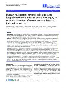
Figure 1 – A) Immunohistochemistry for Cytokeratin-7 (CK-7). Original Magnification 40x. Hepatic Progenitor cells (arrows) within canals of Hering in adult human liver. B) Immunohistochemistry for Neural Cell Adhesion Molecule (NCAM). Original Magnification 40x. Peribiliary glands at the level of hepato-pancreatic ampulla: few small NCAM positive cells can be observed within the acini (arrows) indicating the presence of an undifferentiated stem cell compartment.
Figure 2 – Model for pancreas, liver and biliary development in which the lineages for the liver and for the biliary tree/ pancreas are hypothesized to be separate (see the alternate hypothesis in Figure 3). A common stem/progenitor exists at earlier stages of development in ventral foregut endoderm. This cell could give rise to hepatic stem cells (hHpSCs) and their descendents, hepatoblasts, that mature to liver tissue, whereas the pancreatobiliary progenitors would give rise to the EHBDs and pancreas.
90
Vincenzo Cardinale, Yunfang Wang, Guido Carpino, et al.
Figure 3 – Model for pancreas, liver and biliary development as connected lineages. This model reflects the known events occurring during organogenesis of liver and pancreas in which the endodermal anlage forming the dorsal pancreatic ducts give rises to dorsal pancreas and that forming hepato-pancreatic common duct progress to form the pancreatic ducts for the ventral pancreas, the biliary tree and the liver. This schematic is one modified from a figure from the review by Zaret and Grompe (2008).