Uvidelio F. Castillo,1 Gary A. Strobel,1 Eugene J. Ford,1 Wilford M. Hess,2. Heidi Porter,3 James B. Jensen,3 Heather Albert,3 Richard Robison,3. Margret A. M. Condron,4 David B. ...... Dr Kathleen Donald of the. Manyallaluk Community in the ...
Microbiology (2002), 148, 2675–2685
Printed in Great Britain
Munumbicins, wide-spectrum antibiotics produced by Streptomyces NRRL 30562, endophytic on Kennedia nigriscans Uvidelio F. Castillo,1 Gary A. Strobel,1 Eugene J. Ford,1 Wilford M. Hess,2 Heidi Porter,3 James B. Jensen,3 Heather Albert,3 Richard Robison,3 Margret A. M. Condron,4 David B. Teplow,4 Dennis Stevens5 and Debbie Yaver6 Author for correspondence : Gary Strobel. Tel : j1 406 994 5148. Fax : j1 406 994 7600. e-mail : uplgs!montana.edu
1
Dept of Plant Sciences, Montana State University, Bozeman, MT 59717, USA
2, 3
Dept of Botany and Range Sciences2 and Dept of Microbiology3, Brigham Young University, Provo, UT 84602, USA
4
Brigham and Women’s Hospital and Harvard Medical School, Boston, MA 02115, USA
5
Infectious Diseases Section, Veterans Affairs Medical Center, 500 West Fort St, Boise, ID 83702 and Dept of Medicine, University of Washington School of Medicine, Seattle, WA 98195, USA
6
Novozymes Biotech Inc., 1445 Drew Ave, Davis, CA 95616, USA
Munumbicins A, B, C and D are newly described antibiotics with a wide spectrum of activity against many human as well as plant pathogenic fungi and bacteria, and a Plasmodium sp. These compounds were obtained from Streptomyces NRRL 3052, which is endophytic in the medicinal plant snakevine (Kennedia nigriscans), native to the Northern Territory of Australia. This endophyte was cultured, the broth was extracted with an organic solvent and the contents of the residue were purified by bioassay-guided HPLC. The major components were four functionalized peptides with masses of 1269�6, 1298�5, 1312�5 and 1326�5 Da. Numerous other related compounds possessing bioactivity, with differing masses, were also present in the culture broth extract in lower quantities. With few exceptions, the peptide portion of each component contained only the common amino acids threonine, aspartic acid (or asparagine), glutamic acid (or glutamine), valine and proline, in varying ratios. The munumbicins possessed widely differing biological activities depending upon the target organism. For instance, munumbicin B had an MIC of 2�5 µg mlN1 against a methicillin-resistant strain of Staphylococcus aureus, whereas munumbicin A was not active against this organism. In general, the munumbicins demonstrated activity against Gram-positive bacteria such as Bacillus anthracis and multidrug-resistant Mycobacterium tuberculosis. However, the most impressive biological activity of any of the munumbicins was that of munumbicin D against the malarial parasite Plasmodium falciparum, having an IC50 of 4�5O0�07 ng mlN1. This report also describes the potential of the munumbicins in medicine and agriculture.
Keywords : endophyte, actinomycete, malaria, tuberculosis, biological control
INTRODUCTION
The development of drug resistance in human-pathogenic bacteria such as Staphylococcus, Mycobacterium, Streptococcus, Enterococcus and others has prompted a search for more and better antibiotics, especially as diseases caused by these bacteria now represent a clear and growing threat to world health (National Institutes .................................................................................................................................................
Abbreviation : COSY, correlated spectroscopy. The GenBank accession number for the sequence determined in this work is AY127079. 0002-5548 # 2002 SGM
of Health, 2001 ; Raviglione et al., 1995 ; Pablosmendez et al., 1997). There is also an increasing need for more and better antimycotics, especially as the human population is developing more fungal infections, particularly in HIV\AIDS patients and patients with organ transplants who must take immunosuppressive drugs (Walsh, 1992 ; Walsh & Finberg, 1999 ; Tiphine et al., 1999). In addition, the world’s medicinal arsenal cannot effectively control the increased incidence of parasitic protozoan infections. The most important of these is malaria, caused by Plasmodium spp., which kills up to 1n5–3 million people and produces up to nearly 500 2675
U. Castillo and others
million cases per year (National Institutes of Health, 2001). It is estimated that nearly 40 % of the world’s population is at risk of becoming infected with malaria. Another factor contributing to the increased threat of death caused by malaria is the development of drug resistance in populations of Plasmodium spp. (National Institutes of Health, 2001). In some cases, treatment of malaria and other infectious diseases has been possible by the availability of antibiotics originally derived from soil-borne Streptomyces spp. (Waksman, 1967 ; Arai, 1976). Finally, a call from the public for better and more environmentally sound ways to grow the world’s food indicates that new methods of controlling pests and pathogens are needed (Overton et al., 1996). In the past, the major source of pesticidal agents was organic synthesis. Recently, there has been increased interest in using more environmentally friendly methods in agricultural production. It now appears that a relatively untapped source of microbial diversity for use in agriculture as well as medicine is the microbial endophytes, which live in the interstitial spaces of living plant tissues (Bacon & White, 2000). Some endophytes may produce antimicrobial agents that may be involved in a symbiotic association with a host plant (Yang et al., 1994). A search for specific endophytes that may produce antibiotics is not necessarily a random process. The first objective is to select one or more plants as a source of the endophyte. Usually this selection process is done on the basis of the environment, the age or the natural history of a given plant. Another approach is to select a plant on the basis of its ethnobotanical uses, which was the case in this study. In the Northern Territory of Australia, various Aboriginal groups use the ground-up mass of snakevine (Kennedia nigriscans) to promote the healing of skin wounds and infections. The snakevine, known as ‘ mangerrporlo ’ in Dalabon and Mayali, is harvested as a fresh stem piece, placed on some hot coals for a short time, mashed into a pulp, and then applied as a sticky paste to a cut, wound or infection. Because of the native uses of this plant, it was selected as a source of endophytic micro-organisms with the idea that some of the healing properties of the snakevine may, in fact, be due to the products of one or more endophytes. One of the endophytes isolated from this medicinal plant was a Streptomyces sp. ; as a culture, it was extremely bioactive against a number of test micro-organisms. Interest in this endophyte was further piqued because actinomycetes have not been reported to be endophytic on dicotylendenous plants. However, an endophytic Streptomyces sp. has been reported from the grass Lolium perenne (Guerny & Mantle, 1993). This lolium endophyte produces a weak antibiotic designated as methylalbonoursin, which is a diketopiperazine, condensed from leucine and phenylalanine. Although streptomycetes in general have been an outstanding source of natural products used in medicine, generally they have been isolated from soil. The finding that some Streptomyces spp. have taken up residence in plants opens the 2676
possibility that this may be an entirely untapped source of novel pharmaceuticals and agents for use in agriculture. We report here the isolation, chemical and biological properties of a series of unique wide-spectrum antibiotics designated the munumbicins from an endophytic actinomycete that has been designated Streptomyces NRRL 30562. METHODS Isolation of endophytes. Stems (0n5–1n0 cm in diameter) of
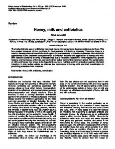
Kennedia nigriscans (Fig. 1) were obtained near the Aboriginal community of Manyallaluk, SE of Katherine, Northern Territory, Australia, at 14 m 16h 352d S and 132 m 49h 750d E. The stems were thoroughly treated with 70 % ethanol and then the outer bark removed with a sterilized sharp blade. The inner pieces of the stem, containing the cambium, phloem and xylem tissues, were plated on water agar in Petri plates. After incubation for 10 or more days at 23 mC, individual fungal and bacterial colonies were removed with a sterile fine-tipped needle and transferred onto potato dextrose [glucose] agar (PDA). Fungal and actinomycete spore formation was encouraged by placing the endophytes onto γ-irradiated carnation leaves and on autoclaved stem and petiole tissues of K. nigriscans. The plates were continuously monitored for spore formation by stereo and light microscopy. The endophytic micro-organism, a streptomycete, was of primary interest, because of its broad antibiotic activities against fungi. This organism has been deposited as Streptomyces NRRL 30562 in the Peoria USDA National Laboratory, and as culture no. 2101 of the Montana State University culture collection. Small PDA pieces, mostly containing spores, were stored in 15 % (v\v) glycerol in water at k70 mC. Microscopy of endophytes. Fruiting structures of the micro-
organism appearing on both carnation leaves and the tissues of the host plant were examined by stereo and light microscopy. These structures were fixed and processed using the standard methods of fixation (Worapong et al., 2001) by placement in 2 % (v\v) glutaraldehye in 0n1 M sodium cacodylate buffer (pH 7n2) and left overnight. The samples
.................................................................................................................................................
Fig. 1. The foliation of snakevine (Kennedia nigriscans) as it exists in a natural field situation near the Manyallaluk Community of Northern Territory, Australia.
Munumbicins – novel antibiotics were then passed through a gradient of ethanol solutions, which was done to discourage the processes of shrivelling which normally occurs in spores and mycelia with rapid dehydration. The samples were then critical-point dried, gold coated with a sputter coater and observed and photographed with a JEOL 6100 scanning electron microscope. Munumbicin isolation procedures. Several small blocks of PDA containing the streptomycete were inoculated into 500 ml PD broth in a 2 l Erlenmeyer flask and incubated for 3 weeks at 23 mC without shaking. The culture filtrate was then extracted three times with half-volumes of methylene chloride. This organic solvent extract was pooled and then taken to dryness under flash evaporation at 40 mC. The yield of dry material per litre was about 250 mg. Approximately 5 mg of this material was subjected to HPLC on a Microsorb 60-8, 250i10 mm Dynamax C-18 column. The elution solvent was 20 % tetrahydofuran (THF)\80 % water programmed for 60 min to a final concentration of 40 % THF\60 % water and then held isocratically for 95 min. The elution stream was continuously monitored at 260 nm with a flow rate of 2 ml min−". Individual fractions were subjected to a bioassay test by placing them on a Petri plate with PDA, drying, and then applying small blocks of agar containing Pythium ultimum. This fungus was chosen for the bioassay because of its rapid growth characteristics and its sensitivity to anticancer compounds such as taxol (Young et al., 1992). The fractions were considered active if inhibition of fungal growth was observed. Each fraction was repurified using the same system and these fractions were weighed and primarily used for bioassay tests. The compounds in these peaks were at least 95 % pure based on HPLC analysis. Final purification for spectroscopic measurements was done by subjecting each compound to HPLC on a 100-5 Microsorb, 250i4n6 C-18 column. The initial elution solvent was 20 % acetonitrile\80 % water for 90 min programmed to a final concentration of 80 % acetonitrile. The retention time of each peak was recorded and the bioactivity of each peak was determined. Disk bioassays on agar Petri plates. The bacterial isolates used for the majority of these tests were from the American Type Culture Collection (ATCC). After primary isolation on the media recommended by ATCC, organisms from a single colony were cultured overnight in 10 ml Mueller–Hinton Broth (MHB) at 35 mC unless otherwise specified with or without 5 % CO . Then, after 12 h incubation, 0n5 ml of the # was added to 4n5 ml pre-warmed MHB bacterial suspension and the solution was incubated at 35 mC to obtain cultures in the exponential phase of growth. The inoculum for the bacterial disk diffusion assays was prepared as described in the protocols of the US National Committee for Clinical Laboratory Standards (NCCLS). Compounds to be tested, including known antibiotics, were dissolved in 2–3 % methanol and applied to sterile (6 mm diameter) paper disks, dried and then applied to the appropriate medium for testing. The plate medium used for testing the disks was MHA for all isolates except Neisseria gonorrhoeae and Streptococcus pneumoniae, for which Bacto GC agar base with 1 % defined growth supplement and MHA with 5 % defibrinated sheep’s blood, respectively, were used according to the NCCLS guidelines. Incubation conditions were 35 mC overnight unless otherwise specified, with or without 5 % CO as recommended. # Minimum inhibitory concentrations (MICs). Microbroth dilution assays of human-associated bacteria and fungi were performed as described in the NCCLS protocols. The bacteria were obtained from the ATCC and the fungi tested were from the microbial collection at Eli Lilly Co, Indianapolis, IN, USA, where those tests were conducted. The assays were performed
in sterile 96-well plates, and the total volume per well was 100 µl. The bacterial inoculum was prepared to give approximately 10%–10& c.f.u. per well and the compounds were tested at concentrations from 0n0625 to 64 µg ml−" in twofold step dilution. The actual number of c.f.u. per well was confirmed by plating onto blood agar. Two wells were inoculated for a given concentration. The plates were incubated for 16–20 h at 35 mC unless otherwise specified, with or without 5 % CO . # The MIC was defined as the minimum concentration of compound resulting in no visible growth of the test organism. Plant-associated micro-organisms were tested for their response to the munumbicins using a microbroth (PD broth) dilution technique carried out in 24-well dilution plates using several 3 mm$ agar blocks containing fungal inoculum placed in 1 ml PD broth. Incubation was for 48 h at 23 mC. A plantpathogenic strain of Pseudomonas syringae was also tested using the same PD broth medium and with concentrations of c.f.u. as given above. The organisms were obtained from the Montana State University plant pathogen culture collection administered by Dr Don Mathre. Inhibitory concentrations (IC50) for Mycobacterium tuberculosis. IC values were determined for multiple-drug-resistant
&! M. tuberculosis (MDR-P) and a standard drug-sensitive strain (H37Rv, ATCC 25618) of this organism. A modified procedure commonly used to test slow-growing bacteria was used to determine the effectiveness of the munumbicins against these bacteria (Isenberg, 1992). The test compounds were dissolved in DMSO and appropriately diluted and placed in Mycobacteria 7H11 agar supplemented with 10 % OADC (oleic acidalbumin-dextrose [glucose]-catalase) enrichment. All experiments were carried out in a biosafety level-3 facility. The IC &! values were calculated by linear regression analysis of plots of recoverable M. tuberculosis against concentration of drug administered. This experiment required relatively large amounts of the munumbicins and was repeated twice.
Antimalarial assay. Cultures of P. falciparum strain CSC-1 (Honduras) were maintained according to the methods of Trager & Jensen (1976) and Trager & Jensen (1978), except that human serum was replaced with Albumax I (Gibco, BRL) ; 6 % (w\v) stock solution in RPMI 1640 medium containing 0n1 mg hypoxanthine ml−" stored at k20 mC. All cultures were maintained in a microaerophilic environment containing 1 % oxygen\5 % CO with the balance being nitrogen. The stock solution was #diluted at a ratio of 5 : 100 (v\v) of the RPMI 1640 to give a final concentration of 0n3 % Albumax I. Stock solutions of the compounds to be tested, including the munumbicins and chloroquine, were initially dissolved in DMSO or water and diluted to final concentrations in Albumax-supplemented RPMI 1640. Control cultures contained the same quantity of DMSO or water as that used in the experimental trials.
Parasite cultures, adjusted to 0n1–0n5 % parasitaemia by addition of freshly washed human erythrocytes, were pipetted into 96-well culture plates by adding 10 µl of a 50 % cell suspension to each well to give a final volume of 5 µl packed erythrocytes per well. The test compounds in RPMI 1640 were added at 95 µl per well to four wells per concentration of test compound. Quadruplicate control wells were treated similarly with DMSO or water added without the test compounds. The cultures were gassed and exposed to [$H]phenylalanine [1 µCi (37 kBq) per well] for the final 24 h of treatment. Cultures were monitored for bacterial contamination and for parasite viability by Giemsa staining of thin films. Ultimately, the cells were harvested onto glass-fibre filters, followed by liquid scintillation counting. The IC for each compound was &! 2677
U. Castillo and others determined by linear regression analysis using 50 % of the control counts as the regression point. The experiment was repeated three times, the data averaged and the standard deviation of the mean determined. Amino acid analysis. HPLC-purified compounds were dissolved in methanol, placed in 6i50 mm glass tubes, dried in vacuo, and then transferred to a hydrolysis vessel (Millipore ; part no. 007603). Approximately 300 µl 6 M HCl was added to the vessel, which was then alternately purged with nitrogen and evacuated three times before being sealed under vacuum. Vapour-phase hydrolysis was performed by heating at 110 mC for 22 h. Separation and quantification of amino acids was carried out on a Beckman model 6300 amino acid analyser. Moles of each amino acid were initially determined using molar absorption values derived from amino acid standards. Other details of these analyses, as performed on a peptide antibiotic from another endophytic organism, have been described (Miller et al., 1998). Each analysis was performed at least three times ; the results are reported as meansp. Mass spectroscopic analyses. A mass spectrum was obtained of each of the HPLC-purified munumbicins. Spectral data were obtained on a Bruker Biflex III MALDI mass spectrometer. The instrument was set on the reflective mode with an accelerating voltage of 19 keV. A nitrogen laser at 337 nm at 3 Hz was used (having a 3 ns pulse width). The number of spectra obtained on any individual sample varied from 10 to 200 shots. The matrix used for crystallization with the munumbicins was α-cyano-4-hydroxycinnamic acid. External calibration to standardize the molecular masses was done with adrenocorticotropic hormone fragment 18–39 (mol. mass 2465n2 Da). Data reported are presumed accurate to the first decimal place. In order to ascertain if any of the peaks in the mass spectra of the munumbicins was sodiated, Na+ was added in excess to the sample prior to crystallization of the matrix and a spectrum was retaken. NMR spectroscopy. NMR spectroscopy was applied to each of
the munumbicins, with the greatest effort given to munumbicin B because of its availability relative to the other munumbicins. Samples were dissolved in 99n95 % deuterated acetonitrile and data obtained in a Bruker 500 MHz instrument. In "H spectroscopy, each sample was subjected to 16 scans with a sweep width of 6024 and 8k real points. A gradient-enhanced COSY ("H–"H coupling) experiment was also conducted with munumbicin B. Materials. All solvents used for HPLC were ‘ HPLC grade ’.
Those used for extraction were ACS grade. All other reagents were obtained from Sigma, including adrenocorticotropic hormone fragment 18–39, standard amino acids and radiolabelled phenylalanine.
RESULTS AND DISCUSSION Isolation and identification of the endophytes
The stems of K. nigriscans (snakevine) yielded at least 24 endophytic micro-organisms. At least three of these were Pestalotiopsis spp., which is common for plants growing in tropical or semi-tropical environments (Li et al., 2001). However, an endophyte designed A11-4C was of greatest interest, because of its potent antimicrobial activity. This organism resembled a Streptomyces sp. and was not isolated as an endophyte from any plants in the near vicinity of K. nigriscans, including Banksia 2678
dentata and Owenia vernisoa. However, it was isolated from K. nigriscans from different individual plants in and around the Manyallaluk Community. This organism was grown on γ-irradiated carnation leaves as well as autoclaved freshly harvested snakevine after placement on water agar. After 10–14 days the cultures were examined for the production of fruiting structures. On carnation leaves, only little or modest spore production occurred (Fig. 2a). However, on the inoculated natural host of this microbe, snakevine, numerous spiral and curved mycelia were produced, some having chains of many spores (Fig. 2b). This observation suggested that this endophytic micro-organism had developed some biochemical relationship with its host plant since it so readily made spores on this plant and not on others, such as carnation leaves. The organism fitted, in all respects, the definition of a Streptomyces sp. It produced slow-growing, erumpent, multisectored and multicoloured colonies on PDA. Whitish mycelia, mixed with areas of spore production that were tan to brownish in colour, occurred primarily in the border regions of 4–5-week-old colonies growing on PDA. Toward the centre and in mid-sections of the colony appeared greasy yellowish-orange raised areas. An examination of the organism by scanning electron microscopy revealed the presence of numerous aerial filaments and smooth, cylindrical spores (0n8 µm in length and 0n7 µm in diameter) that were produced in long curved as well as spiral chains (Fig. 2). Cultures growing on PDA, after 4–5 weeks, began to exude a water-soluble pigment that diffused into the medium, giving a distinctive reddish-orange coloration around the individual colonies. In older cultures, reddish droplets appeared on the mycelium. These droplets were individually collected and dried. After chromatographic separation, munumbicins were the primary compounds present ; they had a yellowish to reddish–orange coloration depending upon the concentration of compound present. The organism has been deposited as Streptomyces NRRL 30562 in the Peoria USDA National Laboratory. The 16S rDNA was PCR-amplified from Streptomyces NRRL 30562 and sequenced, using standard methods (GenBank accession no. AY127079). The sequence was different from all other 16S rDNA entries in GenBank, but over 1500 bp of the sequence was 100 % identical to that of ‘ Streptomyces padanus ’ (GenBank accession no. AF455813), a soil-borne streptomycete (Baldacci et al., 1968). Furthermore the 16S rDNA of Streptomyces NRRL 30562 was 98n5 % homologous to Streptomyces galbus (GenBank accession no. X79325), 98n6 % to Streptomyces sp. NRRL 5799 (GenBank accession no. AJ391814), 97n7 % to Streptomyces eurythermus (GenBank accession no. D63870) and 96n9 % to Streptomyces coelicolor (GenBank accession no. Y00411). It appeared that the closest known relative of Streptomyces NRRL 30562, at the molecular biological level, was ‘ S. padanus ’. Isolate NRRL 30562 was therefore compared to ‘ S. padanus ’ in more detail. ‘ S. padanus ’
Munumbicins – novel antibiotics (a)
(b)
.................................................................................................................................................................................................................................................................................................................
Fig. 2. (a) Growth and limited spore production of Streptomyces NRRL 30562 on carnation leaves. (b) Growth of Streptomyces NRRL 30562 on autoclaved twig pieces of its natural host, K. nigriscans. Note the proliferation of numerous mycelia bearing chains of spores. Bars, 1 µm.
Table 1. Yield values and chromatographic and spectroscopic properties of munumbicins A–D Munumbicin
Yield (mg l−1)
Mol. mass (Da)
Molar absorption coefficients
Retention time (min) Dynamax column
Microsorb column
A
10
1326n5
ε #") ε
23 300 424
126n5
69n9
B
118
1269n6
ε #!) ε #%! ε %#! ε
13 360 54 514 21 200 22 065
116n3
68n5
C
30
1298n5
ε ##! ε #%! ε %"' ε
5929 62 072 55 356 56 785
98n0
67n8
D
15
1312n5
ε ##" ε $"% ε
8442 220 128
80n8
45n0
#%!
%%!
%%!
%!%
possesses long axial filaments with thick verticillate-like branches bearing spores. This is in striking contrast to NRRL 30562, which has no verticils and little branching of its mycelium (Fig. 2a, b). The spores of ‘ S. padanus ’, by electron microscopy, are ovoid (Baldacci et al., 1968) while those of NRRL 30562 are cylindrical (Fig. 2a, b). ‘ S. padanus ’ is reported to produce an antimicrobial substance with a mass of 1150 Da ; this is in contrast to NRRL 30562, which has no substance of this mass based on this study. Other colour differences of the two organisms in various culture media have also been noted. These features, together with its specialized ecological niche as an endophyte in the snakevine, suggest that Streptomyces NRRL 30562 is a unique micro-organism ; further details of its taxonomy will be forthcoming.
Chemical characterization of the antimicrobials produced by NRRL 30562 – munumbicins
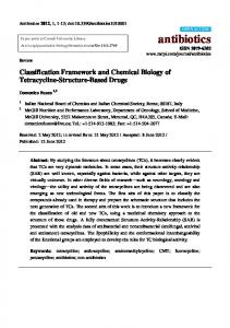
The antimicrobial compounds produced by Streptomyces NRRL 30562 were named munumbicins in memory of R. Munumbi Miller (see acknowledgements). Preparations of the munumbicins made by HPLC were used to chemically characterize these compounds. The major components of the culture medium of Streptomyces NRRL 30562 appeared as peaks A–D, with varying retention times, after the first passages over a Microsorb 60-8 column (Table 1) (Fig. 3). Other smaller peaks with retention times of 45–75 min, which were bioactive, were also observed (Fig. 3). Components in each of these peaks, when assayed on PDA plates 2679
Relative absorbance (260nm)
U. Castillo and others
D
20
40
C
B A
60 80 100 120 Retention time (min)
140
160
180
.................................................................................................................................................
Fig. 3. Elution of the munumbicins, indicated by peaks labelled A–D, from a Dynamax Microsorb 60-8 HPLC column under the conditions described in Methods.
10
9
8
7
6 5 δ (p.p.m.)
4
3
2
1
0
.................................................................................................................................................
Fig. 4. 1H NMR spectrum of munumbicin B taken in 99n95 % deuterated acetonitrile.
inoculated with Pythium ultimum, caused complete inhibition of fungal growth. Repassage of the contents of each peak over the same column gave compounds that were at least 95 % pure, based on numbers of peaks and peak intensity, and were bioactive against P. ultimum ; these preparations were used for bioassay and other tests. Final and total purification of each component was obtained after passage through a Microsorb 100-5 column, ultimately yielding single peaks that have been designated munumbicins A–D. Some indication of the purity of these compounds was also obtained by an NMR-COSY ("H–"H coupling) experiment in which each of the peaks, especially the smaller downfield ones, were coupled to the more intense upfield ones, suggesting that the small peaks in the spectrum are not attributable to contaminating substances (Fig. 4). 2680
Each of the munumbicins reacted with ninhydrin to produce a weakly pinkish product on silica gel plates. This suggested that the compounds contained primary or secondary amino groups. Additionally, the absorbance of each of these compounds in the region of 208 nm suggested the presence of one or more amidochromophoric groups (Table 1) (Silverstein et al., 1991). Finally, the general nature of the "H NMR spectrum of each of the munumbicins suggested that the chemical nature of these compounds is that of a highly functionalized peptide (Fig. 4) (Strobel et al., 1999). The most likely possibility to account for these results was that these compounds were composed of amino acids linked together via peptide bonds. Therefore, each of the munumbicins was subjected to amino acid analysis. The results of these analyses revealed that the munumbicins are peptides which have common compositional features. Each of the four compounds produced chromatographic profiles consistent with the presence of Glx (glutamic acid or glutamine), Pro, Thr and Val (Table 2). The molar ratios of these amino acids were 1 : 2 :1 : 3, respectively, except for munumbicin C, which had an extra proline. In addition, munumbicins A, B and D contained 1 mole of Asx (aspartic acid or asparagine) (Table 2). A unique feature of munumbicin A was the presence of 2 moles of leucine (Table 2). The deduced molar ratios of these amino acids were consistent with the observed molecular masses of the compounds. Three factors affecting the interpretation of amino acid composition are the chemical lability of the amino acid side chains, the acid sensitivity of individual peptide bonds and the presence of unusual amino acids (those not among the basic 20). Our interpretation of the data is parsimonious, and other interpretations are possible. For instance the consistent presence, but relatively low yield, of Glx could indicate that a modified amino acid exists with an identical chromatographic retention time. Unusual amino acids are commonly found in biologically active peptides produced by endophytes (Ballio et al., 1994 ; Strobel et al., 1999). In addition, amino acid acylation, esterification and racemization are commonly observed. Hydrolysis of peptides containing these unusual amino acids usually results in relatively low yields of the unusual residue or in the hydrolytic elimination of the unusual side chain, causing the residue to be misidentified. The data strongly support the partial peptide nature of the munumbicins and their structural relatedness. Further detailed work on these compounds will be necessary to fully elucidate their structural identity. Mass spectroscopy of each of the munumbicins revealed a range of actual masses from 1326n5 Da for munumbicin A to 1269n6 Da for munumbicin B (Table 1). Each of these masses represents the actual mass and not MjNa+ since the addition of Na+ ions to the sample used in the mass spectrometer, on any of the munumbicins, did not give an Mj23 peak. Munumbicins recovered from the second passage through the Microsorb 60-8 column were quite homogeneous as measured by HPLC. However, after mass spectroscopy, munumbicin A still had a
Munumbicins – novel antibiotics Table 2. Amino acid composition of munumbicins A–D .................................................................................................................................................................................................................................................................................................................
Results are presented as the meansp of three individual amino acid analyses. The number in parentheses following each amino acid indicates the tentative number of moles of that residue per mole of the munumbicin. Hydrolysis of glutamine or asparagine results in deamination, producing glutamic acid or aspartic acid, respectively. Therefore, it is possible only to determine the sums of each acid\amide combination, which are referred to as Glx and Asx. The mol % per residue is approximately 10. Amino acid
Asx Glx Leu Pro Thr Val
Molar percentage in : Munumbicin A (1326n5 Da)
Munumbicin B (1269n6 Da)
Munumbicin C (1298n5 Da)
Munumbicin D (1312n5 Da)
12n9p1n4 (1) 5n2p2n3 (1) 20n1p0n7 (2) 17n6p1n1 (2) 7n3p0n5 (1) 32n1p0n7 (3)
14n1p1n5 (1) 9n0p0n2 (1) 0 (0) 21n3p0n4 (2) 12n7p0n3 (1) 41n7p0n7 (3)
2n1p0 (0) 7n0p0n1 (1) 0 (0) 37n6p0n7 (3) 11n3p0n7 (1) 40n9p0n2 (3)
10n0p0n2 (1) 8n6p0n3 (1) 0 (0) 22n9p0n2 (2) 14n2p0n3 (1) 42n9p0n3 (3)
small amount of a compound with a mass of 1266n2 Da associated with it, whereas munumbicin C had an associated component at 1314n5 Da in small quantities. Finally, munumbicin D (1312n5 Da) had a series of components in much lesser amounts at 1328n5, 1314n5 and 1346 Da. These components probably represent derivatives of the major components of the munumbicin series A–D. The phenomenon of a number of peptide derivatives of an antibiotic appearing in a culture broth is not uncommon in nature, especially with the lipopeptides (Ballio et al., 1994). The "H NMR spectrum of the munumbicins (munumbicin B) is certainly compatible with the peptide nature of the molecules ; however, absorbances at 5n5– 6n0 p.p.m. suggest C–C unsaturation, which is not found in the peptide moiety of the molecule (Strobel et al., 1999) (Fig. 4). In addition, the absorbances between 3n5 and 5n0 p.p.m. are suggestive of a sugar moiety. Therefore, each of the munumbicins was analysed for sugar residues after acid hydrolysis, reduction, acetylation and GC-mass spectrometry. There was not a trace of sugars in any of the munumbicin preparations. Thus, the absorption peaks in this portion of the "H NMR spectrum are not those of sugar residue protons, but protons associated with carbons bearing oxygen or nitrogen (Silverstein et al., 1991). The "H NMR spectrum of each of the other munumbicins was related but not identical to munumbin B (Fig. 4). In addition, the relative intensity of absorption peaks in munumbicin B from 0n5 to 2 ppm is consistent with an abundance of methyls and methylenes in the molecule. Certainly, the unsaturated nature of these molecules is also consistent with the strong UV absorbances seen in the UV spectra of the molecules, especially munumbicins B and C (Table 1). These absorbances are contributed by the non-peptide portion of the munumbicins since there is no amino acid present in any of the munumbicins that absorbs in the UV above 230 nm (Table 2). Furthermore, the highly coloured nature of each of the munumbicins
(yellowish–orange–reddish) suggests that the non-peptide portion of the molecule is the contributing pigment. It seems unlikely that the coloration is due to a metal complex with the peptide portion of the molecule since heating an aqueous solution of munumbicin B with EDTA (a metal-chelating reagent) did not eliminate or even reduce the intensity of the coloration. Modifications in the amino acid composition of these compounds account for some of the molecular mass variation that is observed in them (Table 2). Likewise, it is apparent that modifications also occur in the non-peptide portion of the munumbicins, given the slight variation in masses of the minor components that has been noted, e.g. munumbicin D, mass 1312n5 Da, with a minor component at 1314n5 Da, suggesting the difference of one double bond in the molecule. Overall, the peptide portion of these compounds generally contributes about 70–80 % of the mass of each munumbicin and knowledge about the non-peptide moieties of these substances is still lacking. Based on the known molecular masses of the components it appears that the non-peptide component must be in the range of 300–400 Da and the limited spectroscopic data available suggest that it may be a macrolide. Overall, it appears that the munumbicins represent a novel group of bioactive substances since a comprehensive search of the Chapman & Hall Dictionary of Natural Products on CD ROM, 2001, did not reveal complete chemical identity with any previously described products. Compounds with comparable masses and some similarities to the munumbicins are known in the literature. Arbocandin E (Chapman & Hall no. JOX 54-Q-S) has a mass of 1298n5 Da, which is identical to that of munumbin C (Table 1) ; arbocandin F (Chapman & Hall no. JOX 55-R) has a mass of 1312n5 Da, which is identical to that of munumbin D (Table 1). However, the arbocandins, originating from a filamentous fungus, are glucan synthase inhibitors ; they contain certain amino acid residues in quantity and quality not found in 2681
U. Castillo and others Table 3. Inhibitory activity of the munumbicins against various plant-pathogenic fungi and one human-pathogenic bacterium MIC (µg ml−1)
Test organism
Pythium ultimum Rhizoctonia solani Phytophthora cinnamomi Geotrichum candidum Sclerotinia sclerotiorum Pseudomonas syringae
Munumbicin A
Munumbicin B
Munumbicin C
Munumbicin D
2n0 – – – 8n0 15n6
0n2 8n0 6n2 31n2 0n20 2n0
4n0 1n5 1n5 15n6 8n0 15n6
0n4 15n6 15n6 31n2 2n0 0n2
–, Activity not determined.
Table 4. Inhibitory activity of the munumbicins and some standard antibiotics against various bacteria in a disk diffusion assay .................................................................................................................................................................................................................................................................................................................
The test compound was administered on sterile paper disks at the rate of 10 µg per disk ; ciprofloxacin and vancomycin were administered at 5 and 30 µg per disk, respectively. The developing zone of inhibition was measured. Details of the culture conditions are given in Methods. Assays were done on Mueller–Hinton broth agar medium at 35 mC, unless otherwise noted. Bacterium tested*
Pseudomonas aeruginosa 27853 Vibrio fischeri PIC 345 Enterococcus faecalis 29212 Staphylococcus aureus 29213 Acinetobacter sp. 49137 (grown at 30 mC) Neisseria gonorrhoeae 49226† Streptococcus pneumoniae 49619‡ Bacillus anthracis K8902 Escherichia coli 25922§
Diameter of inhibition zone (mm) Mun. A
Mun. B
Mun. C
Mun. D
Ciprofloxacin
Vancomycin
R R R R R 9 R 9n5 R
R 16 18 15 R 14 17 18 R
R 9 R R R 8 7 R R
R 12 16 13 R 9 16 – R
30 – – – 29 – – 16 –
– – 18 15 – 13 20 – –
* The number after the bacterial name is the ATCC or Presque Isle (PIC) acquisition number. † GC agar with 1 % growth supplement, with 5 % CO . # ‡ MHA with 5 % defibrinated sheep’s blood and 5 % CO . # § Resistant to penicillin G applied at 10 units per disk. –, Not determined ; R, resistant.
the munumbicins and these inhibitors are not pigmented. Biological activities of the munumbicins
Each of the munumbicins was active against an array of plant-pathogenic fungi, with the MICs varying from a few tenths of a microgram up to about 30 µg (Table 3). The least active munumbicin, based on the comparative MIC values, appeared to be munumbicin D (Table 3). The fact that these compounds had such remarkable activity against plant-pathogenic fungi and at least one bacterium lends support to the notion that Streptomyces NRRL 30562, as an endophyte, may have some role to 2682
play in protecting the host plant from invading pathogens (Yang et al., 1994 ; Strobel et al., 1997). Since the munumbicins were so active against an array of plant-pathogenic fungi and at least one bacterium, a general disk screening test was applied to a series of human-pathogenic bacteria. The diameter of the zone of inhibition was taken as a relative indication of biological activity of the various munumbicins. Most notable from this testing regimen was the fact that each Grampositive bacterium tested (Bacillus anthracis, Streptococcus pneumoniae, Enterococcus faecalis and Staphylococcus aureus) was sensitive to one or more of the munumbicins (Table 4). In contrast, three of the five Gram-negative bacteria tested (Escherichia coli, Ac-
Munumbicins – novel antibiotics Table 5. MICs of the munumbicins against various human-pathogenic fungi and bacteria MIC (µg ml−1)
Test organism
Cryptococcus neoformans Candida albicans Aspergillus fumigatus Staphylococcus aureus ATCC 33591* Staphyloccus aureus MH II Eli Lilly Co.† Enterococcus faecalis ATCC 51299‡
Mun. A
Mun. B
Mun. C
Mun. D
Amphotericin B
10 – 20 – 8
10 – 20 2n5 –
10 �10 20 0n4 16
10 �10 20 0n4
0n13 1n25 0n62 – – –
* Methicillin-resistant strain. † Sensitive to vancomycin ; MIC 0n06 µg ml−". ‡ Resistant to vancomycin and sensitive to ciprofloxacin. , Not active at any of the concentrations tested ; –, Activity not determined.
Table 6. Effects of the munumbicins on a drug-resistant and a common drug-sensitive strain of M. tuberculosis .................................................................................................................................................................................................................................................................................................................
The data are reported as the values in µg ml−" and were obtained from plots of treated recoverable M. tuberculosis as a function of concentration of drug administered to the culture. Munumbicins A and D were not active at any concentration in this assay against M. tuberculosis. IC50 (µg ml−1) for :
Compound tested
Munumbicin B Munumbicin C Rifampicin
M. tuberculosis MDR-P (drug resistant)
M. tuberculosis H37Rv (ATCC 25618) (drug sensitive)
10 �125 �150
46 �150 1
inetobacter sp. and Pseudomonas aeruginosa) were resistant to the munumbicins at the concentration applied on the disk and then to the assay plate (Table 4). It is interesting that the human-pathogenic pseudomonad appeared to be resistant to all of the munumbicins, whereas a plant-pathogenic pseudomonad was sensitive (Tables 3 and 4). The results of the disk tests led to the more refined method of determining the relative effectiveness of antibiotics – the MIC test against some of the drugsensitive bacteria and fungi pathogenic on humans. In fact, two of the Gram-positive munumbicin-sensitive bacterial strains are common drug-resistant ATCC strains : a methicillin-resistant strain of Staph. aureus (ATCC 33591) and a vancomycin-resistant strain of Ent. faecalis (ATCC 51299) (Table 5). However, Ent. faecalis ATCC 51299 was sensitive to ciprofloxacin (Table 5). MIC values of 2n5 µg ml−" (munumbicin B) and 8n0 µg ml−" (munumbicin A) were observed against drugresistant Staph. aureus and Ent. faecalis, respectively (Table 5). Also, in this MIC test, munumbicin C was
active against Ent. faecalis at 16 µg ml−" (Table 5). In addition, munumbicins C and D had bioactivity (0n4 µg ml−") against a drug-sensitive strain of Staph. aureus (Table 5). Although the munumbicins were active against plant-pathogenic fungi their activity against human-pathogenic fungi was less impressive, with MIC values that did not reach below 10 µg ml−", which is in marked contrast to the striking effects of amphotericin B on these organisms (Table 5). Thus, their potential for use in treating human fungal caused diseases is greatly reduced. One of the most interesting activities of the munumbicins is munumbin B against multiple-drug-resistant (MDR-P) M. tuberculosis, an acid-fast bacterium. It had an IC value of 10 µg ml−" whereas rifampicin was &! inactive against this organism, even at elevated virtually concentrations (Table 6). What is peculiar, however, is the fact that only the MDR-P strain of M. tuberculosis was sensitive to munumbin B whereas the drug-susceptible strain of this organism was less sensitive to it (Table 6). 2683
U. Castillo and others
Most surprising was the outstanding activity of each of the munumbicins against P. falciparum, the most pathogenic plasmodium causing malaria. The IC &! values of the munumbicins, determined by the assay described in Methods, indicated that each was within the range to be pharmacologically interesting : munumbicin A, 175p106 ng ml−" (meanp, nl3) ; munumbicin B, 130p70 ng ml−" ; munumbicin C, 6n5p 2 ng ml−" ; munumbicin D, 4n5p0n7 ng ml−". However, munumbicins C and D were of special interest because of their extremely low IC values. The IC of munum&! &! bicin D is about 50 % below that of chloroquine, the gold-standard antimalarial drug (IC 7n0p0 ng ml−"). &! D did not cause Furthermore, munumbicins B, C and any detectable lysis of human red blood cells up to a concentration of 80 µg ml−". While these results are encouraging, any further development of the munumbicins for human use will have to await data from more extensive cellular and clinical toxicity tests. Presently, there are indications that the munumbicins are active against an array of cancer-cell lines, while having some toxicity, at higher concentrations, to primary human cell lines (D. Yaver, unpublished). Therefore, the ultimate development of these compounds as antimalarial, anticancer or anti-infectious drugs may have to depend upon the synthesis of munumbicin derivatives that have reduced toxicity. This could potentially be achieved by carrying out chemical modifications on the munumbicin of choice in order to reduce toxicity while simultaneously increasing efficacy. This approach has been effectively taken with another antibiotic family, the pseudomycins, obtained from a plant-associated microbe – Pseudomonas syringae (Ballio et al., 1994). A specific pseudomycin has been subjected to modifications by organic synthesis and has yielded a derivative that is no longer toxic to mammalian systems and yet remains effective against human-pathogenic fungi (Zhang et al., 2001a, b). Given the fact that the munumbicins have numerous functional groups, especially those associated with the peptide portion of the molecule, chemical derivatization would unquestionably be possible. Anticipated agricultural applications of the munumbicins and Streptomyces NRRL 30562
Each of the munumbicins demonstrated biological activity against an array of plant pathogens at relatively low MIC values (Table 2). Comparisons were made between Streptomyces NRRL 30562 and Streptomyces griseoviridis, which is included in the formulation for ‘ Mycostop ’, a commercial agricultural product of Butts International Inc., Fairfield, Connecticut, USA, which is used to control plant diseases caused by Alternaria, Fusarium and Phomopsis. Cultures of both were grown for 7–10 days and then challenged with plant pathogens for a 4 day incubation period on plates of PDA. Zones of inhibition were measured and compared. The two streptomycetes had comparable effects on Rhizoctonia solani, Aspergillus sp., Fusarium oxysporum, Botrytis alli and Alternaria helianthi. However, the zones of 2684
inhibition were nearly twice as great with Streptomyces NRRL 30562 as with S. griseovirdis for Pythium ultimum, Phytophthora infestans, Penicillium sp., Sclerotinia sclerotiorum, Erwinia carotovora and Cochliobolus carbonum. It definitely appears that Streptomyces NRRL 30562 has the potential to be considered for development as an agricultural agent, especially as it relates to the control of plant diseases either in or on plants or possibly in soils infested with them. ACKNOWLEDGEMENTS The authors greatly appreciate the help of Dr Joe Sears of the Montana State University Chemistry Department in assisting with the acquisition of mass spectral data. Dr Scott Busse of the same department helped with NMR spectroscopy. Dr Peter Albersheim of the Complex Carbohydrate Laboratory and his colleagues at the University of Georgia did the carbohydrate analyses. Dr Kathleen Donald of the Manyallaluk Community in the Northern Territory of Australia generously acquired the photograph of the snakevine while in full leaf during the wet season. Dr Doug Zechner of Eli Lilly Co. kindly performed the MIC tests on the human fungal pathogens. Financial help for the project was supplied by Novozymes Biotech of Davis, CA, the NSF and the Montana Agricultural Experiment Station. The material in this report is, in part, the result of work supported with resources and the use of facilities at the Boise, VA, Medical Center. Finally, we particularly wish to acknowledge the help and guidance of Mr Reggie Munumbi Miller of Manyallaluk community near Katherine, Australia. He and his assistants graciously identified trees and shrubs of ethnobotanical interest in and around his village for our scientific examination and sampling purposes. These novel antibiotics and this report are presented in his honour and memory.
REFERENCES Arai, T. (1976). Actinomycetes : the Boundary Microorganisms. Singapore : Toppan. Bacon, C. W. & White, J. F. (2000). Microbial Endophytes. New York : Marcel Dekker. Baldacci, E., Farina, G., Piacenza, E. & Fabbri, G. (1968).
Description of Streptomyces padanus sp. nov. and emendation of Streptomyces xantophaeus. Giorn Microbiol 16, 9–16. Ballio, A., Bossa, F., DiGiogio, P., Ferranti, P., Paci, M., Pucci, P., Scaloni, A., Segre, A. & Strobel, G. A. (1994). Structure of the
pseudomycins, new lipodepsipeptides produced by Pseudomonas syringae MSU 16H. FEBS Lett 355, 96–100. Gurney, K. A. & Mantle, P. G. (1993). Biosynthesis of 1-Nmethylalbonoursin by an endophytic Streptomyces sp. J Nat Prod 56, 1194–1198. Isenberg, H. D. (editor) (1992). Clinical Microbiology Procedures Handbook, vol. 1. Washington, DC : American Society for Microbiology. Li, J. Y., Harper, J. K., Grant, D. M., Tombe, B., Bashyal, B., Hess, W. M. & Strobel, G. A. (2001). Ambuic acid, a highly function-
alized cyclohexenone with bioactivity from Pestalotiopsis spp. and Monochaetia sp. Phytochemistry 56, 464–468. Miller, C. M., Miller, R. V., Garton-Kinney, D., Redgrave, B., Sears, J., Condron, M., Teplow, D. & Strobel, G. A. (1998). Ecomycins,
unique antimycotics from Pseudomonas viridiflava. J Appl Microbiol 4, 937–944.
Munumbicins – novel antibiotics National Institutes of Health (2001). NIAID Global Health
Waksman, S. A. (1967). The Actinomycetes. New York : Ronald
Research Plan for HIV\AIDS, Malaria and Tuberculosis. Bethesda, MD : US Department of Health and Human Services. Overton, J. (1996). Ecologically Based Pest Mangement – New Solutions for a New Century. Washington, DC : National Academy of Sciences Press.
Press.
Pablosmendez, A., Raviglione, M. C., Laszlo, A. & 8 other authors (1997). Global surveillance for antituberculosis-drug resistance.
N Engl J Med 338, 1641–1649. Raviglione, M. C., Snider, D. E. & Kochi, A. (1995). Global
epidemiology of tuberculosis : morbidity and mortality of a worldwide epidemic. J Am Med Assoc 273, 220–226. Silverstein, R. M., Bassler, G. C. & Morrill, T. C. (1991). Spectrometric Identification of Organic Compounds. New York : Wiley. Strobel, G. A., Torczynski, R. & Bollon, A. (1997). Acremonium sp. – a leucinostatin A producing endophyte of European yew (Taxus baccata). Plant Sci 128, 97–108. Strobel, G. A., Miller, R. V., Miller, C., Condron, M., Teplow, D. B. & Hess, W. M. (1999). Cryptocandin, a potent antimycotic from
the endophytic fungus Cryptosporiopsis cf. quercina. Microbiology 145, 1919–1926. Tiphine, M., Letscher, V. & Herbrecht, R. (1999). Amphotericin B and its new formulations : pharmacologic characteristics, clinical efficacy, and tolerability. Transplant Infect Dis 1, 273–283. Trager, W. & Jensen, J. B. (1976). Human malaria parasites in continuous culture. Science 193, 673–675. Trager, W. & Jensen, J. B. (1978). Cultivation of malarial parasites. Nature 273, 621–622.
Walsh, T. A. (1992). Inhibitors of β-glucan synthesis. In Emerging
Targets in Antibacterial and Antifungal Chemotherapy, pp. 349–373. Edited by J. A. Sutcliffe & N. H. Georgopapadakou. London : Chapman & Hall. Walsh, T. A. & Finberg, R. W. (1999). Liposomal amphotericin B for therapy in patients with persistent fever and neutropenia. N Engl J Med 340, 764–771. Worapong, J., Strobel, G. A., Ford, E. J., Li, J. Y., Baird, G. & Hess, W. M. (2001). Muscodor albus albus gen. et sp. nov., an endophyte
from Cinnamomum zeylanicum. Mycotaxon 79, 67–79. Yang, X., Strobel, G., Stierle, A., Hess, W. M., Lee, J. & Clardy, J. (1994). A fungal endophyte–tree relationship ; Phoma sp. in
Taxus wallichana. Plant Sci 102, 1–9. Young, D. H., Michelotti, E. J., Sivendell, C. S. & Krauss, N. E. (1992). Antifungal properties of taxol and various analogues.
Experientia 48, 882–885. Zhang, Y. Z., Sun, X., Zeckner, D., Sachs, B., Current, W. & Chen, S. H. (2001a). 8-Amido-bearing pseudomycin B (PSB) analogue :
novel antifungal agents. Bioorg Med Chem Lett 11, 123–126. Zhang, Y. Z., Sun, X., Zeckner, D., Sachs, B., Current, W., Gidda, J., Rodriguez, M. & Chen, S. H. (2001b). Synthesis and antifungal
activities of novel 3-amido bearing pseudomycin analogs. Bioorg Med Chem Lett 11, 903–907. .................................................................................................................................................
Received 25 February 2002 ; revised 10 April 2002 ; accepted 8 May 2002.
2685