[Cell Cycle 8:16, 2509-2517; 15 August 2009]; ©2009 Landes Bioscience
Extra View
Mutant p53 rescue and modulation of p53 redox state Vladimir J.N. Bykov,1 Jeremy M.R. Lambert,1,† Pierre Hainaut2 and Klas G. Wiman1,* 1Department of Oncology-Pathology; Cancer Center Karolinska (CCK); Karolinska Institutet; Stockholm, Sweden; 2International Agency for Research on Cancer (IARC); Lyon, France †Current
address: Apoptosis, Cancer and Development Laboratory-Equipe labellisée ‘La Ligue,’ CNRS UMR 5238; Université de Lyon; Centre Léon Bérard; Lyon, France
Key words: p53, redox regulation, mutant p53 rescue, PRIMA-1, apoptosis, cancer therapy
The p53 tumor suppressor is a key regulator of cell growth and survival upon various forms of cellular stress. p53 is a redoxregulated transcription factor that binds specifically to DNA and activates transcription of target genes. The core domain of p53 holds a zinc atom that protects p53 from oxidation and is critical for DNA binding. A large fraction of human tumors carry p53 mutation, allowing evasion of apoptosis and tumor progression. Restoration of wild type p53 expression triggers rapid elimination of tumors in vivo. This makes mutant p53 an attractive target for novel cancer therapy. Small molecules have been identified that reactivate mutant p53 and induce apoptosis in tumor cells. Interestingly, several of these compounds share the ability to target thiols and affect the redox state of p53, indicating that this is critical for mutant p53 rescue. The identification of a common chemical activity among mutant p53-targeting compounds will facilitate the design of even more potent and selective mutant p53-targeting drugs for improved cancer therapy in the future.
Introduction The p53 tumor suppressor gene encodes a transcription factor that regulates hundreds of genes involved in several pathways including cell cycle control, apoptosis and DNA repair. p53 is normally expressed at low levels but is stabilized and accumulates upon DNA damage or oncogenic stress, resulting in cell cycle arrest, senescence and/or cell death by apoptosis through upregulation of target genes such as p21, BAX and PUMA.1 In addition, recent studies have shown that p53 can induce the proapoptotic miR-34a microRNA.2 p53 can also localize to mitochondria and promote apoptosis via transcription-independent mechanisms.3,4 The p53 gene is mutated in around half of human tumors.5 The majority of p53 mutations are point mutations that cluster within the DNA binding domain (amino acid residue 100 to 300). As a rule, these single amino acid substitutions disrupt p53 *Correspondence to: Klas G. Wiman; Department of Oncology-Pathology; Cancer Center Karolinska (CCK); Karolinska Institutet; Stockholm SE-171 76 Sweden; Tel.: +46.8.51779342; Fax: +46.8.321047; Email:
[email protected] Submitted: 05/08/09; Accepted: 06/29/09 Previously published online as a Cell Cycle E-publication: http://www.landesbioscience.com/journals/cc/article/9382
www.landesbioscience.com
specific DNA binding.6 Moreover, mutant p53 has an increased half life, probably due to failure to transactivate the MDM2 gene whose protein product is an E3 ligase that targets p53 for proteasome-mediated degradation.7 There are two main classes of p53 mutants. The DNA contact mutants carry substitutions of residues directly involved in DNA binding, such as Arg248, whereas the structural mutants have substitutions that affect the overall structure of the core domain, for instance Arg175.8 Certain residues, so called mutation hot spots, e.g., Arg175, Arg248 and Arg273, are frequently mutated in tumors. In particular cases the pattern of mutations is associated with exposure to specific mutagens. This is the case for the Arg to Ser mutation at codon 249 observed in hepatocellular carcinomas linked to the exposure to aflatoxin B1, a mycotoxin produced by food-contaminating fungi of the Aspergillus species.9 Similarly, certain p53 mutation patterns including a prevalence for G to T transversions at some codons are associated with exposure to tobacco smoke.10 Accumulating evidence indicates that p53 mutations not only cause loss of wild type properties but also endow the protein with so called gain of function activities, including promiscuous DNA binding and illegitimate transactivation of genes such as C-MYC, BAG-1, and the MDR1 (multidrug resistance) gene.11,12 Moreover, mutant p53 can acquire the ability to inhibit p53 family members p63 and p73 through oligomerization, thus inhibiting apoptosis.13
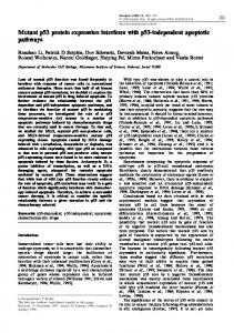
p53 as a Regulator of Intracellular Fluxes of Reactive Oxygen Species The p53 protein lies at the center of a vast network of pathways in which reactive oxygen (ROS) and nitrogen (NOS) species play critical roles (Fig. 1).14,15 Upstream of p53, production of ROS and NOS is a fundamental mechanism of endogenous or exogenous DNA damage and constitutes a trigger for p53 protein activation and stabilization. Downstream of p53, ROS operate as second messengers in signaling apoptosis. Many p53-regulated genes are involved in ROS and NOS metabolism, with contrasted biological consequences. For example, induction of the ROS-generating gene PIG3/NQO1 (encoding a NADPH quinone oxidoreductase) is a pro-apoptotic event. BAX1 and PUMA induce the release of ROS from the mitochondria into the cytoplasm. In contrast, repression
Cell Cycle
2509
Mutant p53 reactivation and redox regulation
2510
Cell Cycle
2009; Vol. 8 Issue 16
Mutant p53 reactivation and redox regulation Figure 1. The p53 redox network. Reactive oxygen species (ROS) and nitrogen species (NOS) induce DNA damage that trigger stabilization and accumulation of p53. This leads to upregulation of p53 target genes and a biological response. In addition, several redox-active factors control p53 folding and DNA binding activity. These factors include Metallothionein (MT), involved in Zn2+-dependent folding, and the complex Thioredoxin (Trx)Thioredoxin reductase (TRR)-APE1/Ref1 (the latter also involved in Base-Excision DNA Repair). Several p53 targets affect cellular redox status. The PIG3/NQO1 protein generates ROS. Similarly, BAX and PUMA can induce release of ROS from mitochondria. In contrast, p53-mediated repression of NOS2 and upregulation of GPX can reduce levels of ROS/NOS in the cell. TIGAR inhibits glycolysis and directs glucose into the pentose phosphate pathway, leading to increased levels of the reductant NADPH and thus contributing to the cellular redox balance. Adapted and modified from ref. 14.
by p53 of NOS2 (nitric oxide synthase 2) and activation of GPX (Glutathione Peroxidase) may contribute to cell detoxification by reducing the levels of damaging reactive species. Yet another system by which p53 may alter intracellular ROS status is the induction of TIGAR (TP53-induced glycolysis and apoptosis regulator) that inhibits glycolysis and promotes redirection of glucose into the pentose phosphate pathway, increasing the production of the reductant NADPH.16 Thus, the role of p53 in ROS metabolism contributes to bridge DNA damage signaling with energy metabolism and signaling of apoptosis. These mechanisms may be critical in p53-induced cellular senescence, a process involving dramatic changes in ROS-dependent signaling.
p53 as a Redox and Metal-Dependent Transcription Factor Given the role of p53 in the regulation of ROS fluxes, it is not surprising that p53 itself is subject to redox effects that provide a regulatory feedback loop in which ROS control basal p53 level and activity. p53 was initially identified as a redoxactive transcription factor when it appeared that high affinity binding to DNA in vitro required a strong reducing environment, equivalent to 10 mM Dithiothreitol (DTT).17,18 It then rapidly emerged that reducing power was important to allow p53 to bind a structural ion of zinc and to fold in the correct conformation for DNA binding. Biochemical and site-directed mutagenesis studies identified that the redox targets were cysteines scattered through the DNA biding domain, 3 of which are involved in Zn2+ binding (Cys176, Cys238 and Cys242).19 In fact, there is a complex interplay between Zn2+ binding and redox regulation of p53. Zinc binds with high affinity to thiols and stabilizes the tertiary folding of p53 by bridging the two loose L1 and L2 loops into a structure that makes optimal contact with p53 Response Elements in DNA. Zinc also protects thiols from becoming oxidized and forming disulfides that crosslink p53 either with itself or with other redox-active proteins. In the absence of zinc and sufficient reducing power, p53 tends to become oxidized and to form disulfide-liked aggregates. This intrinsic redox and metal sensitivity may allow p53 to undergo subtle variation of its level of activity in relation to changes of intracellular redox status.20 Such biochemical properties are not unique to p53. At least two other transcription factors with a wide spectrum of target genes, AP1 and NFkappaB, share similar redox-dependent regulation. In these factors, DNA binding involves direct contact between one or several thiol groups and DNA. Oxidation of these thiols, even partial, prevents contact and abrogates DNA binding. In the case of p53, the mechanism is somewhat different since the critical thiols are involved in maintaining the protein structure that binds to DNA, rather than in binding to DNA directly. www.landesbioscience.com
Mechanisms Regulating p53 Redox Status In Vivo The biochemical properties summarized above have led to the hypothesis that mechanisms regulating intracellular redox status and zinc bioavailability may play control p53 folding and activity. Inside cells, zinc availability and fluxes of ROS are tightly controlled by overlapping sets of effectors. ROS fluxes are regulated by electron transfer reactions between donor and acceptor molecules defining redox cycles and redox signaling pathways. Zinc availability is controlled through metal transfer reactions mediated by metallothioneins, a class of small, cysteine rich proteins that operate as intracellular buffers for Zn2+ and several other divalent metal ions (Cu2+, Ca2+). In recent years, evidence has accumulated that these mechanisms may control p53 status in both physiological and pathological conditions.21 The main mechanism that controls the reduced status of thiol-containing proteins is the thioredoxin (TRX)-thioredoxin reductase (TRR) system. There is evidence that this system regulates p53 redox status in vivo. Trx is a protein of low molecular weight (12 kDa) detected in the cytoplasm, nucleus, membrane and mitochondria. Trx functions as hydrogen donor for ribonucleotide reductase (RNR) providing deoxyribonucleotides for DNA replication and as protein disulfide reductase for many oxidized proteins. Trx is itself reduced by TRR, using NADPH as source of reducing equivalents. Studies in yeasts and in human cells have shown that inhibition of TRR results in the accumulation of oxidized Trx, which in turn induces the formation of disulfides in p53 and inactivate DNA binding.22,23 This interaction may involve APE1/Ref1, a bifunctional enzyme with two separate catalytic domains, a thiol reductase which is recycled by Trx/TRR and an apurinic/apyriminidic endonuclease. APE1/Ref1 provides the second major enzymatic step in base-excision repair (BER), the first one being the elimination of damaged bases by DNA glycosylases. The thiol reductase activity of APE1/Ref1 has been shown to control DNA binding by NFkappaB and AP1. APE1/ Ref1 interacts with p53 and stimulates DNA binding in vitro. In cells, silencing of APE1/Ref1 accelerates the turnover of p53 and decreases basal p53 levels.24 However, it has relatively little impact on p53 activation in response to DNA damage.22 Metallothioneins (MT) are small proteins (7 kDa) containing about 30% of cysteines organized in two structural cluster capable of binding 7 Zn2+ equivalents per MT molecules. About 10 years ago, MTs have been shown to regulate the Zn2+ dependent folding of p53 in vitro. Recent studies on the regulation of p53 by the Homeodomain-interacting protein kinase-2 (HIPK2) have shed light on the physio-pathological relevance of p53 regulation by MTs.25,26 HIPK2 phosphorylates p53 at Ser46, a necessary step to induce p53-dependent apoptosis in response to severe DNA damage. Stable knock-down and silencing of HIPK2 not only
Cell Cycle
2511
Mutant p53 reactivation and redox regulation
prevents Ser46 phosphorylation but also leads to p53 misfolding. This effect is due to enhanced expression of MT2a in response to HIPK2 knockdown and can be rescued either by supplementation with zinc or by silencing MT2a. These observations demonstrate that p53 folding requires a cellular environment in which the availability of zinc is tightly controlled. Given their thiol-rich nature, MTs are important contenders in the control of intracellular redox status. They become oxidized under Zn2+ depletion and can be recycled into their thiol-free form by the Trx/TRR redox cycle. Whereas many aspects of these regulations remain obscure, the emerging mechanism is that redox control of p53 is critical for Zn2+ binding and for correct folding of the DNA-binding domain. By interacting with p53, APE1/Ref-1 may facilitate p53 reduction. Given the bifunctional nature of APE1/Ref-1, the basal redox regulation of p53 may be coupled with DNA damage sensing and repair. APE1/Ref-1 may act not only directly as a p53 reductase, but also indirectly by recruiting other reducing factors such as Trx, thus functioning as a “redox chaperone”. On the other hand, MTs may also become involved in this redox chaperoning, regulating the transfer of Zn2+ into or out of p53 structure, depending upon the availability of this metals as well as upon the oxidation status of MTs. This process may further involve other protein chaperones, such as members of the heat-shock protein family, which may control p53 folding during redox- and metal transfer reaction.
Redox Regulation of Mutant p53 Early studies have pointed out that oxidation of p53 or chelation of Zn2+ turned wild type p53 into a form immunologically and functionally similar to mutant p53.17 More recent structural studies on the stability of the core domain of wild type or mutant p53 have shown that many conformational mutants are only slightly less stable than wild type and may, under appropriate energetic conditions, adopt a form close to wild type. This is the case, for example, for p53 Ser249 (Serine to Arginine at codon 249), a mutant that commonly occurs in hepatocellular carcinoma in some parts of the world.27 Yet, in intact cells, these mutants show a complete disruption of wild type structure with loss of DNA binding activity. This severe phenotype may result from the fact that, in vivo, imperfect folding exposes thiols in a way that favors oxidation, with formation of cross-links that prevent the mutant to revert to wild type form even if energetic conditions may allow this conformation. The His175 mutant (Arginine to Histidine at codon 175) shows an example of how mutation disrupts normal zinc/redox dependent folding.28 This mutant is one of the most common and most severe mutants in cancer. Arg175 is located in the L2 loop, adjacent to Cys176, one of the zinc binding sites. It stabilizes p53 structure by donating a pair of hydrogen bonds to the backbone carbonyl of Met237 on the L3 loop and a bifurcated hydrogen bond to the backbone of Pro191 and the side chain of Ser183 on the L2 loop. Substitution to histidine perturbs the geometry of these hydrogen bonds, although histidine still has the potential to donate two bonds and does not induce major clashes with neighboring residues. Strikingly, two other mutations have been described at Arg175, both predicted to have worse effects on p53 structure than 2512
His175. Gly175 introduces a glycine which is unable to donate hydrogens. Pro175 is even worse since the proline forms a fixed, disallowed angle into the protein backbone. Yet these two mutants have milder functional effects than Arg175. In fact, the severity of Arg175 may be due to a metal-related effect. It has been shown that the His175 mutation substantially accelerates the rate of Zn2+ loss from p53.29 Introducing histidine at position 175 generates an additional metal binding site that perturbs the tetrahedral geometry required for stable binding of Zn2+ into p53 structure. This effect results in an unstable and oxidation-prone mutant protein. Oxidation of mutant p53 may be the biggest biochemical obstacle to the renaturation of mutant p53 into wild type. Thus, keeping mutant p53 in reduced form may enhance its capacity to spontaneously renature into wild type p53, a process further enhanced by scaffolding drugs that may increase p53 stability.
p53 Status and Cancer Therapy The efficacy of radiotherapy and many currently used anticancer drugs, including cisplatin, etoposide, adriamycin and 5-FU relies on the presence of wild type p53.30,31 This is presumably due to induction of wild type p53 by these DNA-damaging agents and elimination of tumor cells by p53-dependent apoptosis or senescence. Absence of p53 expression or expression of mutant p53 has been associated with increased resistance to chemotherapy and radiotherapy.31-33 The type of p53 mutation is important for the degree of therapy resistance. For example, cells expressing the His175 or His179 conformational mutants are more resistant to etoposide than cells expressing the His273 and Trp248 DNA contact mutants, but His175 and His273 mutants show a similar sensitivity to low doses of cisplatin.34 Thus, mutant p53 promotes cancer by stimulating cell growth, inhibiting cell death and increasing drug resistance. Expression of mutant p53 is associated with poor prognosis in several tumors, including breast, colorectal and renal cell carcinomas.35-38 Given the high frequency of p53 mutations in human tumors and the association between mutant p53 and poor prognosis in several tumor types, there is clearly a great need for more efficient treatment of mutant p53-carrying tumors. Recent studies have demonstrated that restoration of wild type p53 expression in tumors in vivo results in rapid elimination of the tumors by apoptosis and/or senescence.39-41 In other words, wild type p53 is capable of triggering tumor cell death even in the context of multiple tumor-promoting genetic alterations. Therefore, it seems a desirable strategy to develop drugs that restore wild type function to mutant p53 and thus trigger massive tumor cell apoptosis (Fig. 2). This would not affect normal cells that express low levels of wild type p53. Indeed, several recent studies have identified peptides42,43 or low molecular weight compounds44-48 that target mutant p53 and induce apoptosis in tumor cells (Fig. 3). This raises hopes for the development of novel anticancer drugs that restore p53 function and p53-dependent apoptosis in tumors.
Mutant p53 Reactivation by PRIMA-1 The low molecular compound PRIMA-1 was identified in a cell-based screen of the Diversity set from the National Cancer
Cell Cycle
2009; Vol. 8 Issue 16
Mutant p53 reactivation and redox regulation
Figure 2. Strategy for treatment of mutant p53-carrying tumors. The aim is to specifically target mutant p53, often overexpressed in cancer cells, restore its wild type functions, and thus trigger massive apoptosis that eradicates the tumor. Normal cells expressing low levels of wild type p53 should not be affected and therefore this therapeutic approach is not expected to have severe side effects.
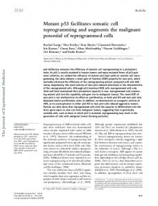
Figure 3. Chemical structures of mutant p53-reactivating compounds. PRIMA-1 and the more potent analog PRIMA-1MET are converted to MQ. MIRA-1, STIMA-1, CP31398 and MQ share a common chemical activity as Michael acceptors that can potentially modify cysteines in p53. WR1065 (the active form of amifostine) has a reactive SH group. www.landesbioscience.com
Cell Cycle
2513
Mutant p53 reactivation and redox regulation
Figure 4. Mechanism for PRIMA-1-mediated elimination of tumor cells. PRIMA-1 can be regarded as a prodrug that is converted to the active compound MQ. MQ modifies mutant p53 covalently, restoring wild type function and triggering cell death by apoptosis. MQ can probably also modify other cellular proteins, which might synergize with mutant p53 reactivation and thus potentiate apoptosis.
Institute. PRIMA-1 restores wild type conformation and function to mutant p53 proteins and triggers massive apoptosis in tumor cells.46 Moreover, systemic treatment with PRIMA-1 and the structural analog PRIMA-1MET inhibits tumor growth in mice.46,49,50 Henceforth we shall use the name PRIMA-1 for both PRIMA-1 and PRIMA-1MET. PRIMA-1 induces activation of caspase-2 and upregulation of pro-apoptotic p53 target genes such as BAX, PUMA and NOXA.51 A PRIMA-1 analog is currently being evaluated in a phase I clinical trial. Despite a wealth of data on the cellular events triggered by PRIMA-1, the exact molecular mechanism of action has been unclear. However, we noted that PRIMA-1 is not stable under physiological conditions but is converted to several different compounds. One of these, methylene quinuclidone or MQ, has a reactive double bond that can potentially participate in Michael addition reactions. Thus, MQ is a Michael acceptor, raising the possibility that it reacts covalently with cysteines in mutant p53. Mass spectrometry confirmed covalent binding to the p53 core domain.52 Moreover, we confirmed binding to mutant p53 in living cells by treatment with 14C-labelled PRIMA-1 and immunoprecipitation of mutant p53. Importantly, the anti-oxidant N-acetyl cysteine (NAC) completely inhibited PRIMA-1-induced apoptosis, and formed adducts with MQ. A critical role of conversion of PRIMA-1 to MQ was further supported by our finding that the PRIMA-1 analog PRIMA-D that cannot be converted to MQ is completely inactive.52 However, we do not exclude that other conversion products of PRIMA-1 play a role in mutant p53 reactivation. We also asked whether modification of mutant p53 per se is sufficient to trigger apoptosis in tumor cells. Introduction of PRIMA-1-treated mutant p53 protein into p53 null tumor cells using a protein transfer reagent induced expression of the proapoptotic p53 targets BAX, PUMA and NOX, as well as caspase activation, followed by massive apoptosis. Importantly, efficient induction of apoptosis required preincubation of PRIMA-1 to allow conversion to reactive products. These findings indicate that 2514
covalent modification of mutant p53 per se by MQ is responsible for mutant p53-dependent apoptosis induced by PRIMA-1. Thus, PRIMA-1 can be considered a prodrug that is converted to its active metabolite MQ in vivo. It is conceivable that conversion occurs largely intracellularly. This would allow the reactive MQ to reach mutant p53 in tumor cells rather than proteins in the extracellular environment (Fig. 4). The p53 core domain contains ten cysteine residues. At this stage, we do not know which of these are targeted by PRIMA-1, and the exact molecular mechanism of mutant p53 reactivation upon cysteine modification is unknown. However, it is plausible that thiol modification by MQ may inhibit the formation of both intramolecular and intermolecular disulfide bonds that could otherwise prevent proper folding of the core (Fig. 5) and/or result in mutant p53 aggregation. Furthermore, adducts might promote correct folding through their hydrophobic nature, or create additional protein-DNA bridges, thus stabilizing DNA binding. We found that MQ can also bind covalently to wild type p53. This is expected if cysteine residues are exposed on the surface of the core domain. Interestingly, our experiments revealed that the higher the degree of wild type p53 unfolding, the higher the binding of MQ. Thus, incorrectly folded wild type p53 can also be reactivated by PRIMA-1. These observations imply that unfolded wild type p53 in tumor cells could be a target for PRIMA-1, resulting in p53-dependent apoptosis.
Targeting Cysteines as a Common Property of Mutant p53Reactivating Compounds Several compounds that were identified based on their ability to selective target mutant p53-expressing cells, or promote wild type conformation of mutant p53, are Michael acceptors that can potentially modify thiol groups. The compound MIRA-1 is an informative case in point. MIRA-1 was identified in the same cellular screening of the NCI Diversity set that led to the identification of PRIMA-1.46,47 MIRA-1 and its active structural analogs induce apoptosis with a similar selectivity for mutant p53-expressing
Cell Cycle
2009; Vol. 8 Issue 16
Mutant p53 reactivation and redox regulation
c isplatin preferentially kills wild type p53-expressing tumor cells whereas mutant p53-expressing tumor cells are less affected,34,63,64 or even protected from cisplatin-induced apoptosis.49,65 This reinforces the notion that the recently identified mutant p53-targeting compounds act through entirely different mechanisms than conventional drugs such as cisplatin. Modification Figure 5. Unfolding of mutant p53 may result in exposure of cysteines and the formation of intramolecular of free cysteines seems critical for disulfide bonds that lock the core domain in an inactive conformation. Modification of cysteines in mutant restoration of wild-type activity to p53 by MQ may prevent such disulfide bonds and thus promote proper folding and restoration of wild type mutant p53, but it is likely that function. other chemical groups in mutant p53-targeting compounds will cells as PRIMA-1, but with faster kinetics.47 MIRA-1 belongs to contribute to their biological effect and mutant p53 selectivity, for the maleimides, a group of compounds that are known Michael instance by bringing parts of the collapsed protein structure together acceptors with ability to modify thiols. Interestingly, all biologi- or by creating additional bridges between p53 and DNA. Also, the cally active mutant p53-selective MIRA analogs contain a reactive ability of compounds to target specific cysteines in mutant p53 and double bond, strongly suggesting that thiol modification is critical other proteins could be restricted by steric hindrance. It remains for their activity. In addition, other mutant p53-reactivating to be determined which other structural features would define compounds such as STIMA-1,53 and CP-31398,44 also contain mutant p53 specificity apart from thiol modification. Studies of reactive carbon-carbon double bonds located in direct proximity to the maleimide MIRA-1 that has promising properties in vitro but an electron withdrawing group, a characteristic feature of Michael is toxic in vivo47 indicate that too strong reactivity with thiols will acceptors. Thus, all these compounds could potentially participate produce toxic side effects. The optimal mutant p53-reactivating in the reactions of nucleophilic additions. In other words, they are compound is perhaps a prodrug like PRIMA-1 which is converted potent electrophils that may readily react with nucleophilic groups to active metabolites in vivo, preferentially inside cells to minimize unspecific toxicity. such as thiols. The compound WR1065 is an active metabolite of amifostine, an agent used as chemo- and radioprotector to counteract some Conclusions side effects of chemotherapy.54 WR1065 promotes wild type Mutant p53 is an important target for novel cancer therapy. conformation of M272 temperature-sensitive mutant p53 and Reactivation of mutant p53 should eliminate tumor cells by restores its transcription transactivation capacity.45 Interestingly, massive apoptosis. However, pharmacological reactivation of it has been shown that WR1065 directly binds to p53.55 Since mutant p53 is a greater challenge than for example inhibition WR1065 has a thiol group, it might bind to mutant p53 via of a protein tyrosine kinase receptor, the active site of which formation of disulfide bridges between cysteines on the surface of can be targeted by high-affinity, small competitive inhibitory p53 and the thiol in WR1065. Adducts between protein thiols and compounds. Yet several small molecules that preferentially target glutathione have been described56 and such reaction was reported mutant p53-expressing tumor cells and restore at least some wild for p53.57 Therefore it is plausible that WR1065 could react cova- type properties to mutant p53 have been identified. Interestingly, lently with cysteines in mutant p53 in a similar manner. these compounds appear to share a common chemical property, Ellipticine was identified in an NCI-led large chemical i.e., the ability to modify thiol groups in p53 and other proteins. screening program as a compound that preferentially suppresses This is consistent with studies indicating a critical role of cellular growth of mutant p53-expressing cells.58 The analog 9-hydroxyelredox regulation for p53 conformation and DNA binding. Further lipticine induces apoptosis in tumor cells expressing mutant studies by NMR and X-ray crystallography will be required to p53.59 Ellipticine is metabolised to several products, including identify structural consequences of thiol modification of the p53 13-hydroxyellipticine and N2-oxide of ellipticine,60 both of which core domain by compounds such as MQ. This may ultimately can form DNA adducts with deoxyguanosine.60,61 These reacprovide the basis for rational design of more efficient and selective tive metabolites can potentially react with nucleophiles and it is unlikely that only nucleophilic centers on deoxyguanosine will be anticancer drugs that reactive mutant p53. targets. The other likely substrates for this type of reaction are thiol groups in proteins, possibly including mutant p53. The ability to reactivate mutant p53 and/or selectively target mutant p53-expressing cells is clearly not a general feature of all thiol-modifying compounds. Cisplatin, for instance, is a currently used chemotherapeutic drug known to alkylate thiols.62 Yet www.landesbioscience.com
Acknowledgements
The authors thank the Swedish Cancer Society (Cancerfonden), Cancerföreningen, Karolinska Institutet, Magn. Bergvalls Stiftelse, and the EU 6th framework program (FP6) for financial support. This publication reflects the author’s views and not necessarily those of the EC. The information in this document is provided as
Cell Cycle
2515
Mutant p53 reactivation and redox regulation
is and no guarantee or warranty is given that the information is fit for any particular purpose. The user thereof uses the information at its sole risk and liability. The Community is not liable for any use that may be made of the information contained herein. Work on p53 regulation at IARC is supported by the French Association for Research against Cancer (ARC). K.G.W. and V.J.N.B. are cofounders and shareholders of Aprea AB, and K.G.W. is a member of its board. References 1. Riley T, Sontag E, Chen P, Levine A. Transcriptional control of human p53-regulated genes. Nat Rev Mol Cell Biol 2008; 9:402-12. 2. Raver-Shapira N, Oren M. Tiny actors, great roles: microRNAs in p53’s service. Cell Cycle 2007; 6:2656-61. 3. Mihara M, Erster S, Zaika A, Petrenko O, Chittenden T, Pancoska P, et al. p53 has a direct apoptogenic role at the mitochondria. Mol Cell 2003; 11:577-90. 4. Green DR, Kroemer G. Cytoplasmic functions of the tumour suppressor p53. Nature 2009; 458:1127-30. 5. Olivier M, Eeles R, Hollstein M, Khan MA, Harris CC, Hainaut P. The IARC TP53 database: new online mutation analysis and recommendations to users. Hum Mutat 2002; 19:607-14. 6. Cho Y, Gorina S, Jeffrey PD, Pavletich NP. Crystal structure of a p53 tumor suppressorDNA complex: understanding tumorigenic mutations. Science 1994; 265:346-55. 7. Vousden KH, Lu X. Live or let die: the cell’s response to p53. Nat Rev Cancer 2002; 2:594-604. 8. Soussi T, Beroud C. Assessing TP53 status in human tumours to evaluate clinical outcome. Nat Rev Cancer 2001; 1:233-40. 9. Bressac B, Kew M, Wands J, Ozturk M. Selective G to T mutations of p53 gene in hepatocellular carcinoma from Southern Africa. Nature 1991; 350:429-31. 10. Pfeifer GP, Denissenko MF, Olivier M, Tretyakova N, Hecht SS, Hainaut P. Tobacco smoke carcinogens, DNA damage and p53 mutations in smoking-associated cancers. Oncogene 2002; 21:7435-51. 11. Scian MJ, Stagliano KE, Ellis MA, Hassan S, Bowman M, Miles MF, et al. Modulation of gene expression by tumor-derived p53 mutants. Cancer Res 2004; 64:7447-54. 12. Sigal A, Rotter V. Oncogenic mutations of the p53 tumor suppressor: the demons of the guardian of the genome. Cancer Res 2000; 60 6788-93. 13. Gaiddon C, Lokshin M, Ahn J, Zhang T, Prives C. A subset of tumor-derived mutant forms of p53 down-regulate p63 and p73 through a direct interaction with the p53 core domain. Mol Cell Biol 2001; 21:1874-87. 14. Hainaut P, Mann K. Zinc binding and redox control of p53 structure and function. Antioxid Redox Signal 2001; 3:611-23. 15. Liu B, Chen Y, St. Clair DK. ROS and p53: a versatile partnership. Free Radic Biol Med 2008; 44:1529-35. 16. Bensaad K, Tsuruta A, Selak MA, Vidal MN, Nakano K, Bartrons R, et al. TIGAR, a p53-inducible regulator of glycolysis and apoptosis. Cell 2006; 126:107-20. 17. Hainaut P, Milner J. Redox modulation of p53 conformation and sequence-specific DNA binding in vitro. Cancer Res 1993; 53:4469-73. 18. Hupp TR, Meek DW, Midgley CA, Lane DP. Regulation of the specific DNA binding function of p53. Cell 1992; 71:875-86. 19. Rainwater R, Parks D, Anderson ME, Tegtmeyer P, Mann K. Role of cysteine residues in regulation of p53 function. Mol Cell Biol 1995; 15:3892-903. 20. Verhaegh GW, Richard MJ, Hainaut P. Regulation of p53 by metal ions and by antioxidants: dithiocarbamate downregulates p53 DNA-binding activity by increasing the intracellular level of copper. Mol Cell Biol 1997; 17:5699-706. 21. Meplan C, Richard MJ, Hainaut P. Metalloregulation of the tumor suppressor protein p53: zinc mediates the renaturation of p53 after exposure to metal chelators in vitro and in intact cells. Oncogene 2000; 19:5227-36. 22. Seemann S, Hainaut P. Roles of thioredoxin reductase 1 and APE/Ref-1 in the control of basal p53 stability and activity. Oncogene 2005; 24:3853-63. 23. Pearson GD, Merrill GF. Deletion of the Saccharomyces cerevisiae TRR1 gene encoding thioredoxin reductase inhibits p53-dependent reporter gene expression. J Biol Chem 1998; 273:5431-4. 24. Jayaraman L, Prives C. Covalent and noncovalent modifiers of the p53 protein. Cell Mol Life Sci 1999; 55:76-87. 25. Puca R, Nardinocchi L, Bossi G, Sacchi A, Rechavi G, Givol D, et al. Restoring wtp53 activity in HIPK2 depleted MCF7 cells by modulating metallothionein and zinc. Exp Cell Res 2009; 315:67-75. 26. Puca R, Nardinocchi L, Gal H, Rechavi G, Amariglio N, Domany E, et al. Reversible dysfunction of wild-type p53 following homeodomain-interacting protein kinase-2 knockdown. Cancer Res 2008; 68:3707-14. 27. Friedler A, DeDecker BS, Freund SM, Blair C, Rudiger S, Fersht AR. Structural distortion of p53 by the mutation R249S and its rescue by a designed peptide: implications for “mutant conformation”. J Mol Biol 2004; 336:187-96.
2516
28. Martin AC, Facchiano AM, Cuff AL, Hernandez-Boussard T, Olivier M, Hainaut P, et al. Integrating mutation data and structural analysis of the TP53 tumor-suppressor protein. Hum Mutat 2002; 19:149-64. 29. Bullock AN, Henckel J, DeDecker BS, Johnson CM, Nikolova PV, Proctor MR, et al. Thermodynamic stability of wild-type and mutant p53 core domain. Proc Natl Acad Sci USA 1997; 94:14338-42. 30. Lowe SW, Ruley HE, Jacks T, Housman DE. p53-dependent apoptosis modulates the cytotoxicity of anticancer agents. Cell 1993; 74:957-67. 31. Vasey PA, Jones NA, Jenkins S, Dive C, Brown R. Cisplatin, camptothecin and taxol sensitivities of cells with p53-associated multidrug resistance. Mol Pharmacol 1996; 50:1536-40. 32. Bunz F, Hwang PM, Torrance C, Waldman T, Zhang Y, Dillehay L, et al. Disruption of p53 in human cancer cells alters the responses to therapeutic agents. J Clin Invest 1999; 104:263-9. 33. Weller M. Predicting response to cancer chemotherapy: the role of p53. Cell Tissue Res 1998; 292:435-45. 34. Blandino G, Levine AJ, Oren M. Mutant p53 gain of function: differential effects of different p53 mutants on resistance of cultured cells to chemotherapy. Oncogene 1999; 18:477-85. 35. Berns EM, van Staveren IL, Look MP, Smid M, Klijn JG, Foekens JA. Mutations in residues of TP53 that directly contact DNA predict poor outcome in human primary breast cancer. Br J Cancer 1998; 77:1130-6. 36. Borresen-Dale AL, Lothe RA, Meling GI, Hainaut P, Rognum TO, Skovlund E. TP53 and long-term prognosis in colorectal cancer: mutations in the L3 zinc-binding domain predict poor survival. Clin Cancer Res 1998; 4:203-10. 37. Girgin C, Tarhan H, Hekimgil M, Sezer A, Gurel G. p53 mutations and other prognostic factors of renal cell carcinoma. Urol Int 2001; 66:78-83. 38. Petitjean A, Achatz MI, Borresen-Dale AL, Hainaut P, Olivier M. TP53 mutations in human cancers: functional selection and impact on cancer prognosis and outcomes. Oncogene 2007; 26:2157-65. 39. Ventura A, Kirsch DG, McLaughlin ME, Tuveson DA, Grimm J, Lintault L, et al. Restoration of p53 function leads to tumour regression in vivo. Nature 2007; 445:661-5. 40. Xue W, Zender L, Miething C, Dickins RA, Hernando E, Krizhanovsky V, et al. Senescence and tumour clearance is triggered by p53 restoration in murine liver carcinomas. Nature 2007; 445:656-60. 41. Martins CP, Brown-Swigart L, Evan GI. Modeling the Therapeutic Efficacy of p53 Restoration in Tumors. Cell 2006; 127:1323-34. 42. Selivanova G, Iotsova V, Okan I, Fritsche M, Strom M, Groner B, et al. Restoration of the growth suppression function of mutant p53 by a synthetic peptide derived from the p53 C-terminal domain. Nat Med 1997; 3:632-8. 43. Friedler A, Hansson LO, Veprintsev DB, Freund SM, Rippin TM, Nikolova PV, et al. A peptide that binds and stabilizes p53 core domain: chaperone strategy for rescue of oncogenic mutants. Proc Natl Acad Sci USA 2002; 99:937-42. 44. Foster BA, Coffey HA, Morin MJ, Rastinejad F. Pharmacological rescue of mutant p53 conformation and function. Science 1999; 286:2507-10. 45. North S, Pluquet O, Maurici D, El-Ghissassi F, Hainaut P. Restoration of wild-type conformation and activity of a temperature-sensitive mutant of p53 (p53(V272M)) by the cytoprotective aminothiol WR1065 in the esophageal cancer cell line TE-1. Mol Carcinog 2002; 33:181-8. 46. Bykov VJ, Issaeva N, Shilov A, Hultcrantz M, Pugacheva E, Chumakov P, et al. Restoration of the tumor suppressor function to mutant p53 by a low-molecular-weight compound. Nat Med 2002; 8:282-8. 47. Bykov VJ, Issaeva N, Zache N, Shilov A, Hultcrantz M, Bergman J, et al. Reactivation of mutant p53 and induction of apoptosis in human tumor cells by maleimide analogs. J Biol Chem 2005; 280:30384-91. 48. Zache N, Lambert JMR, Rökaeus N, Shen J, Hainaut P, Bergman J, et al. Mutant p53 targeting by the low molecular weight compound STIMA-1. Mol Oncol 2008; 2:7080. 49. Bykov VJ, Zache N, Stridh H, Westman J, Bergman J, Selivanova G, et al. PRIMA1(MET) synergizes with cisplatin to induce tumor cell apoptosis. Oncogene 2005; 24:3484-91. 50. Zache N, Lambert JM, Wiman KG, Bykov VJ. PRIMA-1MET inhibits growth of mouse tumors carrying mutant p53. Cell Oncol 2008; 30:411-8. 51. Shen J, Vakifahmetoglu H, Stridh H, Zhivotovsky B, Wiman KG. PRIMA-1MET induces mitochondrial apoptosis through activation of caspase-2. Oncogene 2008; 27:6571-80. 52. Lambert JM, Gorzov P, Veprintsev DB, Soderqvist M, Segerback D, Bergman J, et al. PRIMA-1 reactivates mutant p53 by covalent binding to the core domain. Cancer Cell 2009; 15:376-88. 53. Zache N, Lambert JM, Rokaeus N, Shen J, Hainaut P, Bergman J, et al. Mutant p53 targeting by the low molecular weight compound STIMA-1. Mol Oncol 2008; 2:70-80. 54. Kouvaris JR, Kouloulias VE, Vlahos LJ. Amifostine: the first selective-target and broadspectrum radioprotector. Oncologist 2007; 12:738-47. 55. Shen H, Chen ZJ, Zilfou JT, Hopper E, Murphy M, Tew KD. Binding of the aminothiol WR-1065 to transcription factors influences cellular response to anticancer drugs. J Pharmacol Exp Ther 2001; 297:1067-73.
Cell Cycle
2009; Vol. 8 Issue 16
Mutant p53 reactivation and redox regulation 56. Thomas JA, Poland B, Honzatko R. Protein sulfhydryls and their role in the antioxidant function of protein S-thiolation. Arch Biochem Biophys 1995; 319:1-9. 57. Sun XZ, Vinci C, Makmura L, Han S, Tran D, Nguyen J, et al. Formation of disulfide bond in p53 correlates with inhibition of DNA binding and tetramerization. Antioxid Redox Signal 2003; 5:655-65. 58. Shi LM, Fan Y, Myers TG, O’Connor PM, Paull KD, Friend SH, et al. Mining the NCI anticancer drug discovery databases: genetic function approximation for the QSAR study of anticancer ellipticine analogues. J Chem Inf Comput Sci 1998; 38:189-99. 59. Sugikawa E, Hosoi T, Yazaki N, Gamanuma M, Nakanishi N, Ohashi M. Mutant p53 mediated induction of cell cycle arrest and apoptosis at G1 phase by 9-hydroxyellipticine. Anticancer Res 1999; 19:3099-108. 60. Stiborova M, Sejbal J, Borek-Dohalska L, Aimova D, Poljakova J, Forsterova K, et al. The anticancer drug ellipticine forms covalent DNA adducts, mediated by human cytochromes P450, through metabolism to 13-hydroxyellipticine and ellipticine N2-oxide. Cancer Res 2004; 64:8374-80. 61. Stiborova M, Arlt VM, Henderson CJ, Wolf CR, Kotrbova V, Moserova M, et al. Role of hepatic cytochromes P450 in bioactivation of the anticancer drug ellipticine: studies with the hepatic NADPH:cytochrome P450 reductase null mouse. Toxicol Appl Pharmacol 2008; 226:318-27. 62. Pasetto LM, D’Andrea MR, Brandes AA, Rossi E, Monfardini S. The development of platinum compounds and their possible combination. Crit Rev Oncol Hematol 2006; 60:59-75. 63. Bykov VJ, Issaeva N, Selivanova G, Wiman KG. Mutant p53-dependent growth suppression distinguishes PRIMA-1 from known anticancer drugs: a statistical analysis of information in the National Cancer Institute database. Carcinogenesis 2002; 23:2011-8. 64. Weinstein JN, Myers TG, O’Connor PM, Friend SH, Fornace AJ Jr, Kohn KW, et al. An information-intensive approach to the molecular pharmacology of cancer. Science 1997; 275:343-9. 65. Matas D, Sigal A, Stambolsky P, Milyavsky M, Weisz L, Schwartz D, et al. Integrity of the N-terminal transcription domain of p53 is required for mutant p53 interference with drug-induced apoptosis. EMBO J 2001; 20:4163-72.
www.landesbioscience.com
Cell Cycle
2517