May 27, 1986 - JOHN F. THOMPSON,1 DARYLE WAECHTER-BRULLA,2 RICHARD I. GUMPORT,3 JEFFREY F. GARDNER,2. LINA MOITOSO DE VARGAS,1 ...
Vol. 168, No. 3
JOURNAL OF BACTERIOLOGY, Dec. 1986, p. 1343-1351
0021-9193/86/121343-09$02.00/0 Copyright C 1986, American Society for Microbiology
Mutations in an Integration Host Factor-Binding Site: Effect on Lambda Site-Specific Recombination and Regulatory Implications JOHN F. THOMPSON,1 DARYLE WAECHTER-BRULLA,2 RICHARD I. GUMPORT,3 JEFFREY F. GARDNER,2 LINA MOITOSO DE VARGAS,1 AND ARTHUR LANDYl* Division of Biology and Medicine, Brown University, Providence, Rhode Island 02912,1 and Department of Microbiology2 and Department of Biochemistry and College of Medicine,3 University of Illinois, Urbana, Illinois 61801 Received 27 May 1986/Accepted 4 September 1986
The manner in which integration host factor (IHF) regulates lambda site-specific recombination has been analyzed by examining the behavior of both wild-type and mutant DNAs in integrative and excisive recombination as well as in protein binding. While integrative recombination of an attP with two base changes in the Hi site required 8-fold more IHF than did wild type, binding to this site was lowered at least 500-fold, suggestive of cooperative interactions. A mutant attP with nine base changes did not integrate at all in vitro, with the defect being less severe in vivo. IHF inhibition of excisive recombination was relieved by both mutations in vitro and in vivo. These results imply that occupancy of the Hi site is critical for determining the direction of recombination. It is proposed that IHF inhibition of excision provides a monitor of the strength of the induction stimulus and the nutritional state of the cell; this would allow the prophage to excise selectively in conditions which favor successful completion of the lytic cycle.
During the lysogenic mode of growth, bacteriophage lambda DNA is integrated into the bacterial genome and most phage genes are repressed. Induction of the bacterial SOS system leads to cleavage of lambda repressor followed by prophage excision and lytic gene expression. Integrative and excisive recombination represent alternative biological decisions which affect gene expression over a relatively long time span and therefore must be carefully regulated and coordinated with gene expression in both the viral and host chromosomes. The manner in which the Escherichia coli protein integration host factor (IHF) is involved in these processes is described in this report. The integration of the phage DNA (attP) into the bacterial DNA (attB) requires the phage protein Int as well as the E. coli protein IHF, whereas the excision reaction, which regenerates attP and attB from attL and attR, requires the phage protein Xis in addition to Int and IHF (reviewed in references 35 and 48). Int does the actual cutting and ligation of DNA while bound to sites at the edge of the common core region (a 15-base-pair [bp] region of homology present in all four att sites) (7, 25, 33). In addition to binding these core-type recognition sequences, Int can also bind to another class of sites with a different recognition sequence, the arm-type sites (37). IHF binds to three sites in attP (8) and is normally required for both integrative and excisive recombination. However, it is also capable of specifically inhibiting the in vitro excision reaction when added at higher concentrations (4). In addition to its role in the actual mechanism of recombination, IHF influences the lysis/lysogeny decision at another level, by increasing expression of cIT protein (19, 28, 29, 36). Elevated levels of cII lead to the establishment of lysogeny by activating the PI, PE, and PaQ promoters that lead respectively to the expression of Int, repressor, and an antisense RNA that assists in repressing late genes (6, 17, 18, 22, 38, 41, 45). cIT prevents abortive lysogeny by coupling repression of lytic gene expression with the presence of proteins required for integration. IHF is an integral part of *
this coupling because of its effect on both clI synthesis and recombination (29). IHF, in addition to its role in lambda development, plays a role in regulating several processes in E. coli (11, 23, 31). We studied the role of IHF in the regulation of phage development by examining wild-type and mutant DNAs for their behavior in protein binding and recombination, both in vivo and in vitro. In particular, the Hi site has been analyzed because it is in a region known to have strong cooperative protein-protein interactions which play a role in controlling directionality (4, 5). MATERIALS AND METHODS Construction of H1- and H1= mutants. The H1- mutant was generated by oligonucleotide-directed mutagenesis of M13mp9::attP (2) which contains the 1,594-bp RsaI-HaeIIattP fragment of X. A mutagenic primer 5' (CTATGAAT TCACTACTT) 3' synthesized by the solid-phase phosphatephosphotriester method (20) was used for mutagenesis, according to the procedure of Gillam and Smith (14) as modified by Bauer et al. (3). This construction, in which the T-G at positions -117 and -116 was changed to G-A (introducing an EcoRI site while removing the Hinfl site), was made by using M13 as a template for a doubly mismatched oligonucleotide primer and will be referred to in this paper as the Hl- site. The H1= mutant was made in a manner analogous to the linker spanning method. pWB55-38, a plasmid containing an attR shortened in the P arm to -111 (4), contains a normal HindIII linker as well as an aberrant linker with an extra cytidine residue which cannot be cut by HindIII. The presence of these two linkers generates a new BalI site, the position of which coincides with the DdeI site normally present in attR. The presence of the extra linker and the fortuitous location of the BalI site allowed us to insert 9 bp of heterologous DNA in place of the Hi site. To do this, pWB55-38 was cut with Ball and BamHI and the fragment containing the att DNA was purified by using low-melting agarose. A plasmid containing the intact attR site in the HindIII-BamHI sites of pBR327 was cut with DdeI in 200 ,ug
Corresponding author. 1343
1344
THOMPSON ET AL.
of ethidium bromide per ml-150 mM NaCi-10 mM MgCl2-50 mM Tris hydrochloride (pH 7.5) at 25°C for 2 h. This procedure yields full-length linear DNA, with cleavage occurring at any of the six DdeI sites. This DNA was then purified on low-melting agarose, after which the singlestranded overhang was filled in by using the Klenow fragment of DNA polymerase. After being cut with BamHI, the 3,050-bp fragment was purified and ligated to the 890-bp Bal-BamHI fragment of pWB55-38. This construction, in which nine bases were changed between -122 and -111 (introducing a StyI site and removing both the DdeI and Hinfl sites), will be referred to as the H1= site. Manipulation of mutant sites. The H1= site was placed in M13mp9: :attP by cutting both attPs with BamHI and HindIll and religating. The attP for the H1= and the attR for the Hi- were made by in vitro recombination as previously described (4). Plasmids containing only the Hi IHF-binding site with H2 and H' deleted were made by cutting the three attP constructions with NdeI and BamHI, filling in the overhangs with DNA polymerase, and ligating. These constructions also contain the P1 and P2 Int sites. Transfer of the mutant attP sites into A was accomplished via homologous recombination between the M13mp9: :attP mutants and X biol6A as previously described (2). Purified phage DNA was digested with EcoRI or StyI to ensure that the recombination event generated phages carrying the Hi- or H1= mutation. In vivo recombinations. To measure lysogenization frequencies, an overnight culture of E. coli MO (16) was diluted 1:25 into fresh LB broth and incubated at 37°C with vigorous aeration. At predetermined cell densities representing different phases of the growth curve, 2.5 ml of culture was removed to a chilled sterile tube and held on ice for 10 min. Cells were pelleted, suspended in an equal volume of 10 mM MgSO4, and infected at a multiplicity of infection (MOI) of either 1 or 10 (estimated from the growth curve). Immediately after preadsorption, cells were diluted and spread on EMBO plates (15) with or without A h80A9 c and incubated at 30°C overnight. Lysogenization frequency is calculated as the number of colonies on plates with selective phage divided by the number of colonies on plates with no phage. Temperature-sensitive X lysogens of strain MO were isolated from colonies generated in lysogenization assays. Monolysogens were identified with the virulent strain A cI90 c17 by using the protocol of Sly and Rabideau (44) as modified by R. Weisberg (personal communication). Lysogens were suspended in top agar and then spotted with a series of 1:2 dilutions of the phage stock (starting with a titer greater than 1010 PFU per ml). The lysogens were compared with a known monolysogen and polylysogen. The dilution at which each strain gave clear plaques indicated that all the lysogens generated by us contained a single prophage. Derivatives resistant to A vir were isolated by spotting a drop of A vir (109 PFU) onto a lawn of each lysogen. After 24 h of incubation at 30°C, colonies within the cleared area were picked and purified. These derivatives were checked for a Mal- phenotype on MacConkey maltose plates. Resistance to A vir was confirmed by plating X vir onto a lawn of cells. The monolysogens were used for heat-pulse curing by a modification of the procedure described by Shimada et al. (40). All curing experiments were done with wild-type and mutant lysogens exactly in parallel to ensure identical conditions. An overnight culture of each lyosgen was diluted 100-fold into LB broth at 30°C. Cells were grown to an optical density at 650 nm of 0.3 to 0.4 or 1.5. Short pulses of
J. BACTERIOL.
induction (3 to 8 min) were obtained by diluting cells 50-fold into prewarmed broth at 39°C and then by diluting again into broth at 30°C. Because 39°C is just slightly above that needed for repressor denaturation, temperature had to be carefully controlled. After the second dilution, cells were grown for 3 to 5 h at 30°C and plated at 30 and 42°C. Growth curves were done to ensure that exponential-phase cells were out of lag phase and that cells entering stationary phase were out of exponential phase. To obtain comparable levels of curing, longer induction periods were required for stationary-phase cells than those required for exponential-phase cells (see also Discussion). Phage generated by heat curing were analyzed by digestion of their DNA with EcoRI and StyI to ensure that normal integration and excision reactions had occurred. In vitro recombination. In vitro recombinations were carried out in 20 ,ul with 25 mM Tris hydrochloride (pH 7.9)-6 mM spermidine-5 mM EDTA-2 mM dithiothreitol-1 mg of bovine serum albumin per ml-75 mM NaCl. DNA and protein concentrations are as described in the figure legends. After the buffer mixture was added to the DNA, proteins were added in the order IHF, Int, and Xis. Proteins and DNAs were purified as described previously (4). Nuclease protection and gel mobility shift experiments were also performed as described previously (5). RESULTS Protein binding. The Hi-binding site on the P arm of the phage DNA was mutated in the consensus binding sequence to generate new sites that would not bind IHF (Fig. 1). The expected sequences were confirmed by DNA sequencing and by assaying for the introduction or deletion of the predicted restriction endonuclease sites. After the original mutations were made, each was incorporated into attP and attR sequences in both pBR327 and X. Additionally, for each mutant and for wild type, a region of the P arm containing only the Hi region was inserted into pBR327. These constructions made it possible to characterize both mutant and wild-type sites for their protein-binding properties and their in vitro and in vivo recombination efficiencies. IHF binding to the mutated sites was determined by monitoring nuclease protection (12) and gel mobility shifts (10, 13). The nuclease digestion profile of the H1- attP is shown relative to wild type in the presence and absence of IHF (Fig. 2). While 3 U of IHF was sufficient for partial protection of wild-type Hi, even 300 U yielded no protection of H1-. When more IHF is used, no additional information is gained because protection of all sites is lost. Nuclease protection experiments showed no binding of IHF to the H1= site (data not shown). Even though the changes in H1= border the P2 Int consensus-binding sequence, no change in the affinity of Int for either P2 or P1 was observed. Neither Int nor Xis could cooperatively aid in the binding of IHF to Hl- or HF1 as assayed by nuclease protection (data not shown). Nuclease protection is not very sensitive to the presence of a small fraction of bound sites because protein binding is measured by the loss of intensity from an already present band. In contrast, the technique of gel mobility shifts measures binding by the appearance of discrete, new bands, thus providing a sensitive assay for the detection of very small amounts of bound DNA. When IHF is bound to DNA, it produces a large change in the mobility of DNA in a polyacrylamide gel, suggesting that IHF is bending the DNA or altering its conformation in some other way (49). In either
VOL. 168, 1986 VOL. 1986 IN AN IHF-BINDING SITE ~~~~~~~~MUTATIONS 168,
attP or attR DNA, the other IHF-binding sites would obscure any small degree of binding to the mutant sites, so the wild-type and mutant Hi sites were analyzed on restriction fragments containing no other IHF-binding sites. As with other specific DNA-binding proteins, IHF, when a'dded at high concentrations, will bind nonspecifically to any DNA. To circumvent this problem, a 2,000-fold excess of salmon sperm carrier DNA was added to the binding mixture. Under these conditions, IHF binding to Hl' could be detected at 0.25 U, whereas no equivalent band was observed with either mutant even with 128 U of IHF (Fig. 3). At the highest concentrations of IHF, the DNA smeared to lower mobility, indicative of nonspecifically bound IHF. The smear resulted from the loss of a weakly bound IHF molecule during the course of electrophoresis. Because the protein was lost at different points in time, the DNA was retarded to different extents. The Hi- fragment smeared at a slightly lower concentration and to a greater degree than did the Hl1-- fragment. This may mean that IHF was binding specifically but very weakly to the HlI site. In any event, the binding was at least 500-fold worse for both mutants relative to that for wild-type Hi. The gel binding assay was also used to compare the affinity of the Hi site with the affinity of the IHF-binding site in the croc/cI operon (this will be referred to as the cll site). When two DNA fragments, each containing one of these IHF sites, were labeled, mixed, and run together in the presence of
Hl+
1345
Hl1 3
3
03
Po 1-3 H'
Cs
H2
.3
I
2
x 1 m
P2
~. 1- I- p~
.T
HI
P1
attP
Pop,
-f* 0
.
qm. .
BOB' attB
IHF
Integration
IHF~~t
Excision
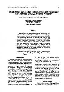
Nuclease protection pattern of Hl' and Hi- attPs. protection was performed on DNA fragments labeled with polynucleotide kinase at the HindlIl site. The attP fragments were digested with neocarzinostatin in the presence of 30, 3, or 0 U of IHF (Hl', lanes 1 to 3) or 300, 30, or 0 U (Hi- lanes 4 to 6) as previously described (5). The locations of the different proteinbinding sites are shown to the right of the autoradiogram. The IHF sites (LI) are identified to the left while the core-type (C and C') and arm-type (P1, P2, P' 1-3) Int (U) and Xis sites (Z) are identified to the right. FIG.
2.
Nuclease
attL
attR
IHF, lower concentrations of Consensus
Hi'
PyAA
TTGATTA
ctAAgtagTTT
T~~Uc
binding
to the Hi
mo'bility
FIG.
1.
c[TAgt agTgIsATTc
Hi'
cTAAccaag
tTgg
Schematic of lambda
sites involved in the
site-specific recombination. The att integration and excision reactions as well as the
proteins required for each are shown. The locations of the binding sites for Int (0), IHF (LI), and Xis (K0) have been deduced by nuclease protection (8, 37, 51). The IHF consensus sequence (27) and coordintes relative to the center of the overlap region (25) are shown
over
consensus
the sequences of the different Hi
sites. Matches to the
in uppercase letters.
Diagnostic restriction
sequence
are
endonuclease sites for each site
are
indicated.
were
sufficient to observe
cll fragment suggested that it IHF-binding site. Sequence inspection of this fragmhent reveals only one region that resembl'es the IHF consensus sequence (in addition to the site previously characterized [8]). This site is located a few nucleotides upstream and in the opposite orientation from the documented site. Nuclease protection studies on this fragment suggested that binding to this site may be occurring bands found with the
may have
H1-
IHF
site than to the clI site. The two lower-
with
a
more
than
o'ne
small fraction
of DNA molecules but most DNA
molecules contain IHF bound to the
major
IHF site
(data
not
shown). In vitro
r'ecombination.
The functional
mutant att sites were characterized
tion reactions of
12 P-labeled
properties of the by in vitro recombina-
DNA substrates with
varying
1346
THOMPSON ET AL.
J. BACTERIOL.
A) H1IHF
4 8 18 32 64 128 4
bound Hi -
unbound Hi-
8 16 32 84 128
I *
B)
IHF bound unbound
1121
18 32 64 128
2 4 8
4--*o
-_ _ _
a
OM
H1+
H 1=
I_.
-
18 114
112
1
2
4
B
croXcI
cro/c3l-
bound HIunbound
Hi-
_
.4
FIG. 3. Gel mobility shifts induced by IHF binding to the Hi+, Hl-, H1=, and clI sites. (A) DNA fragments were labeled with the Klenow fragment of DNA polymerase and [a-32P]dATP at the HindIII site in each. The labeled DNA was then cut with HaeIII to generate a 180-bp fragment and to remove the other unwanted labeled end. The binding was carried out with 5 x 10-'3 M labeled DNA-100 ,ug of salmon sperm DNA per ml-100 mM NaCi-10 mM MgCl2-50 mM Tris hydrochloride (pH 7.9) and the indicated amount of IHF. Ficoll was added to a final concentration of 1% before electrophoresis on 5% polyacrylamide gels. (B) The H1+ DNA was labeled as above. The clI DNA was labeled with the Klenow fragment of DNA polymerase and [Ot-32P]dCTP at the AvaI site of pKMLH2 (plasmid constructed by K. Madden containing the X tRl terminator region). The labeled DNA was cut with NruI and gel purified as previously described (5). IHF binding was done as described above except 5 ptg of salmon sperm DNA per ml was used. The two DNA fragments were mixed before IHF addition.
40-, -
-
0
[IHF]
2.5n ---80n
[Int]
25nM
Hi- ° H1+ *
concentrations of purified proteins. DNA concentrations were kept below the apparent KD of all three proteins so that stoichiometric requirements for a protein would not mask any inherent differences in binding affinity. The behavior of the H1- and H1= attRs in excision in vitro was identical, so the two will be discussed interchangeably in this regard. The dependence of excisive recombination on the Xis concentration is shown (Fig. 4). At low IHF concentrations, the Hl+ and Hl- attR DNAs had virtually identical recombination efficiencies at all concentrations of Xis. However, at high IHF concentrations, they were clearly different. While the Hl+ attR was depressed in excision at all Xis concentrations, the Hl- attR excision remained unchanged at high Xis concentrations and actually was better than low IHF concentrations at low Xis concentrations. The effect of IHF inhibition on excision can be accentuated, as shown in Fig. 5 where IHF concentration was varied at lower Int concentration. Both attR DNAs initiated&recombination at the same IHF concentration but the Hl+ attR became completely inhibited at IHF concentrations where the HY= attR remained fully competent. This specific inhibition by IHF was also very dependent on the concentration of Int (Fig. 6). At low Int concentrations, the selective inhibitory effect of IHF on H1+ attR was very pronounced while, at high Int concentrations, there was no apparent inhibition of excisive recombination. Analysis of the Hi- attP for the ability to perform integrative recombination yielded ambiguous results. Although it could integrate as well as H1+ attP under some conditions, it required eightfold higher concentrations of IHF to reach maximal levels of integration (Fig. 7). If only H2 and H' are required for integration, the IHF dependence of the two attP DNAs should be identical while, if Hi is required for the reaction, there should be no recombination under these conditions. Examination of the H1= attP clarified this situation; it did not integrate in any conditions in vitro (data not shown). These results establish that Hi is indeed necessary for integration. Similar results have been obtained with other mutations in the Hi site (J. F. Gardner and H. A. Nash, J. Mol. Biol., in press).
30 30- O-
E~~~~~~~ /* // 0
20
T
[Xis] (nM)
FIG. 4. Dependence of excisive recombination on Xis concentration. In vitro recombinations were performed as described in Materials and Methods with 1.25 x 10⁻¹¹ M attR and 5 x 10-"1 M supercoiled attL. The fraction of each attR which recombined is plotted versus the concentration of Xis (logarithmic scale). Two curves for each attR are shown: , low IHF concentration; --- -, high IHF concentration.
1347
MUTATIONS IN AN IHF-BINDING SITE
VOL. 168, 1986
[Int]
4u
lu Int
[Xis] 15OnM
hnt
6nM
IHF ....
1/2
1
2
4
8
1/2
1
2
4
8
0 c
E 0
H
D
H1- product
0.CD
I+ product -
attB
1.2
.62
5.0
2.5
Hl+ attP
[IHF] (nM)
FIG. 5. Dependence of excisive recombination on IHF concentration. In vitro recombinations were performed as described in the legend to Fig. 4, with H1+ autR (0) and Hl= attR (0). The fraction of each attR which recombined is plotted versus IHF concentration.
In vivo recombination. The in vitro recombination results have been essential for deciphering the complex interplay between the DNAs and the three proteins, but it is also interesting to determine how the reactions might be modulated by in vivo parameters -not present in the purified in vitro system. Lysogenization with an MOI of 1 to 10 showed no difference between the Hi+ and Hl- attPs, whereas the wt H1
*
0
[IHF] lOnM [Xis] 150nM
50