Nano-Structures & Nano-Objects 16 (2018) 120–126
Contents lists available at ScienceDirect
Nano-Structures & Nano-Objects journal homepage: www.elsevier.com/locate/nanoso
Highly sensitive detection and removal of mercury ion using a multimodal nanosensor Soumitra Satapathi a,c, *, Vishal Kumar a , Mrinmoy Kumar Chini a , Rajesh Bera b , Krishna Kanta Halder b , Amitava Patra b a b c
Department of Physics, Indian Institute of Technology Roorkee, Roorkee, Haridwar, 247667, India Department of Materials Science Indian Association for the Cultivation of Science, Kolkata 700032, India Centre of Nanotechnology, Indian Institute of Technology Roorkee, Roorkee, Haridwar, 247667, India
highlights
graphical abstract
• A highly sensitive and novel multimodal nanosensor encompassing magnetic and fluorescent functionality is designed. • The design of the multimodal nanosensor employed for the simultaneous detection and removal of mercury ion. • Significant fluorescence quenching is observed with surprisingly low limit of detection. • A novel facile route towards field based mercury sensor development.
article
info
Article history: Received 12 April 2017 Received in revised form 18 April 2018 Accepted 16 May 2018 Keywords: Multimodal nanosensor Mercury detection and removal Fluorescence quenching Chemical sensors
a b s t r a c t A highly sensitive and environment friendly multimodal nanosensor encompassing magnetic and fluorescent functionality is designed for the simultaneous detection and removal of mercury ion in water. A significant fluorescence quenching is observed with the increasing concentration of Hg2+ with surprisingly low limit of detection. The detected analyte is successfully removed with the help of a bar magnet leaving no residual secondary pollution. The details mechanism of sensing is also investigated. The simple and elegant nanochemistry reported here provides a facile route towards field based mercury sensor development in future. © 2018 Published by Elsevier B.V.
1. Introduction Recently, the sensitive and selective detection of mercuric ions (Hg2+ ) has attracted considerable attention because of its deleterious effects on the environment and human health [1]. Mercury is a
*
Corresponding author at: Department of Physics, Indian Institute of Technology Roorkee, Roorkee, Haridwar, 247667, India. E-mail address:
[email protected] (S. Satapathi). https://doi.org/10.1016/j.nanoso.2018.05.006 2352-507X/© 2018 Published by Elsevier B.V.
highly toxic heavy metal and its exposure can cause serious damage to the brain, kidneys, and nervous system [2–6]. The United States Environmental Protection Agency (EPA) has set a maximum Hg2+ contaminant level in drinking water at 0.002 mg L−1 (10.0 nM L−1 ) [7]. Commonly, mercury contamination comes from a variety of sources, both natural (e.g., volcanic activity) and industrial (e.g., mining, fossil fuels) [8,9]. Ionic mercury is easily converted into the neurotoxic monomethyl mercuric cation which then builds up in aquatic system and enters the food chain [10].
S. Satapathi et al. / Nano-Structures & Nano-Objects 16 (2018) 120–126
It is thus very important to monitor its level in water and develop a simple yet environmentally friendly sensor with high selectivity and sensitivity. In the past few years, several optical sensor systems have been developed for the detection of Hg2+ in the aqueous environment [11]. This includes small organic molecules (fluorophores or chromophores), biomolecules (proteins, antibodies, oligonucleotides, DNAzymes, etc.), polymer [12], inorganic materials and the nanoparticles [13–18]. Yan and co-workers reported 4-4′ -bis(carboxyl phenyl)-dibenzo-18-crown-6 dye and its application for colorimetric detection of Hg2+ [19]. Song et al. have reported a ‘‘Turn-off’’ fluorescent sensor incorporating benzothiadiazole moiety for the Hg2+ detection [20]. Li and co-workers have synthesized a naphthalimide derivative based fluorescent ‘‘Turn-on’’ chemosensor for aqueous phase detection of Hg2+ [21]. Milan Balaz and co-workers developed a Pyridylporphyrin–DNA Conjugates for Hg2+ recognition [22]. Recently, we have designed a fluorescent resonance energy transfer (FRET) based chemosensor which was highly selective to a variety of analytes [23]. Several reports based on fluorescent semiconducting nanomaterial, CdTe quantum dots (CdTe-QDs), have been reported for the Hg2+ detection due to their unique optical, electronic properties [23–26]. Paramanik et al. has developed a single probe of Au–NC–CdTe QD nanocomposite by using tripeptide-capped CdTe quantum dots (QDs) and BSA-conjugated Au25 nanocluster (NC) [27]. The conjugation was formed through an H-bonding interaction between tripeptideglutathione (GSH) and BSA protein-capped NC. The limit of detection of Hg2+ is detected to be 7 nM by using photoluminescence quenching. Although many of these systems, are capable of detecting Hg2+ with high sensitivity, they are constrained with respect to simplicity, sensitivity and selectivity, or limited in practical applications (e.g. incompatible with aqueous environment) [28–30]. Furthermore, these chemosensors are inconvenient for the removal of the target species which would cause secondary pollution. Towards this end, the incorporation of a fluorescent and magnetic functionality in a single nanocomposite particle would be a promising alternative [31]. Previously, magnetic nanoparticles have widely been used in many fields such as drug delivery, protein resistant, bio-separation and magnetic resonance imaging for their attractive properties of superparamagnetism and the size tenability [32–34]. It can be attracted to the target zone in the presence of an external magnet or an implanted permanent magnet. However, the strong photoluminescence quenching of fluorescent moiety in the presence of magnetic nanoparticles possesses a serious challenge to develop this kind of nanocomposite. To address this issue, various molecular interactions, such as electrostatic and hydrophobic interactions, covalent linkage and hydrogen bonding such as quantum dots (QDs) deposited onto the surface of polymer-coated Fe2 O3 beads through thiol chemistry, gold nanoparticles immobilized on the surface of various materials including Fe2 O3 nanoparticles, silica microspheres via electrostatic interactions have been developed [35,36]. In this paper, we have used a nano-chemistry to synthesize a multilevel, hierarchically built nanoparticle, which we define as a multimodal nanosensor, for efficient detection and removal of Hg2+ ion in aqueous media. This nanosensor produced by multistep synthesis process, consists of a thin silica shell encapsulating magnetic (Fe2 O3 ) nanoparticles, a fixed spacer arm and fluorescent quantum dot for simultaneous detection and removal of detected mercury. The surface of this prototype nanosensor is functionalized with a (triethoxysilyl propyl–carbamoyl) butyric acid to make it negatively charged. The cysteamine capped positively charged cadmium telluride (CdTe) QD is electrostatically adsorbed on the surface. Previously, various chemical syntheses were adopted to develop fluorescently coated magnetic beads for biomedical applications like MRI contrast agent and multiphoton imaging. However, to the best of our knowledge, the synthetic route developed here to synthesize multimodal
121
nanoparticles and also their use as aqueous mercury sensor has not been reported in the literatures before. These nanosensor are capable of detecting Hg2+ in nano-molar level with limit of detection (LOD) of 1 nm. The detected analyte was also removed with the use of an external bar magnet leaving no residual pollution. 2. Experimental section 2.1. Synthesis of multimodal nanosensor A multistep process was involved for the fabrication of multimodal nanosensor. Fig. 1 represents the schematic diagram of synthetic protocol. The superparamagnetic Fe2 O3 nanoparticles were prepared by the chemical coprecipitation method. Initially, the pristine Fe2 O3 NPs tend to aggregate into large clusters, and thus lose the special magnetic properties associated with single domain. Citric acid was employed as a surfactant to create an electrostatic double layer, thereby, reducing their tendency to aggregate. Coating Fe2 O3 nanoparticles with silica was carried out at room temperature by a modified Stöber method through a sol–gel approach involving the hydrolysis and condensation of tetraethyl orthosilicate (TEOS). The surface of the colloidal Fe2 O3 NPs has a strong affinity to silica, thus silica can be directly deposited on the Fe2 O3 NPs without other pretreatment. 3-(Triethoxylsilylpropyl– carbamoyl) butyric acid (spacer arm) was prepared by incubating 3-(triethoxylsilyl) propylamine (4.5 mmol) with 4.5 mmol of succinic anhydride in 1 mL of anhydrous DMF. The resulting product was dispersed in DMF containing 10 mg of particles. After the solution was stirred for 24 h at room temperature, the particles were repeatedly washed with DMF and dried. The COOH-coated nanoparticles (2 mg) were dispersed in 1 mL of H2 O using sonication. Finally, the multimodal nanosensor were electrostatically attached with the cysteamine capped CdTe QDs. 2.2. Synthesis of cysteamine capped CdTe QD Colloidal solution of CdTe QD was synthesized using the reaction between CdCl2 and NaHTe solutions with some modification. At first, NaHTe was prepared by adding 0.53 mmol of NaBH4 (Merck) and 0.18 mmolTe powder (Aldrich) in a round-bottom flask, containing 2 mL of Milli-Q water in inert atmosphere. On the other hand 0.50 mmol CdCl2 • 2.5H2 O (Aldrich) and 1 mmol of cysteamine (Aldrich) were dissolved in 70 mL of Milli-Q water. The pH of the cysteamine solution was maintained to 5.5 by adding required amount of HCl. The mixture was bubbled by N2 for 45 min to make O2 free. Then 0.06 mmol NaHTe was immediately injected into the mixture under vigorous stirring for 30 min followed by refluxing at 110 ◦ C in aerial atmosphere. A solution of different size was taken out from the reaction mixture with the growth time of 15, 30, and 50 min. These nanocrystals are stable for more than three months when stored at 10 ◦ C. 2.3. Characterizations Room temperature optical absorption spectra were taken using a UV–Vis spectrophotometer (Shimadzu). Room temperature photoluminescence spectra were taken by a Fluoromax-P (Horiba JOBIN YVON) photoluminescence spectrophotometer. For the time correlated singlephoton counting (TCSPC) measurements, the samples were excited at 440 nm using a nanosecond diode laser (IBH Nanoled) in an IBH Fluorocube apparatus. The typical full width at half-maximum (FWHM) of the system response using a liquid scatter was about 40 ps. The repetition rate was 1 MHz. The fluorescence decays were analyzed using IBH DAS6 software. The transmission electron microscopy (TEM) image was taken by using a JEOL-JEM-2100F transmission electron microscope.
122
S. Satapathi et al. / Nano-Structures & Nano-Objects 16 (2018) 120–126
Fig. 1. Schematic of the multimodal nanosensor synthesis.
Fig. 2. TEM image of Fe2 O3 NP (a) silica coated Fe2 O3 NP (b) and multimodal nanosensor (c).
3. Results and discussions 3.1. Design of multimodal nanosensor The superparamagnetic Fe2 O3 nanoparticles were prepared by the chemical co-precipitation method. Initially, the pristine Fe2 O3 NPs tend to aggregate into large clusters, and thus lose the special magnetic properties associated with single domain. Citric acid was employed as a surfactant to create an electrostatic double layer, thereby, reducing their tendency to aggregate. The TEM image of the citric acid-stabilized Fe2 O3 NPs is shown in Fig. 2(a). The surface of the magnetic nanoparticles was functionalized with an inert silica layer to prevent their aggregation in liquid, to improve their chemical stability and to attach various functional groups capable of sensing analytes. This coating stabilizes the magnetite nanoparticles in two different ways. One is by shielding the magnetic dipole interaction with the silica shell. On the other hand, the silica nanoparticles are negatively charged. Therefore, the silica coating enhances the Coulomb repulsion of the magnetic nanoparticles. The coating of silica on Fe2 O3 nanoparticles was carried out at room temperature by a modified Stöber method through a sol–gel approach involving the hydrolysis and condensation of tetraethyl orthosilicate (TEOS) [37]. The surface of the colloidal Fe2 O3 NPs
has a strong affinity to silica, thus silica can be directly deposited on the Fe2 O3 NPs without other pretreatment. Fig. 2(b) shows the TEM image of SiO2 /Fe2 O3 nanoparticles. The core–shell structural particles were rather spherical and uniform, even though some of them had no magnetic core. The thickness of silica shell can be changed from a few to several hundred nanometers simply by varying the initial amount of TEOS. The growth of silica on Fe2 O3 NPs was also monitored through SEM as shown in Fig. S1 in SI. Upon formation of the silica layer the brownish colored magnetic nanoparticles dispersion turned slightly milky. 3(Triethoxylsilylpropylcarbamoyl) butyric acid (spacer arm) was prepared by incubating 3-(triethoxylsilyl)propylamine (4.5 mmol) with 4.5 mmol of succinic anhydride in 1 mL of anhydrous DMF. The resulting product was dispersed in DMF containing 10 mg of iron oxide nanoparticles. After the solution was stirred for 24 h at room temperature, the particles were repeatedly washed with DMF and dried. Then, the nanoparticles (2 mg) were dispersed in 1 mL of H2 O using sonication. Finally, the multimodal nanosensor was fabricated by electrostatically attaching COOHcoated nanoparticles with the cysteamine capped CdTe QDs. Fig. 2(c) shows the TEM image of multimodal nanosensor. The thiol group of cysteamine was used to confine the growth of CdTe QDs, and the amino group was linked to the free carboxyl group in
S. Satapathi et al. / Nano-Structures & Nano-Objects 16 (2018) 120–126
123
Scheme 1. Design of multimodal nanosensor.
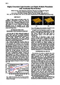
spacer arm, which was already bonded to the silica surface. The measurement of z-potential was used to monitor this stage of the preparation process of nanosensor. The silica coated Fe2 O3 particles were negatively charged after surface carboxyl-termination and the z-potential was −38.2 mV. The potential has changed to +14.3 mV for the final products, indicating that CdTe QDs are successfully deposited on the silica surface. Scheme 1 shows the design of the multimodal nanosensor. 3.2. Magnetic characterizations of multimodal nanosensor Magnetic characterization for Fe2 O3 NPS is carried out in a Quantum Design SQUID magnetometer. Hysteresis loops M(H) are measured under a maximum applied field of ±60 kOe at 10 K (Fig. 3(a)). As depicted in Fig. 3(a), the magnetization saturates at a field intensity of 20 kOe which is quite high and might be attributed to the lack of surface spin disorder as reported in the literature. The synthesized particles are right at the fringe of the superparamagnetic limit. Ferromagnetic rather than superparamagnetic behavior could emerge from multi-domain structures in the particles and/or because of interparticle interaction within the magnetic fluid volume. The size dependent switch from superparamagnetic to ferromagnetic response in the magnetic ferrofluid has been previously reported by many research groups [38–41]. The saturation magnetization also depends on stabilizing agent. Shafi et al. reported that phosphonate-coated particles are strikingly different from the ones coated with sulfonate and undecenoate, though it is not clear the role of capping agent from their study. The ample rotational degree of freedom within the magnetic fluid is attributed to the cause of high saturation magnetization in our case (oleic acid capping). Zero-field-cooling (ZFC) and fieldcooling (FC) magnetization curves are measured at 100 Oe in the temperature range 0–325 K (Fig. 3(b)). Typically, the ZFC curve first increases with temperature as the magnetic field partially aligns the magnetization of the NP, reaching a maximum at a temperature TM , thus indicating that the magnetic moment of each particle is fixed along its favorable magnetization direction at a temperature T , which depends on the particle volume, anisotropy, and orientation. Because the NPs are constituted of a random assembly of
crystallites that presents a certain volume distribution, f (V ), each crystallite is blocked at a different temperature TB . As a result, we observe a distribution of blocking temperatures F (TB ) yielding a broad peak in the ZFC curve. TM is related to the mean blocking temperature TB as TM ≈ 2TB , when the particle size distribution is log-normal and in the absence of relevant dipolar interactions. Besides, the ZFC magnetization strongly decreases below the peak at TM , because the NPs become superparamagnetic. In contrast, the FC curve monotonically increases as 1/T (Curie law) in an assembly of noninteracting particles, whereas strong dipolar interparticle interactions yield a flat FC curve at temperatures below TM . 3.3. Mercury (Hg2+ ) sensing with fluorescence quenching Furthermore, the synthesized multimodal nanosensor is employed to detect Hg2+ ion in aqueous environment. The increasing concentration of Hg2+ reduces the PL emission intensity of nanosensor. A significant signal off of the nanosensor is observed in the presence of increasing concentrations of Hg2+ . Fig. 4a depicts the in PL intensity of nanosensor with changing the concentration of Hg2+ . Quantitatively, the PL quenching sensitivity can be correlated to the Stern–Volmer constant (KSV ), determined by the equation, I0 /I = 1 + KSV
(1)
where, I0 is the initial PL intensity, I is the resulting PL intensity upon addition of the analyte, and Q is the analyte concentration [41]. A larger KSV value represents a Higher sensitivity of the fluorophore toward the analyte. The Stern–Volmer constant is calculated to be 0.14 × 10⁹ M−1 (Fig. 4b). We performed the sensing experiments with the different Hg2+ concentration and found the lowest detection limit to be 0.49 nm which is within the range set by USEPA [Fig. S9]. This also reveals that this multimodal nanosensor probe is more sensitive than the previously reported sensing probes in the literature. As a standard comparison, we also confirmed the behavior of bare CdTe QDs to Hg2+ ion to establish the superiority of our approach. Interestingly, the bare CdTe QD is not effective like the multimodal nanosensor as confirmed by its lower Stern–Volmer value (Fig. S2 and S3). Along
124
S. Satapathi et al. / Nano-Structures & Nano-Objects 16 (2018) 120–126
Fig. 3. M(T ) curve of Fe2 O3 NPs (a) and M(H) curve of Fe2 O3 NPs (b).
Fig. 4. (a) PL quenching of MM nanosensor with 0 nM (a) 0.49 nM (b) 0.99 nM (c) 2.9 nM (d) and 4.8 nM (e) Hg2+ conc; (b) Stern–Volmer plot.
Fig. 5. PL decay of MM nanosensor (a) lifetime vs. quencher concentration (b).
with this, we had also performed the Hg2+ quenching studies at different pH using cysteamine capped CdTe QDs based multimodal nanosensor (Fig. S10) [42,43]. The quenching mechanism of Hg2+ on the fluorescence of multimodal nanosensor can be attributed to three main reasons. Firstly, Hg2+ has a strong affinity to N atom. Consequently, Hg2+ absorbs onto the surface of QDs through functional amide groups in cysteamine which facilitates the electron transfer from QDs to Hg2+ as confirmed by the lifetime data. We have observed shortening of the decay time in presence of the Hg2+ ion of various concentrations (Fig. 5a). The average decay time of pure
multimodal nanosensor is 0.795 ns. It has three components. The slowest component is 2.57 ns (84%). In the presence of 0.49 nM Hg2+ ion, the slowest component remains almost the same to 2.5 ns (69%) with an average lifetime of 0.615 ns. However, the average lifetime decreases to 0.506 ns in the presence of 0.99 nM Hg2+ ion with the slowest component becomes 2.17 ns (67%). The average lifetime further decreases to 1.75 ns (57%) with average lifetime 0.361 ns when the Hg2+ concentration increases to 2.9 nM. Table 1 in SI summarizes all the lifetime data. Fig. 5b shows the lifetime vs. analyte concentrations. It shows an exponential decrease. Secondly, HgTe has solubility approximately 20 times
S. Satapathi et al. / Nano-Structures & Nano-Objects 16 (2018) 120–126
125
References
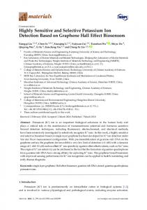
Fig. 6. MM Nanosensor before adding Hg2+ (a) after adding Hg2+ (b) and after removing with a magnet (c).
slower than that of QD. This drives the substitution of Cd2+ in CdTe QDs by Hg2+ and leads to the formation of alloyed Cdx Hg1−x Te. These very small particles quench the recombination fluorescence of CdTe nanoparticles by facilitating nonradiative recombination of excited electrons (e−) in the conduction band and holes (h+) in the valence band. The quenching process is similar to the effect of Cu2+ on CdS QDs investigated by Isarov and Chrysochoos. Finally, according to Pendyala’s theory, the band alignment between the core and the surface band structures is one of the reasons for PL enhancement or quenching. The lower bandgap of HgTe (−0.41 eV) than that of CdTe (1.47 eV) is responsible for this quenching behavior as well. The selectivity of the multimodal nanosensor with different analytes like arsenic, copper, and lead, were performed and depicted in Fig. S5, S6, and S7 respectively in SI. The corresponding histogram is shown in Fig. S9 in SI which clearly demonstrates the high selectivity of the designed nanosensor towards Hg2+ . The adsorbed Hg2+ on multimodal nanosensor as Cdx Hg1−x Te alloy was efficiently removed with the help of a bar magnet because of the presence of iron core (Fig. 6). The Inductively coupled plasma mass spectrometry (ICP-MS) was performed to quantify the amounts of Cd, Te, and Hg before and after the magnetic separation and shown in the supporting information Fig. S4. It shows the amounts of Cd, Te, and Hg decrease after magnetic separation as they form alloyed Cdx Hg1−x Te by substitution of Cd2+ with Hg2+ in the CdTe QDs. Moreover, we have already proved the paramagnetic properties of multimodal nanosensor which facilitate the removal. 4. Conclusions In summary, a multimodal nanosensor probe was developed for the simultaneous detection and removal of Hg2+ ion. A turn off (PL quenching) phenomenon is observed in presence of Hg2+ ions, which is due to the efficient electron transfer, ultra small quantum alloy formation and band alignment mechanism. This simple and elegant approach provides a route towards field based mercury sensory devices in future. Acknowledgment SS acknowledges the SERB grant (Grant No. YSS/2015/000061). Appendix A. Supplementary data Supplementary material related to this article can be found online at https://doi.org/10.1016/j.nanoso.2018.05.006.
[1] L.M. Campbell, D.G. Dixon, R.E. Hecky, A review of mercury in Lake Victoria, east Africa: implications for human and ecosystem health, J. Toxicol. Environ. Health Part B. 6 (2003) 325–356. [2] J. Lee, H. June, J. Kim, Selective and sensitive detection of melamine by intra/inter liposomal interaction of polydiacetylene liposomes, Adv. Mater. 21 (2009) 3674–3677. [3] I. Hoyle, R.D. Handy, Dose-dependent inorganic mercury absorption by isolated perfused intestine of rainbow trout, Oncorhynchus mykiss, involves both amiloride-sensitive and energy dependent pathways, Aquat. Toxicol. 72 (2005) 147–159. [4] P.B. Tchounwou, W.K. Ayensu, N. Ninashvili, D. Sutton, Environmental exposure to mercury and its toxicopathologic implications for public health, Environ. Toxicol. 18 (2003) 149–175. [5] A.B. Kobal, M. Horvat, M. Prezelj, A.S. Briski, M. Krsnik, T. Dizdarevic, D. Mazej, I. Falnoga, V. Stibilj, N. Arneric, D. Kobal, J. Osredkar, The impact of longterm past exposure to lemental mercury on antioxidative capacity and lipid peroxidation in mercury miners, J. Trace Elem. Med. Biol. 17 (2004) 261–274. [6] S. Vupputuri, M.P. Longnecker, X. Guo, J.L. Daniels, D.P. Sandler, Blood mercury level and blood pressure among US women: results from the National Health and Nutrition Examination Survey 1999-2000, Environ. Res. 97 (2005) 195– 200. [7] EPA (US Environmental Protection Agency) established the mercury (II) limit on 2 ppb, 2006, EPA-HQ-OPPT-2005-0013. [8] G. Qiu, X. Feng, S. Wang, L. Shang, Environmental contamination of mercury from Hg-mining areas in Wuchuan, northeastern Guizhou, China, Environ. Pollut. 142 (2006) 549–558. [9] Q. Wang, D. Kim, D.D. Dionysiou, G.A. Sorial, D. Timberlake, Sources and remediation for mercury contamination in aquatic systems—a literature review, Environ. Pollut. 131 (2004) 323–336. [10] D.W. Boening, Ecological effects, transport, and fate of mercury: a general review, Chemosphere 40 (2000) 1335–1351. [11] M.K. Nazeeruddin, D.Di. Censo, R. Humphry-Baker, M. Grat¨ zel, Highly selective and reversible optical, colorimetric and electrochemical detection of mercury (II) by amphiphilic ruthenium complexes anchored onto mesoporous oxide films, Adv. Funct. Mater. 16 (2006) 189–194. [12] R. Muthivhi, S. Parani, B. May, O.S. Oluwafemi, Green synthesis of gelatin-noble metal polymer nanocomposites for sensing of Hg2+ ions in aqueous media, Nano-Struct. Nano-Objects 13 (2018) 132–138. [13] J. Xie, Y. Zheng, J.Y. Ying, Highly selective and ultrasensitive detection of Hg(2+) based on fluorescence quenching of Au nanoclusters by Hg(2+)-Au(+) interactions, Chem. Commun. 46 (2010) 961–963. [14] C. Diez-Gil, R. Martinez, I. Ratera, A. Tarraga, P. Molina, J. Veciana, Nanocomposite membranes as highly selective and sensitive mercury (II) detectors, J. Mater. Chem. 18 (2008) 1997–2002. [15] J.S. Lee, M.S. Han, C.A. Mirkin, Colorimetric detection of mercuric ion (Hg2+) in aqueous media using DNA-functionalized gold nanoparticles, Angew. Chem. Int. Ed. 46 (2007) 4093–4096. [16] H.Z. He, D.S.H. Chan, C.H. Leung, D.L. Ma, A highly selective G-quadruplexbased luminescent switch-on probe for the detection of gene deletion, Chem. Commun. 48 (2012) 9462–9464. [17] N. Chen, Y. Zhang, H. Liu, X. Wu, Y. Li, L. Miao, Z. Shen, A. Wu, High-performance colorimetric detection of Hg2+ based on triangular silver nanoprisms, ACS Sens. 1 (2016) 521–527. [18] S. Chandra, A.R. Chowdhuri, T.K. Mahto, D. Laha, S.K. Sahu, Sulphur and nitrogen doped carbon dots: A facile synthetic strategy for multicolour bioimaging, tiopronin sensing, and Hg2+ ion detection, Nano-Struct. Nano-Objects 12 (2017) 10–18. [19] Z. Yan, H. Lei, N. Li, L. Hong, Preparation of 4,4′ -bis-(carboxyl phenylazo)dibenzo-18-crown-6 dye and its application on ratiometric colorimetric recognition to Hg2+, Spectrochim. Acta Part A 79 (2011) 661–665. [20] S.Y. Kuo, H.H. Li, P.J. Wu, C.P. Chen, Y.C. Huang, Y.H. Chan, Dual colorimetric and fluorescent sensor based on semiconducting polymer dots for ratiometric detection of lead ions in living cells, Anal. Chem. 87 (2015) 4765–4771. [21] M. Li, H.Y. Lu, R.L. Liu, J.D. Chen, C.F. Chen, Turn-on fluorescent sensor for selective detection of Zn(2+), Cd(2+), and Hg(2+) in water, J. Org. Chem. 77 (2012) 3670–3673. [22] K. Ueno, S. Juodkazis, V. Mizeikis, K. Sasaki, H. Misawa, Clusters of closely spaced gold nanoparticles as a source of two-photon photoluminescence at visible wavelengths, Adv. Mater. 20 (2008) 26–30. [23] P. Taya, B. Maiti, V. Kumar, P. De, S. Satapathi, Design of a novel FRET based fluorescent chemosensor and their application for highly sensitive detection of nitroaromatics, Sens. Actuators, B 255 (2018) 2628–2634. [24] D. Saikia, P. Dutta, N.S. Sarma, N.C. Adhikary, CdTe/ZnS core/shell quantum dotbased ultrasensitive PET sensor for selective detection of Hg (II) in aqueous media, Sensors Actuators, B 230 (2016) 149–156. [25] T. Gong, J. Liu, X. Liu, J. Liu, J. Xiang, Y. Wu, A sensitive and selective sensing platform based on CdTe QDs in the presence of L-cysteine for detection of
126
[26]
[27]
[28]
[29] [30] [31] [32]
[33] [34]
[35]
S. Satapathi et al. / Nano-Structures & Nano-Objects 16 (2018) 120–126 silver, mercury and copper ions in water and various drinks, Food Chem. 213 (2016) 306–312. J. Pei, H. Zhu, X. Wang, H. Zhang, X. Yang, Synthesis of cysteamine-coated CdTe quantum dots and its application in mercury (II) detection, Anal. Chim. Acta 757 (2012) 63–68. B. Paramanik, S. Bhattacharyya, A. Patra, Detection of Hg2+ and F− Ions by using fluorescence switching of quantum dots in an Au-Cluster–CdTe QD nanocomposite, Chem. Eur. J. 19 (2013) 5980–5987. D.L. Ma, H.Z. He, K.H. Leung, H.J. Zhong, D.S. Chan, C.H. Leung, Label-free luminescent oligonucleotide-based probes, Chem. Soc. Rev. 42 (2013) 3427– 3440. H. Xu, M. Hepel, Molecular beacon -based fluorescent assay for selective detection of glutathione and cysteine, Anal. Chem. 83 (2011) 813–819. Y. Wu, R.Y. Lai, ‘‘Signal-on’’ electrochemical DNA sensor with an oligo-thymine spacer for point mutation detection, Chem. Commun. 49 (2013) 3422–3424. L. LaConte, N. Nitin, G. Bao, Magnetic nanoparticle probes, Mater. Today 8 (2005) 32–38. M.L. Steigerwald, L.E. Brus, Synthesis, stabilization, and electronic structure of quantum semiconductor nanoclusters, Annu. Rev. Mater. Sci. 19 (1989) 471– 495. A.H. Lu, E.L. Salabas, F. Schüth, Magnetic nanoparticles: synthesis, protection, functionalization, and application, Angew. Chem. 46 (2007) 1222–1244. N.A. Frey, S. Peng, K. Cheng, S. Sun, Magnetic nanoparticles: synthesis, functionalization, and applications in bioimaging and magnetic energy storage, Chem. Soc. Rev. 38 (2009) 2532–2542. T. Neuberger, B. Schopf, H. Hofmann, M. Hofmann, B. Von Rechenberg, Superparamagnetic nanoparticles for biomedical applications: Possibilities and limitations of a new drug delivery system, J. Magn. Mater. 293 (2005) 483– 496.
[36] K.T. Yong, I. Roy, M.T. Swihart, P.N. Prasad, Multifunctional nanoparticles as biocompatible targeted probes for human cancer diagnosis and therapy, J. Mater. Chem. 19 (2009) 4655–4672. [37] C. Liu, Z.J. Zhang, Size-dependent superparamagnetic properties of Mn spinel ferrite nanoparticles synthesized from reverse micelles, Chem. Mater. 13 (2001) 2092–2096. [38] W. Zheng, G.F. Strouse, Involvement of carriers in the size-dependent magnetic exchange for Mn:CdSe quantum dots, J. Am. Chem. Soc. 133 (2011) 7482–7489. [39] B. Issa, I.M. Obaidat, A.B. Albiss, Y. Haik, Magnetic nanoparticles: surface effects and properties related to biomedicine applications, Int. J. Mol. Sci. 14 (2013) 21266–21305. [40] J. Zhu, Z.-J. Zhao, J.-J. Li, J.-W. Zhao, CdTe quantum dot-based fluorescent probes for selective detection of Hg (II): The effect of particle size, Spectrochim. Acta, Part A 177 (2017) 140–146. [41] S. Satapathi, A. Bheemaraju, S.K. Surampudi, D. Venkataraman, J. Kumar, Enhanced sensory response of quaterthiophene bearing 1, 2, 3-triazole moiety to explosives, IEEE Sens. J. 14 (2013) 4334–4339. [42] A.P.S. Paim, S.S.M. Rodrigues, D.S.M. Ribeiro, G.C.S. de Souza, J.L.M. Santos, A.N. Araujo, C.G. Amorim, E. Teixeira-Neto, V.L. da Silvad, M.C.B.S.M. Montenegro, Fluorescence probe for mercury(II) based on the aqueous synthesis of CdTe quantum dots stabilized with 2-mercaptoethanesulfonate, New J. Chem. 14 (2017) 3265–3272. [43] X. Ding, L. Qu, R. Yang, Y. Zhoua, J. Lia, A highly selective and simple fluorescent sensor for mercury (II) ion detection based on cysteamine-capped CdTe quantum dots synthesized by the reflux method, Lumin. 30 (2015) 465–471.