NanoArrays, The Next Generation Molecular Array Format for High Throughput Proteomics, Diagnostics and Drug Discover. Curtis Mosher, Michael Lynch, Saju Nettikadan, Will Henderson, Asrun Kristmundsdottir, 1Michael W. Clark and Eric Henderson BioForce Laboratory, Inc., 2901 South Loop Drive, Ames, IA 50010 To whom correspondence should be addressed:
[email protected],
[email protected]
1
B
iomolecular array technology is an invaluable tool for rapid screening of nucleic acid mixtures. This approach has been tremendously successful both in its breadth of application and its commercial value. Entire genomes, including the human genome, have been screened by molecular array techniques. Arrays are a rapid and now routine method for analysis of expression patterns and their association with physiological states. Such a rapid, high throughput analysis of cellular expression is key to the expansion of our basic knowledge of the relationship between gene expression and organismal function, as well as to the understanding of the genetic component of disease states and the predisposition to disease. Despite the success of array technology for nucleic acid applications, a similar trend for proteins has not occurred. Due, in part, to the difficulties involved in production and labeling of proteins for solid state analysis, solid state arrays of proteins are not widely utilized. Protein function and interaction have been traditionally addressed by the combination of 2D gel electrophoretic separation and mass spectrometry to examine individual protein spots, a slow, tedious and expensive process. Another approach uses in vivo methods for examining protein-protein interactions by the two-hybrid system in yeast and mammalian cells [1]. Although the two-hybrid system has shown some success in finding new interaction between proteins in important cellular pathways, it is far more difficult, costly and time consuming than the solid state methods used for nucleic acids. BioForce Laboratory, Inc., has developed a solid state method for examining the interaction between a wide range of molecules in an array format. This technology involves several key technological innovations. NANOARRAYS AND THE NANOARRAYER™
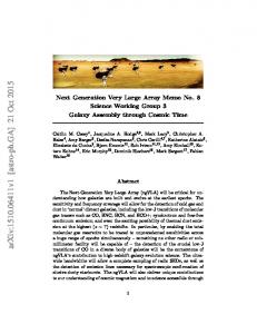
An essential element of this new technological platform is termed a NanoArray, an array of biological molecules deposited in micron or sub-micron spatial addresses. The NanoArrayer™, also developed by BioForce Laboratory, is used to construct NanoArrays as shown in Figure 1. In the figure, a 10 x 10 spot NanoArray of the yeast replication protein RPC19 is compared to the size of a single state-of-the-art microarray spot. One hundred molecular binding tests can be placed in the area occupied by a single conventional microarray spot.
Figure 1. Comparison of state-of-art microget spot (~30 µm dia., left) with a 10 x 10 NanoArray of protein domains generated inour Phase I feasibility study (right). The NanoArray spot domains shown here are less than 1 µm in diameter.
Moreover, the NanoArray shown here is far from the density and spot size limits for the NanoArrayer. The first generation NanoArrayer is capable of reproducibly creating spot sizes as small as 500 nm in diameter, with a spot to spot pitch of 2-3 microns. The next generation NanoArrayer, currently under construction and testing, will be capable of spot sizes of a few 10s of nanometers and a pitch on the order of 40 nanometers. It is important to note that this capability exceeds the requirements for the most demanding miniaturization requirements for biological assays. At this level the number of molecules in a spot domain is so small that a statistical sampling of events at each molecular address is not achieved. Therefore, from a practical point of view, the highest density commercial NanoArrays will have molecular spot domains on the order of 250 nm in diameter, corresponding to a few hundred average protein molecules per spot, with a pitch of about one micron. The NanoArrayer uses microfabricated deposition tools and careful control of environmental conditions to facilitate reproducible deposition of biological materials in NanoArrays. Deposition tool positioning is highly automated and controlled via a user friendly graphical user interface. The details of the NanoArrayer construction and operation are the subject to a pending patent and will be described in detail elsewhere. NANOREADER™
Due to their ultraminiaturization, NanoArrays require novel approaches for readout. Our favored approach is to use a dedicated NanoReader™ atomic force microscope (AFM), for this purpose.
The AFM [2] is a member of a family of instruments called scanning probe microscopes (SPM). Atomic Force Microscopy has become widely accepted in a variety of fields, and the AFM and its relatives have been described in detail elsewhere [3, 4]. Briefly, in the AFM a sharp probe is scanned over a surface while probe/surface interactions are recorded (Fig. 2). These interactions can be any of a wide variety, the simplest being surface topography. First generation NanoArrays rely on this relatively simple measurement to report the formation of a molecular complex or pairing between a molecule in a NanoArray and one introduced in solution. It is noteworthy that the AFM operates in liquids, making it possible to observe molecular binding events during the scanning process. An entire NanoArray can be evaluated in the time it takes to accumulate a full scan, usually 5-10 minutes. A new generation of “fast-scan” AFMs is just emerging and will increase this scan rate by a factor of 10 or more. Another key point is that the AFM directly observes the formation of a molecular complex by reporting a change in the height of the topography. This molecular detection process is label-free, that is, there is no requirement for a fluorescent, radioactive, enzymatic or other molecular taggant. Thus, this approach is applicable to a broad range of molecular species, many of which are difficult to label with conventional taggants, including proteins and drug candidate molecules. Table I compares the features of the NanoArray technology with some of the most commonly utilized methods. An example of an immunodiagnostic analysis by AFM is shown in Figure 3. In this test the antigen (a protein), was deposited on a sur-
Sensitivity
Speed
NanoArrays
Single molecule
PCR ELISA
Multiplexes
Automation
Problems
1-10 minutes per Variable depending on scan type of assay1.
Yes, 10 to thousands per chip
Almost complete
New technology. Background must be low.
A few molecules
1-2 hours
Personnel and supply costs high
No
Substantial
Error prone. Only reveals nucleic acids2 .
Millions of molecules
Several hours
Personnel, instrumentation and supply costs high
No
Substantial
Insensitive. Slow.
Immunoprecipitation Millions of
Several hours
Personnel and supply costs high
No
Minor
Insensitive. Slow.
Wet Lab Methods
Millions of molecules
Hours to days
Personnel and supply costs high
Can be
Minor
Insensitive. Slow.
Cell Culture
Single cell/virus
Days
Personnel and supply costs high
No
Minor
Very slow. Not always dependable.
Particle Assays
Thousands of molecules
Hours
Moderate
Ñot usually (one assay per particle)
Substantial
Particles must be prepared slowly.
Minutes
Expensive
Yes, 10 to 1000s per chip
Substantial
Low spatial resoulution. Difficult to apply to systems other than nucleic acids.
molecules
Flourescence assays Can be single molecule, but usually thousands or more
Costs
Figure 2. Schematic of the AFM. As the tip is scanned over the sample it interacts with the sample surface. The sum of the forces acting on the tip result in bending of the cantilever. The degree of bending is reported by an optical lever system. This signal can be interpreted as a topographical image of the sample. Cantilever bending can activate a feedback circuit that moves the sample relative to the tip to minimize the applied vertical force.
face along with a second, non-antigenic protein. The simple NanoArray was then incubated in the presence of an antibody directed against one of the proteins while simultaneously monitoring the chip via AFM. A clear change of height was observed only for the antigenic domains, indicating that antibody had bound there, but not to the non-antigenic protein domain. This real time assay took 10 minutes (signal first observed in 5 minutes, the time for a single scan), and used an antibody concentration of 6.7 nM. There are several thousand molecules in the smallest spots in this array. We believe that a robust signal can be obtained with just a few 10s of molecules per spot. Thus, another advantage of the NanoArray approach is the limited requirement for molecular or signal amplification. In addition to the very high detection sensitivity, the ultraminiaturized format of the NanoArray makes the sample quantity required far lower than is now commonly used. The advantages of the NanoArrays are listed in Table II. CURRENT AND FUTURE DIRECTIONS
Figure 3. Solid state immunoassay read out using an AFM. The left and right hand panels show AFM scans of 2-3 µ dia. protein domains on a surface before and after the addition of a soluble antibody directed against one of the proteins. The assay took about 10 minutes and was carried out in solution using contact mode AFM. The change in height of the domain for the target protein (middle panels, labeled "antigen") is about 3 nanometers while the change in height for the control (top panels, labeled "non-antigen") is negligible. In the top view images (top four panels), the height change is color coded, making the data resemble conventional fluorescent microarray analysis. However, it is important to note that no molecular taggant is needed for AFM based NanoArray analyses. The bottom two panels show a 3D rendering of the target domain before and after antibody binding to illustrate the actual change in topography of the domain.
The current emphasis at BioForce is to develop NanoArray assays for three target areas: proteomics (protein-protein interaction screening), drug discovery (solid state evaluation of immobilized molecular libraries), and diagnostics (fast and cheap multiplexed assays for pathogenic viruses and spores, and for disease specific antigens). Table III details the impact the NanoArray technology will have in particular areas of research. In the longer term view, the ability to position and manipulate molecules at the nanometer spatial scale is a key element to the “top down” approach to the emerging field of nanotechnology. Indeed, this nano-fabrication ability is also key to the bottom up approach to nanotechnology since precisely placed molecules can seed self-assembling processes. NanoArrays are the next step in the progression of high throughput, fully automated, solid state bioaffinity binding assays and bridge the gap between the two industries predicted to be
Feature
Advantage
Direct molecular operation
Reduced cost, reduced waste, reduced effort, improved stereochemistry
Sub-micron spatial resolution
Higher density screens
Single molecule detection capability
Cost savings and higher density screens
Minimal perturbation of native molecular structure
More biologically relevant hits
Operation in physiological environments
Biologically relevant binding information
Real time data acquisition and analysis
More rapid data acquisition for diagnostics and drug discovery
Facile automation
Reduction in instrument size, screening time and personnel costs
Diagnostics
Small, multiplexed assays for clinical diagnoses. By putting multiple related assays on a single chip the AFM-based approach will reduce the cost and time required to diagnose a variety of physiological states. For example, screening for the presence of antibodies to defined circulating antigens in patients with symptoms of infection will be reduced to a single, rapid and inexpensive assay.
Proteomics
Larger assays for the analysis of the spectrum of proteins expressed from a given genome (proteome) under various sets of conditions. These data will buttress and corroborate gene level expression screens, and reveal genes that are transcribed but not expressed at the protein level. Again, miniaturized and multiplexed assays that do not require labeling probe molecules with a reporter system are highly cost effective, rapid, and do not intoduce additional stereochemical constraints.
Drug Discovery
Large scale screens of molecular interactions in high throughput drug screening. In this scenario a large number of target molecules could be immobilized and allowed to interact with soluble potential partner molecules. This has the potential to reduce current room-sized robotic efforts to microminiaturi9zed desktop robotic screening, with the associated savings in reagent and personnel costs.
Table III. Areas of impact for NanoArray technology.
the primary economic movers in the new century: biotechnology and nanotechnology. LITERATURE CITED
1. Martzen, M.R., S.M. McCraith, S.L. Spinelli, F.M. Torres, S. Fields, E.J. Grayhack, and E.M. Phizicky, A biochemical genomics approach for identifying genes by the activity of their products. Science, 1999. 286(5442): p. 1153-1155.
2. Binnig, G., C.F. Quate, and C. Gerber, Atomic force microscope. Physical Review Letters, 1986. 56: p. 930-933. 3. Bottomley, L., Scanning Probe Microscopy. Anal. Chem., 1998. 70: p. 425-476. 4. Engel, A., Y. Lyubchenko, and D. Meuller, Atomic force microscopy: a powerful tool to observe biomolecules ar work. Trends Cell Biol., 1999. 9: p. 77-80.
If you are interested in seeing more articles like this in JALA, please email
[email protected], and simply put the code (5511) in the subject line.