May 22, 2012 - Neural Bases for Anticipation Skill in Soccer: An fMRI Study. Daniel T. Bishop,1 Michael J. Wright,1 Robin C. Jackson,1 and Bruce Abernethy2.
JOURNAL OF
SPORT EXERCISE PSYCHOLOGY
Journal of Sport & Exercise Psychology, 2013, 35, 98-109 © 2013 Human Kinetics, Inc.
Official Journal of NASPSPA www.JSEP-Journal.com ORIGINAL RESEARCH
Neural Bases for Anticipation Skill in Soccer: An fMRI Study Daniel T. Bishop,1 Michael J. Wright,1 Robin C. Jackson,1 and Bruce Abernethy2 1Brunel
University; 2University of Queensland
The aim of this study was to examine the neural bases for perceptual-cognitive superiority in a soccer anticipation task using functional magnetic resonance imaging (fMRI). Thirty-nine participants lay in an MRI scanner while performing a video-based task in which they predicted an oncoming opponent’s movements. Video clips were occluded at four time points, and participants were grouped according to in-task performance. Early occlusion reduced prediction accuracy significantly for all participants, as did the opponent’s execution of a deceptive maneuver; however, high-skill participants were significantly more accurate than their low-skill counterparts under deceptive conditions. This perceptual-cognitive superiority was associated with greater activation of cortical and subcortical structures involved in executive function and oculomotor control. The contributions of the present findings to an existing neural model of anticipation in sport are highlighted. Keywords: cognitive, expert, oculomotor, perceptual, sport In interceptive sports such as soccer, experts’ advantage over their lesser skilled counterparts is due in part to superior anticipation ability (Reilly, Williams, Nevill, & Franks, 2000); they are more adept at picking up early movement information, enabling them to execute an appropriate response in a timely manner (Savelsbergh, Van der Kamp, Williams, & Ward, 2005; Williams, Ford, Eccles, & Ward, 2011). The temporal occlusion paradigm has enabled researchers to identify the points at which information pickup is greatest: Participants view video clips of an opponent performing an action such as the tennis serve; these clips are foreshortened at various points relative to racket–ball contact so as to provide varying degrees of visual information. Experts consistently detect kinematic information at very early, precontact levels of occlusion to successfully determine not only the direction of a projectile, but also the force with which it is struck (Abernethy & Russell, 1987; Abernethy, Zawi, & Jackson, 2008; Jones & Miles, 1978). The expert anticipatory advantage at early levels of occlusion also extends to the detection of deceptive bodily movements. Jackson, Warren, and Abernethy (2006) asked skilled and less-skilled rugby football
Daniel T. Bishop is with the Centre for Cognition and Neuroimaging and the Centre for Sports Medicine and Human Performance, Brunel University, London, UK. Michael J. Wright is with the Centre for Cognition and Neuroimaging, Brunel University, London, UK. Robin C. Jackson is with the Centre for Sports Medicine and Human Performance, Brunel University, London, UK. Bruce Abernethy is with the School of Human Movement Studies, University of Queensland, Queensland, Australia.
98
players to respond to video clips that depicted one-onone tackle situations: An attacking player ran toward the participant (acting as the defending player) before obliquely changing direction, as if to pass the defender on the left or right. In deceptive trials the player effected a contralateral “side step” maneuver before direction change. Low-skill players were more susceptible to this deception than were skilled players, who could accurately predict the intended direction change even when viewing early-occluded sequences. Such expert sensitivity and novice susceptibility to deceptive movements have been found in boxing (Ripoll, Kerlirzin, Stein, & Reine, 1995), handball (Cañal-Bruland & Schmidt, 2009), and basketball (Kunde, Skirde, & Weigelt, 2011)—but experts may still require directional information such as ball flight to move substantially beyond chance performance (Rowe, Horswill, Kronvall-Parkinson, Poulter, & McKenna, 2009). Although the accumulation of perceptual experience underpins many explanations for anticipation skill superiority, others have suggested that because action perception and execution share common neural origins (Prinz, 1997), then it is motor expertise, be it in deception or otherwise, that determines the extent of this advantage. This notion is corroborated by investigations of the mirror neuron system (MNS), a parieto-frontal network of neurons that are similarly active when individuals perform, imagine, or witness an action within their own repertoire (Rizzolatti & Maddalena Fabbri, 2007). Subtle differences in this MNS motor resonance when viewing and predicting sporting actions are manifest in behavioral (Knoblich & Flach, 2001) neuroimaging (Calvo-Merino, Grèzes, Glaser, Passingham, & Haggard, 2006) and psychophysiological (Aglioti, Cesari, Romani, & Urgesi, 2008) data.
Neural Bases for Soccer Anticipation 99
The roles of neural systems in sport anticipation have been investigated more extensively in recent years. Wright and Jackson (2007) employed the temporal occlusion paradigm to examine novice tennis players’ cortical fMRI activation when predicting an opponent’s serve direction. Action prediction significantly activated MNS regions when contrasted with a passive observation condition. Wright, Bishop, Jackson, and Abernethy (2010) subsequently found stronger activations for earlythan for late-occluded sequences of a badminton shot, notably in premotor MNS regions and in medial frontal cortex. Experts also exhibited greater frontal MNS and medial frontal cortex activation than did novices when viewing the early-occluded sequences. To assess the relative contribution of kinematic information to these differences, Wright, Bishop, Jackson, and Abernethy (2011) compared expert, intermediate, and novice badminton players’ responses to normal video and point-light displays of opponents in a badminton prediction task. Activations were highly similar for both video formats, reinforcing the prominence of kinematic information; moreover, greater frontal activity was apparent in experts when viewing early-occlusion sequences. There was also evidence for suppression of low-level, task-irrelevant stimuli in experts, suggesting greater attentional efficiency. However, experts’ comparatively high levels of activation in anticipation tasks stands in sharp contrast to that witnessed during imagery of a self-paced sport: Milton, Solodkin, Hluštík, and Small (2007) compared the neural activity of expert and novice golfers as they mentally prepared for a hypothetical putt shot. The authors found almost ubiquitously stronger activation in novices, in areas of the brain associated with motor planning and execution—most notably the basal ganglia; this collection of nuclei are pivotally involved in decision making and subsequent action selection, making reciprocal connections with motor and premotor areas of the cortex. Milton et al. interpreted the comparatively lower activity in experts as a reduction in the complexity of dynamic motor control, thereby promoting greater movement consistency. Milton et al.’s (2007) findings contrast with the very active role for the basal ganglia proposed by Yarrow, Brown, and Krakauer (2009) in their affordance competition model of motor preparation and decision making, based on Cisek’s (2007) affordance competition hypothesis. Yarrow et al. propose a complex cortico-subcortical network comprising not only regions of the MNS, but also prefrontal cortex, ventral and dorsal visual pathways, and two subcortical structures—the basal ganglia and the cerebellum. In this model, visual inputs are transformed into motor plans, which may be manifested in commonly observed MNS resonance, before the basal ganglia behaviorally bias the best possible motor action, by encoding the difference between expected and actual reward of a given course of action (Stocco, Lebiere, & Anderson, 2010)—ultimately leading to action execution; the hours of deliberate practice accrued by expert performers (Ericsson, Krampe, & Tesch-Römer, 1993) may potentiate this
function of the basal ganglia. According to the model, the cerebellum is primarily involved in transforming visual input into motor plans. However, activation in the culmen, a region of the cerebellum, has been correlated with low response time variability in children performing a go/ no-go task (Simmonds et al., 2007), which indicates a potential role for this region also in biasing the correct response. Yarrow et al. propose that the basal ganglia and cerebellum serve important functions in generating and selecting motor plans. Accordingly, we might expect greater activation in superior anticipators, in both of these subcortical structures, which is contrary to neural activity witnessed in golf putting (Milton et al.) and in previous fMRI studies of anticipation skill in sport (Wright et al., 2010, 2011). The primary aim of this study was to provide an insight into those neural mechanisms identified in the affordance competition model (Yarrow et al., 2009) that may differentiate those demonstrating superior anticipation skill from their lesser skilled counterparts, using rapidly occurring and unpredictable stimuli (see Mann, Williams, Ward, & Janelle, 2007); this is a novel step for an fMRI study in sport. A second aim was to uncover a neural basis for the previously identified expert advantage when confronted with deceptive actions, as this has hitherto received no attention in neuroimaging studies of sport anticipation thus far. In accordance with existing sport anticipation fMRI data (Wright et al., 2010, 2011) and research into deception in sport (e.g., Jackson et al., 2006; Kunde et al., 2011), we propose four primary hypotheses: (1) That high-skilled anticipators’ superiority will be greatest when viewing early-occluded sequences and when viewing deceptive footage; (2) that this group disparity will be greatest when participants view deceptive footage at the earliest point of occlusion; (3) that there will be comparatively higher levels of MNS and medial frontal cortex activation in high-skilled anticipators when predicting an oncoming opponent’s actions; and (4) that the differences in MNS activation will be greater still under combined early occlusion and deceptive conditions. Yarrow et al.’s (2009) affordance competition model provides us with a useful basis for predictions, grounded as it is in an extensive corpus of experimental and behavioral research; hence, we also cautiously predict increased activation, in superior anticipators, of basal ganglia and cerebellar nuclei.
Methods Participants A convenience sample1 of 41 male participants was recruited on the basis of their competitive experience in soccer: Experiences ranged from none to regular semiprofessional competition. The study was approved by the Brunel University Research Ethics Committee in accordance with the Declaration of Helsinki, and all participants gave their informed consent before partici-
100 Bishop et al.
pation. Two participants’ data were excluded from the analysis due to a z-plane drift in excess of 2 mm from their original position during fMRI data acquisition. Soccer playing expertise is a concatenation of many attributes, one of which is anticipation skill (Reilly et al., 2000). Therefore, to specifically examine the neural mechanisms underpinning anticipation skill in the present task, overall prediction accuracy was used to categorize participants; this criterion has recently been advocated as a valid means by which differences in sport anticipation skill can be investigated (Huys et al., 2009; Roca, Williams, & Ford, 2012; Vaeyens, Lenoir, Williams, & Philippaerts, 2007; Williams & Ericsson, 2005; Williams & Ford, 2008). Consequently, the remaining 39 participants (Mage = 22.5 years, SD = 3.73 years) were classified post hoc into three groups differing in anticipation skill: low-skill anticipators (chance-level performance or below, n = 11; mean competitive experience [Mexp] = 2.4 years, SD = 4.1 years), intermediate-skill anticipators (51–59% accuracy; n = 14; Mexp = 10.2 years, SD = 6.0 years) and high-skill anticipators (≥ 60% accuracy, n = 14, Mexp = 13.2 years, SD = 3.1 years).
Stimuli We filmed sequences of three junior international-level soccer players dribbling toward a video camera (NV GS400; Panasonic Corporation, Secaucus, NJ) placed at a distance of 11.5 m from the start of the players’ run, in an indoor sports hall. The actors ran toward the camera and then moved obliquely in a predetermined direction (left/right), as they would when attempting to evade a defending player’s interception. They performed a deceptive maneuver known as a stepover in 50% of runs immediately before direction change; for the remaining 50% of prediction trials no deception was performed. Video clips were edited using video editing software (Pinnacle Studio Pro v. 11.0, Pinnacle Systems, Mountain View, CA) to create four levels of temporal occlusion for each video format: at the point of direction change (t0), 160 ms before t0 (hereafter, –160 ms), 80 ms before t0 (–80 ms), and 80 ms after t0 (+80 ms). Forty-eight experimental video clips (3 actors × 2 directions × 2 levels of deception × 4 iterations) and 24 control clips of the same soccer players walking casually across the field of view with the ball were created and presented on six occasions each, yielding a total of 432 stimuli. No anticipation was required in the control clips, which enabled a contrast with experimental clips, for levels of MNS activation.
fMRI Data Acquisition We acquired functional and structural images on a Trio 3T MRI scanner (Siemens, Erlangen, Germany) via an eight-channel array head coil. For each functional run, a standard, whole-brain, echo planar gradient-echo imaging sequence was used to acquire 41 transverse slices (3 mm in thickness; TR, 3000 ms; TE, 31 ms; flip angle = 90°). Whole-brain anatomical data were collected using
a 176-slice, 1-mm3 voxel size, MP-RAGE T1-weighted sequence.
Experimental Procedure Participants were familiarized with both the experimental protocol and the scanner environment before commencing the study. Each participant lay in the supine position in the scanner while viewing back-projected video stimuli via an overhead mirror. For experimental stimuli, they were required to press one of two buttons on an MRIcompatible response box (LUMItouch; Photon Control, Inc., Burnaby, BC, Canada) to indicate the direction in which they believed the video clip actor would move (left/right); they pushed a third button to indicate control footage. Participants were asked to respond as quickly and accurately as possible. Prediction accuracy and response time were collected via experiment generator software (E-Prime v. 2.0, Psychology Software Tools, Inc., Pittsburgh, PA). Stimuli were blocked according to level of occlusion; the order in which blocks were viewed was partially counterbalanced across all participants. Presentation of the three video clip types (deceptive/nondeceptive/control) was automatically randomized within each block. A total of 108 clips, each lasting approximately 2 s, were presented in each of the four occlusion blocks. All clips were followed by a blank gray screen lasting 1.7 s, during which participants registered their response. Participants performed a simple visual cognition task for 1 min between blocks. Thus, each block lasted approximately 400 s. On-screen instructions gave additional guidance to the participants. Brain imaging data were acquired throughout.
Data Analysis Response Data. Response data were analyzed not
only to confirm the validity of the within-task criterion for group formation, but also to investigate the extent to which performance was mediated by factors such as level of occlusion and deception; hence, a mixed Group (high, intermediate, and low skill) × Occlusion (–160 ms, –80 ms, t0, +80 ms) × Condition (control, deception, no deception) factorial MANOVA was applied to the data. Due to a button box fault, one high-skill participant did not contribute response data. All analyses were performed using PASW Statistics 18 (v 18.0; IBM, Armonk, NY). Where significant main effects or interactions were detected, simple main effects analysis followed using one-way ANOVA and Tukey’s post hoc test, or dependent t tests where appropriate. Significance was accepted at p < .05.
fMRI Data. Brain imaging data were analyzed using SPM8 (http://www.fil.ion.ucl.ac.uk/spm/). Functional images were spatially realigned to the first image in the series then co-registered with the T1 image. Images were normalized to the Montreal Neurological Institute (MNI) template and then smoothed using a Gaussian kernel of 7 mm full-width half-maximum. The design matrix convolved the experimental design
Neural Bases for Soccer Anticipation 101
with a hemodynamic response function. The model was estimated using proportional scaling over the session to remove global effects, and with a high-pass filter of 128 s. Contrasts were computed to assess the change from the implicit baseline in each combination of experimental conditions, for each participant. Random effects analysis was performed by entering the contrast images derived into SPM’s full factorial model. For each experimental contrast, significantly activated voxels were to be defined as those within the whole-brain smoothed gray matter mask that satisfied a family-wise error (FWE) rate of p < .05 and exceeded an extent threshold of 20 voxels. We labeled brain locations of the peaks of activation with reference to anatomical landmarks and Brodmann areas (BAs) using WFU PickAtlas (Maldjian, Laurienti, Kraft, & Burdette, 2003).
Results Response Data Analyses revealed significant main effects of group, Wilks’s lambda (.27), F(4,68) = 15.93, ηp2 = .48, p < .001; occlusion, Wilks’s lambda (.08), F(6,30) = 56.10, ηp2 = .92, p < .001; and condition, Wilks’s lambda (.02), F(4,32) = 324.57, ηp2 = .98, p < .001. Univariate tests, pairwise comparisons, and descriptive statistics for all main effects are shown in Table 1.2 There were significant interactions for Group × Condition, Wilks’s lambda (.39), F(8,64) = 4.90, ηp2 = .38, p < .001 and Occlusion × Condition, Wilks’s lambda (.09), F(12,24) = 19.25, ηp2 = .91, p < .001. Follow-up univariate tests revealed that differences in prediction
Table 1 Univariate F Tests and Pairwise Comparisons for All Main Effects Factor
Skill Level
DV
Group
Low
Prediction Accuracy (%)*
SD
66.3
39.8
Intermediate
72.7
32.8
High
79.5
26.6
Low
a
M
2156.4
187.7
Intermediate
Response Time (ms)**
2175.9
199.7
High
2063.8
210.9
*F(2,35) = 38.43, p < .001, ηp2 = .69; High > Intermediate > Low, p < .001.
**F(2,35) = 1.05, p > .05, ηp2 = .06. Occlusion
–160 ms
Prediction Accuracy (%)*
–80 ms
65.3
46.4
65.8
41.6
t0
76.2
32.1
+80 ms
86.3
18.3
–160 ms
Response Time (ms)**
–80 ms
2199.5
157.0
2153.6
183.6
t0
2095.7
221.4
+80 ms
2069.1
259.0
b
*F(3,105) = 111.16, ηp2 = .76, p < .001; +80 ms > t0 > –80 ms, –160 ms, p < .001.
a
**F(3,105) = 4.84, ηp2 = .12, p < .005; –160 ms > –80 ms > t0, p < .05.
Condition
Control
96.4
0.2
Deception
33.5
23.9
No Deception
90.2
6.5
Control
Prediction Accuracy (%)*
1895.9
105.3
Deception
Response Time (ms)**
2270.9
12.9
No Deception
2221.6
67.7
b
*F(2,70) = 947.49, ηp = .96, p < .001; Control > No Deception > Deception, p < .001.
b
**F(2,70) = 40.47, ηp2 = .54, p < .001; Deception > No Deception > Control, p < .001.
2
aTukey’s
HSD. corrected for multiple comparisons.
bBonferroni
102 Bishop et al.
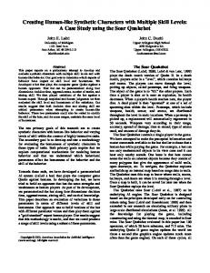
accuracy accounted for the observed Group × Condition interaction, F(4,315) = 20.84, ηp2 = .54, p < .001; however, paired t tests showed that all participants were significantly more accurate when viewing control footage than in the experimental conditions, and when viewing nondeceptive, as compared with deceptive, footage p < .005. Differences in both prediction accuracy, F(6,210) = 59.47, ηp2 = .63, p < .001 and response time, F(6,210) = 5.07, ηp2 = .13, p < .001 accounted for the Occlusion × Condition interaction: Paired t tests showed that prediction accuracy was greater for the control condition than for predictive conditions at the three earliest levels of occlusion, p < .001, but not at t +80 ms, p > .05. In addition, participants took significantly longer to respond to deceptive footage than they did to nondeceptive footage at the two later levels of occlusion, p < .001. Group × Occlusion and Group × Occlusion × Condition interactions did not reach significance, p > .05. The simple main effects of group for prediction accuracy at each level of condition and occlusion are displayed in Figure 1.
fMRI Data There were significant main effects of group, occlusion, and condition (FWE corrected p < .05). On closer scrutiny, some contrasts contributed more strongly than others to these effects; these activations, which met the stringent threshold criteria, are shown in Table 2.3 Activation in cerebellum (pyramis, culmen), inferior visual cortex,
Figure 1 — Main simple effects of group, by occlusion and condition.
superior temporal gyrus, and precuneus differentiated high-skill anticipators from their intermediate- and lowskill counterparts when seeking to predict an opponent’s movements. Further, when visual information was most restricted (i.e., at the earliest level of occlusion), there was also activation of a combination of cortical and subcortical structures—basal ganglia (lentiform nucleus in Table 2), thalamus, and cingulate/supplementary eye field. In addition, the greatest activation differences in high-skill participants occurred between the two earliest levels of occlusion—160 ms and 80 ms before the opponent’s direction change; the foci were in the superior temporal gyrus, superior and inferior parietal lobules, and superior frontal gyrus. Figure 2 shows the loci of activations in high-skill anticipators for each of three contrasts, in cerebellum (pyramis), basal ganglia (lentiform nucleus), and anterior cingulate cortex (ACC) (this figure is in color in the PDF [online] version of this article). The data from the Prediction > Control contrast did not show any significant foci at the original display threshold criterion (p < .05, FWE corrected for multiple comparisons), which may be the result of a diminished contrast-to-noise ratio for these rapidly alternating stimuli. However, at a lowered voxel-wise threshold of p < .005 (uncorrected), activation patterns were similar to those found for both novices and experts in earlier studies of badminton (Wright et al., 2010, 2011), in which prediction and control conditions were separately blocked. Areas included precuneus, premotor cortex, extrastriate
Neural Bases for Soccer Anticipation 103
Figure 2 — (This figure is in color in the PDF [online] version.) Greater cerebellar, basal ganglia, and right anterior cingulate cortex activation for experts when predicting opponents’ movements.
cortex, inferior frontal gyrus, superior frontal gyrus, and supplementary eye fields (SEF). Loci of significant activations at the new threshold, but at an extent threshold of 60 voxels, are shown for all participants combined in Table 3. Figure 3 illustrates the activations witnessed for the same contrast (prediction vs. control) for each of the three groups separately (this figure is in color in the PDF [online] version of this article).
Discussion The foremost contribution of this study was to identify potential neural bases for anticipation skill superiority in soccer. Two additional novel developments on previous fMRI-based studies of anticipation in sport (Wright et al., 2010, 2011) were (i) the introduction of video clips in which the actor was performing a deceptive maneuver and (ii) the randomized interspersing of these deceptive
stimuli with nondeceptive and control clips so as to reduce predictability—and therefore the potential for intask learning. As per our first hypothesis, the high-skill anticipators were significantly better than lesser skilled participants at predicting opponents’ actions in the deceptive condition—although this did not vary according to level of occlusion, contrary to our second prediction. The understanding of others’ actions was reflected somewhat in brain activations, in line with our third hypothesis: There was evidence of stronger activation of MNS (e.g., inferior parietal lobule, BA6) and related areas in highskill participants when compared with the intermediates, who in turn exhibited greater MNS activation than did the low-skill group, when predicting an opponent’s actions (see Figure 3; cf. Wright et al., 2010, 2011)—albeit only when deceptive and nondeceptive conditions were examined conjointly; there was also no apparent threeway interaction (i.e., differences in MNS activations were
Table 2 Loci of Activation for Experimental Contrasts, Determined at a Family-Wise Error–Corrected Display Threshold p < .05 and Extent Threshold k > 20 Region
BA
Size (Voxels)
p(FWE)
Z
x
y
z
(a) High-Skill > Intermediate, Low-Skill for Prediction (Deception + No Deception), All Occlusion Levels R Pyramis
—
525
.001
6.05
6
–79
–26
R Culmen
—
525
.001
5.37
30
–40
–35
R IOG
18
192
.001
5.44
39
–85
–2
R STG
39
192
.026
4.56
60
–61
22
R SPL
19
62
.049
4.4
33
–76
49
(b) High-Skill > Intermediate, Low-Skill for Prediction at –160 ms L Lentiform Nucleus
—
49
.009
5.79
–18
5
-8
L SFG
6
22
.018
5.58
–9
32
61
L SEF
6
29
.001
5.26
–6
–7
58
L Cingulate Gyrus
24
29
.02
4.63
–15
2
46
R Thalamus
—
25
.002
5.12
3
–10
1
.003
5.06
9
17
19
(c) High-Skill > Intermediate, Low-Skill for Deception at –160 ms R ACC
33
22
(d) –160 ms > –80 ms for Prediction, High-Skill Participants L STG
22
340
0.003
5.07
–60
–16
1
R Precuneus
7
240
0.01
4.78
18
–55
70
L IPL
7
160
0.013
4.73
–45
–64
52
L IPL
39
160
0.015
4.69
–51
–61
46
R SFG
6
95
0.022
4.6
24
2
70
Note. In Montreal Neurological Institute coordinates. ACC = anterior cingulate cortex; BA = Brodmann area; IOG = inferior occipital gyrus; IPL = inferior parietal lobule; MFG = middle frontal gyrus; SEF = supplementary eye field; SPL = superior parietal lobule; STG = superior temporal gyrus.
Figure 3 — (This figure is in color in the PDF [online] version.) Mirror neuron system activations for all participants (red = high skill; green = intermediate; blue = low skill).
104
Neural Bases for Soccer Anticipation 105
Figure 3 — (continued)
Table 3 Loci of Activation for Prediction > Control; Determined at a Trend-Level Display Threshold p < .005 and Extent Threshold k > 60 Region
BA
Size (Voxels)
Z
x
y
z
All participants L SEF
6
520
3.00
–24
–4
67
R SEF
6
376
3.16
21
–10
67
R SPL
7
210
3.92
36
–46
58
L MFG
6
148
4.06
–24
–7
61
L premotor
6
148
2.79
–39
–1
61
L premotor
6
148
2.88
–51
2
43
R IOG
18
136
3.42
24
–94
–8
L IFG
9
77
3.11
–45
8
25
L SFG
6
72
3.14
–3
17
49
Note. In Montreal Neurological Institute coordinates. BA = Brodmann area; IFG = inferior frontal gyrus; IOG = inferior occipital gyrus; MFG = middle frontal gyrus; SEF—supplementary eye field; SFG = superior frontal gyrus; SPL = superior parietal lobule; STG = superior temporal gyrus.
not magnified when participants viewed early-occluded deceptive footage). Also in keeping with our predictions, differences between the high-skilled and lower-skilled participants were most clearly manifest in both behavioral and fMRI data when early-occluded sequences were viewed (i.e.,
when the least information was available), but the most robust differences in neural activation—which included cortical and subcortical areas identified in the affordance competition model—occurred consistently between high-skill and intermediate/low-skill participants combined; there was negligible difference between the latter two groups, which is noteworthy when considering that the intermediates had still accrued considerably more competitive experience, on average, than their novice counterparts (t[23] = 3.58, p < .005). Thus, the brain activation differences witnessed may correspond to not only the surpassing of a threshold for hours accumulated in practice/competition to become sufficiently expert (see Ericsson et al., 1993), but also the quality of such practice. The strongest activation of MNS regions that correspond to those found in badminton (Wright et al., 2010, 2011) were witnessed only in high-skill participants, when –160 ms was contrasted with –80 ms (Table 2[d]). This is a somewhat unanticipated finding, because we might expect greater MNS activation when an increased amount of familiar visual information is presented, but this may simply reflect an increased level of engagement with the more challenging brief stimulus duration. Indeed, this is consistent with the notion that early occlusion actually increases participants’ attention (Wright et al., 2010). When novices’ data were considered in isolation (Figure 3), they did not exhibit significant MNS activation when viewing the prediction sequences, relative to baseline, which is consistent with their comparative lack of experiences in soccer and thus lack of familiarity with the actions performed, be they deceptive or otherwise.
106 Bishop et al.
Similar to findings in tennis (Rowe et al., 2009), but in contrast to findings from rugby (Jackson et al., 2006), the high-skill participants’ performance in the presence of deception did not move above chance level until t0— the point of direction change; however, intermediates’ performance did not do so until the opponent’s final direction of movement was visible (+80 ms), and lowskill participants never rose above chance level. Thus, while the high-skill participants were still being deceived regularly at the two earliest stages of occlusion, there was clear behavioral (Figure 1) and neuroimaging evidence (Table 2) of their superiority. Mirror neuron system activation was not clearly apparent when the deceptive condition was considered in isolation, contrary to our hypotheses. However, this may have been a function of a low signal-to-noise ratio in the data, derived from rapid alternating presentation of video stimuli; this is a novel step for such neuroimaging studies, but it is an important one if real-world conditions faced are to be approximated. Nonetheless, there was highly robust evidence (p < .05, FWE corrected) for activity in high-skill participants of a cortico-subcortical network of structures comprising cerebellum, thalamus, basal ganglia, and ACC—a network that has been implicated not only in executive function (Heyder, Suchan, & Daum, 2004; Kim, Kroger, & Kim, 2011; Lütcke, Gevensleben, Albrecht, & Frahm, 2009), but also in oculomotor control (Heyder et al., 2004; Tanaka & Kunimatsu, 2011). Moreover, the cerebellar and basal ganglia activations are consistent with the predictions of the affordance competition model (Yarrow et al., 2009). The single activation that discriminated high-skilled players from both intermediates and low-skill participants when viewing deceptive maneuvers arose in a finite region of right ACC (x = 9, y = 17, z = 19; cluster size = 22 voxels). We previously found right ACC (rACC) activation in badminton experts relative to novices, when they were required to respond to point-light representations of opposing players’ actions (Wright et al., 2011); further fMRI data using point-light displays will help us to better understand the informativeness of opponent kinematics, as opposed to other cues (e.g., opponent’s gaze), with regard to deception. The ACC has consistently been identified as an important structure in the monitoring of response conflict, specifically when a motor response is required (Turken & Swick, 1999)—and right-lateralized activation reflects the processing of visuospatial stimuli (K. E. Stephan et al., 2003). Highly comparable activation has been shown in a similarly focalized and rightlateralized region of ACC (x = 5, y = 21, z = 34) when participants either correctly rejected, or failed to reject, incorrect stimuli in a go/no-go task (Lütcke & Frahm, 2008); similar activation was found in rACC (x = 9, y = 16, z = 32) when participants were required to manage competing response alternatives in a Stroop interference task (Kim et al., 2011). Thus, the rACC activation witnessed in the deceptive condition may represent not only the suppression of the high-skill anticipators’ prepotent responses to the deceptive maneuver—to anticipate/move
in the direction of the deception—but also to monitor any incorrect decisions made; this is comparable to the role proposed for the basal ganglia in assessing the “reward value” of potential response options (Yarrow et al., 2009). Given the absence of any response accuracy differences at –160 ms, the latter rACC function is the more likely of the two, for the present data. Such inhibition is highly adaptive in situations for which the cost of not doing so may be high; for example, the tendency of handball goalkeepers to perceive opponents’ movements as deceptive may stem from a cost–benefits analysis that ultimately favors caution (Cañal-Bruland & Schmidt, 2009). It is also noteworthy that—peculiarly—all participants’ performance in the control condition was still not at 100% accuracy, irrespective of level of occlusion, which suggests that key press errors occurred. Performance for all participants in the nondeceptive condition was not only high, but also largely equivalent, except at occlusion level t0–80 ms (see Figure 1), suggesting that the actors’ movement intentions were easy to predict in the absence of deception. Hence, the ability to perceive, and then inhibit a prepotent response to, an opponent’s deception could be a key factor that discriminates perceptual-cognitively skilled soccer players from those not so skilled. High-skill anticipators’ activations at the earliest stage of occlusion comprised regions similar to those previously identified as supplementary eye fields (SEF), regions of the frontal lobes that are involved in the planning and control of saccadic eye movements (Amiez & Petrides, 2009; Grosbras, Laird, & Paus, 2005; PierrotDeseilligny, Milea, & Müri, 2004) and of a network comprising striatal (lentiform nucleus), thalamic, and cingulate areas identified as coacting in executive control processes (Heyder et al., 2004; Lütcke et al., 2009). Not only do the ventroanterior region of the thalamus and the basal ganglia appear to play important roles in the generation of volitional saccades (Tanaka & Kunimatsu, 2011), but the latter also plays a key role in biasing the correct motor response selection (Yarrow et al., 2009). The greater cerebellar activations in the high-skill anticipators are also consistent with the notion of increased oculomotor activity and motor preparation (Simmonds et al., 2007; Yarrow et al., 2009) and working memory-driven saccades (cf. Nitschke et al., 2004; T. Stephan et al., 2005). These activations collectively suggest that skilled participants’ performance incorporated better preparation of intentional saccades, through biasing oculomotor activity, which relates well to the commonly observed efficiency of expert visual search patterns (Gegenfurtner, Lehtinen, & Säljö, 2011; Mann et al., 2007). Some of the activations observed are pertinent to the shifting of attention, rather than saccadic activity, such as that observed in the lentiform nucleus (see Grosbras et al., 2005). The precuneus, an important part of the dorsal visual stream identified in the affordance competition model (Yarrow et al., 2009) that plays an integral role in orientation of attention (Cavanna & Trimble, 2006) and execution of voluntary saccades (Grosbras et al.,
Neural Bases for Soccer Anticipation 107
2005), was more active in high-skilled participants as they viewed the shortest occlusion condition footage (–160 ms), when contrasted with the next shortest (–80 ms), suggesting a change in attentional strategy when confronted with very limited visual information. There is also evidence for superior shifting of attention in highskill anticipators across all levels of occlusion, in the activation of superior parietal lobule. Almost identical activation has been found for exogenously controlled shifts of attention (Molenberghs, Mesulam, Peeters, & Vandenberghe, 2007). If this is also the case for our data, then high-skill participants’ visual search/attentional strategy was predominantly determined by features of the stimulus (e.g., the opponent’s movements), not by a preconceived plan as to which sections of the display would be most informative. Given the complex, naturalistic qualities of the stimuli used in the current study, the extent to which our data parallel those from the studies cited above, in which simple experimental stimuli were used, is very encouraging. However, there was a notable absence of coactivation of some structures, when we might reasonably have expected it, at the strict FWE threshold; this may be a function of the experimental design. Further analyses from protocols comprising longer blocks (~20 s) of deceptive stimuli may produce data that yield this coactivation; however, the imperative to reduce predictability remains (see Mann et al., 2007). Functional connectivity analyses would confirm/disconfirm the proposed operations of the affordance competition model (Yarrow et al., 2009); the present data depict many robust activations predicted by this model, but cannot tell us about interrelations between the different regions. Trial-by-trial feedback would help us to clarify the role of ACC in the recognition of conflict between outcome and reward (reward in this case would be correct prediction). To our knowledge, this is the first study to identify activity in brain regions comprising a cortico-subcortical network, over-and-above putative attentional and MNS systems, that may underpin perceptual-cognitive superiority in sport anticipation tasks. Consistent with our predictions, high-skill anticipators were more attuned to both early kinematic information and deceptive movements than were their less-skilled counterparts; neuroimaging data also showed greater activation of MNS and related structures in this group. The advantage was most profound when viewing deceptive footage, but this was irrespective of occlusion—contrary to our predictions. There was also neuroimaging evidence for changes in high-skilled participants’ allocation of attention when visual information was constrained, whether these shifts were stimulus or goal driven. Although Yarrow et al.’s (2009) affordance competition model has provided a suitable foundation for the predictions made, some activations—most notably those in basal ganglia and cerebellum—have been conspicuously lacking in previous studies (e.g., Wright et al., 2010, 2011). However, there was robust evidence for greater activation of these structures in the present data. In addition, there was evi-
dence for thalamic activation in high-skill participants when viewing early-occluded footage, and evidence of conflict monitoring (ACC) when viewing opponents’ deceptive actions. Hence, we tentatively propose that these two highly interconnected structures (see Heyder et al., 2004) may be added to the affordance competition model, which would then more comprehensively illustrate the interactions of diverse cortical and subcortical neural systems that characterize superior anticipation skill in sport.
Notes 1. This sample size was recruited according to (a) power calculations based on preliminary analysis of the response data and (b) threshold sample sizes previously established as appropriate for such fMRI designs (Desmond & Glover, 2002; Zandbelt et al., 2008). 2. The main effect of anticipation skill is not meaningful per se, because the groups were formed on this basis. However, these data are presented in Table 1 to confirm the reliability of the classification used; additionally, Figure 1 elucidates the extent to which overall performance was moderated by level of occlusion and deception (i.e., whether high-skill anticipators were superior uniformly, or only under specific conditions). 3. There were a large number of highly significant activations across all contrasts, even with stringent corrections applied to p values. Therefore, to aid interpretability and informativeness, activations were only included for group contrasts when they (a) satisfied the imposed threshold criteria (FWE) and (b) related to the performance differences.
Acknowledgments We extend our gratitude to Jasmine Field and Adrian Williams for their assistance with fMRI data preprocessing and Matlab programming, respectively.
References Abernethy, B., & Russell, D.G. (1987). Expert-novice differences in an applied selective attention task. Journal of Sport Psychology, 9, 326–345. Abernethy, B., Zawi, K., & Jackson, R.C. (2008). Expertise and attunement to kinematic constraints. Perception, 37, 931–948. PubMed doi:10.1068/p5340 Aglioti, S.M., Cesari, P., Romani, M., & Urgesi, C. (2008). Action anticipation and motor resonance in elite basketball players. Nature Neuroscience, 11, 1109–1116. PubMed doi:10.1038/nn.2182 Amiez, C., & Petrides, M. (2009). Anatomical organization of the eye fields in the human and non-human primate frontal cortex. Progress in Neurobiology, 89, 220–230. PubMed doi:10.1016/j.pneurobio.2009.07.010 Calvo-Merino, B., Grèzes, J., Glaser, D.E., Passingham, R.E., & Haggard, P. (2006). Seeing or doing? Influence of visual and motor familiarity in action observation.
108 Bishop et al.
Current Biology, 16, 1905–1910. PubMed doi:10.1016/j. cub.2006.07.065 Cañal-Bruland, R., & Schmidt, M. (2009). Response bias in judging deceptive movements. Acta Psychologica, 130, 235–240. PubMed doi:10.1016/j.actpsy.2008.12.009 Cavanna, A.E., & Trimble, M.R. (2006). The precuneus: A review of its functional anatomy and behavioural correlates. Brain, 129, 564–583. PubMed doi:10.1093/brain/awl004 Cisek, P. (2007). Cortical mechanisms of action selection: the affordance competition hypothesis. Philosophical Transactions of the Royal Society B. Biological Sciences, 362, 1585–1599. PubMed doi:10.1098/rstb.2007.2054 Desmond, J.E., & Glover, G.H. (2002). Estimating sample size in functional MRI (fMRI) neuroimaging studies: Statistical power analyses. Journal of Neuroscience Methods, 118, 115. PubMed doi:10.1016/S0165-0270(02)00121-8 Ericsson, K.A., Krampe, R.T., & Tesch-Römer, C. (1993). The role of deliberate practice in the acquisition of expert performance. Psychological Review, 100, 363–406. doi:10.1037/0033-295X.100.3.363 Gegenfurtner, A., Lehtinen, E., & Säljö, R. (2011). Expertise Differences in the Comprehension of Visualizations: a Meta-Analysis of Eye-Tracking Research in Professional Domains. Educational Psychology Review, 23, 523–552. doi:10.1007/s10648-011-9174-7 Grosbras, M.H., Laird, A.R., & Paus, T. (2005). Cortical Regions Involved in Eye Movements, Shifts of Attention, and Gaze Perception. Human Brain Mapping, 25, 140–154. PubMed doi:10.1002/hbm.20145 Heyder, K., Suchan, B., & Daum, I. (2004). Cortico-subcortical contributions to executive control. Acta Psychologica, 115, 271 10.1016/j.actpsy.2003.12.010. PubMed doi:10.1016/j. actpsy.2003.12.010 Huys, R., Cañal-Bruland, R., Hagemann, N., Beek, P.J., Smeeton, N.J., & Williams, A.M. (2009). Global Information Pickup Underpins Anticipation of Tennis Shot Direction. Journal of Motor Behavior, 41(2), 158–171. PubMed doi:10.3200/JMBR.41.2.158-171 Jackson, R.C., Warren, S., & Abernethy, B. (2006). Anticipation skill and susceptibility to deceptive movement. Acta Psychologica, 123, 355–371. PubMed doi:10.1016/j. actpsy.2006.02.002 Jones, C.M., & Miles, T.R. (1978). Use of advance cues in predicting the flight of a lawn tennis ball. Journal of Human Movement Studies, 4, 231–235. Kim, C., Kroger, J.K., & Kim, J. (2011). A functional dissociation of conflict processing within anterior cingulate cortex. Human Brain Mapping, 32, 304–312. PubMed doi:10.1002/hbm.21020 Knoblich, G., & Flach, R. (2001). Predicting the Effects of Actions: Interactions of Perception and Action. Psychological Science, 12, 467–472. PubMed doi:10.1111/14679280.00387 Kunde, W., Skirde, S., & Weigelt, M. (2011). Trust my face: Cognitive factors of head fakes in sports. Journal of Experimental Psychology. Applied, 17, 110–127. PubMed doi:10.1037/a0023756 Lütcke, H., & Frahm, J. (2008). Lateralized anterior cingulate function during error processing and conflict monitoring
as revealed by high-resolution fMRI. Cerebral Cortex, 18, 508–515. PubMed doi:10.1093/cercor/bhm090 Lütcke, H., Gevensleben, H., Albrecht, B., & Frahm, J. (2009). Brain Networks Involved in Early versus Late Response Anticipation and Their Relation to Conflict Processing. Journal of Cognitive Neuroscience, 21, 2172–2184. PubMed doi:10.1162/jocn.2008.21165 Maldjian, J.A., Laurienti, P.J., Kraft, R.A., & Burdette, J.H. (2003). An automated method for neuroanatomic and cytoarchitectonic atlas-based interrogation of fMRI data sets. NeuroImage, 19, 1233. PubMed doi:10.1016/S10538119(03)00169-1 Mann, D.T.Y., Williams, A.M., Ward, P., & Janelle, C.M. (2007). Perceptual-Cognitive Expertise in Sport: A MetaAnalysis. Journal of Sport & Exercise Psychology, 29, 457–478. PubMed Milton, J., Solodkin, A., Hluštík, P., & Small, S.L. (2007). The mind of expert motor performance is cool and focused. NeuroImage, 35, 804–813. PubMed doi:10.1016/j.neuroimage.2007.01.003 Molenberghs, P., Mesulam, M.M., Peeters, R., & Vandenberghe, R.R.C. (2007). Remapping attentional priorities: Differential contribution of superior parietal lobule and intraparietal sulcus. Cerebral Cortex, 17, 2703–2712. PubMed doi:10.1093/cercor/bhl179 Nitschke, M.F., Binkofski, F., Buccino, G., Posse, S., Erdmann, C., Kömpf, D., . . .. (2004). Activation of Cerebellar Hemispheres in Spatial Memorization of Saccadic Eye Movements: An fMRI Study. Human Brain Mapping, 22, 155–164. PubMed doi:10.1002/hbm.20025 Pierrot-Deseilligny, C., Milea, D., & Müri, R.M. (2004). Eye movement control by the cerebral cortex. Current Opinion in Neurobiology, 17, 17–25. PubMed doi:10.1097/00019052-200402000-00005 Prinz, W. (1997). Perception and action planning. The European Journal of Cognitive Psychology, 9, 129–154. doi:10.1080/713752551 Reilly, T., Williams, A.M., Nevill, A., & Franks, A. (2000). A multidisciplinary approach to talent identification in soccer. Journal of Sports Sciences, 18(9), 695–702. PubMed doi:10.1080/02640410050120078 Ripoll, H., Kerlirzin, Y., Stein, J-F., & Reine, B. (1995). Analysis of information processing, decision making, and visual strategies in complex problem solving sport situations. Human Movement Science, 14, 325–349. doi:10.1016/0167-9457(95)00019-O Rizzolatti, G., & Maddalena Fabbri, D. (2007). Understanding actions and the intentions of others: The basic neural mechanism. European Review (Chichester, England), 15, 209–222 doi:10.1017/S1062798707000221. Roca, A., Williams, A.M., & Ford, P.R. (2012). Developmental activities and the acquisition of superior anticipation and decision making in soccer players. Journal of Sports Sciences, 30, 1643–1652. doi:10.1080/02640414.2012.701761. Rowe, R., Horswill, M.S., Kronvall-Parkinson, M., Poulter, D.R., & McKenna, F.P. (2009). The Effect of Disguise on Novice and Expert Tennis Players’ Anticipation Ability. Journal of Applied Sport Psychology, 21, 178–185. doi:10.1080/10413200902785811
Neural Bases for Soccer Anticipation 109
Savelsbergh, G.J.P., Van der Kamp, J., Williams, A.M., & Ward, P. (2005). Anticipation and visual search behaviour in expert soccer goalkeepers. Ergonomics, 48, 1686–1697. PubMed doi:10.1080/00140130500101346 Simmonds, D.J., Fotedar, S.G., Suskauer, S.J., Pekar, J.J., Denckla, M.B., & Mostofsky, S.H. (2007). Functional brain correlates of response time variability in children. Neuropsychologia, 45, 2147–2157. PubMed doi:10.1016/j. neuropsychologia.2007.01.013 Stephan, K.E., Marshall, J.C., Friston, K.J., Rowe, J.B., Ritzl, A., Zilles, K., . . .. (2003). Lateralized cognitive processes and lateralized task control in the human brain. Science, 301, 384–386. PubMed doi:10.1126/science.1086025 Stephan, T., Deutschländer, A., Nolte, A., Schneider, E., Wiesmann, M., Brandt, T., . . .. (2005). Functional MRI of galvanic vestibular stimulation with alternating currents at different frequencies. NeuroImage, 26, 721–732. PubMed doi:10.1016/j.neuroimage.2005.02.049 Stocco, A., Lebiere, C., & Anderson, J.R. (2010). Conditional routing of information to the cortex: A model of the basal ganglia’s role in cognitive coordination. Psychological Review, 117, 541–574. PubMed doi:10.1037/a0019077 Tanaka, M., & Kunimatsu, J. (2011). Contribution of the central thalamus to the generation of volitional saccades. The European Journal of Neuroscience, 33, 2046–2057. PubMed doi:10.1111/j.1460-9568.2011.07699.x Turken, A.U., & Swick, D. (1999). Response selection in the human anterior cingulate cortex. Nature Neuroscience, 2, 920–924. PubMed doi:10.1038/13224 Vaeyens, R., Lenoir, M., Williams, A.M., & Philippaerts, R.M. (2007). Mechanisms Underpinning Successful Decision Making in Skilled Youth Soccer Players: An Analysis of Visual Search Behaviors. Journal of Motor Behavior, 39, 395–408. PubMed doi:10.3200/JMBR.39.5.395-408 Williams, A.M., & Ericsson, K.A. (2005). Perceptual-cognitive expertise in sport: Some considerations when applying the expert performance approach. Human Move-
ment Science, 24(3), 283–307. PubMed doi:10.1016/j. humov.2005.06.002 Williams, A.M., & Ford, P.R. (2008). Expertise and expert performance in sport. International Review of Sport and Exercise Psychology, 1(1), 4–18. doi:10.1080/17509840701836867 Williams, A.M., Ford, P.R., Eccles, D.W., & Ward, P. (2011). Perceptual-cognitive expertise in sport and its acquisition: Implications for applied cognitive psychology. Applied Cognitive Psychology, 25, 432–442. doi:10.1002/acp.1710 Wright, M.J., Bishop, D.T., Jackson, R.C., & Abernethy, B. (2010). Functional MRI reveals expert-novice differences in brain activation during sport-related anticipation. Neuroreport, 21, 94–98. PubMed doi:10.1097/ WNR.0b013e328333dff2 Wright, M.J., Bishop, D.T., Jackson, R.C., & Abernethy, B. (2011). Cortical fMRI activation to opponents’ body kinematics in sport-related anticipation: Expert-novice differences with normal and point-light video. Neuroscience Letters, 500, 216–221. PubMed doi:10.1016/j. neulet.2011.06.045 Wright, M.J., & Jackson, R.C. (2007). Brain regions concerned with perceptual skills in tennis: An fMRI study. International Journal of Psychophysiology, 63, 214–220. PubMed doi:10.1016/j.ijpsycho.2006.03.018 Yarrow, K., Brown, P., & Krakauer, J.W. (2009). Inside the brain of an elite athlete: the neural processes that support high achievement in sports. Nature Neuroscience Reviews, 10, 585–596. PubMed doi:10.1038/nrn2672 Zandbelt, B.B., Gladwin, T.E., Raemaekers, M., van Buuren, M., Neggers, S.F., Kahn, R.S., . . .. (2008). Within-subject variation in BOLD-fMRI signal changes across repeated measurements: Quantification and implications for sample size. NeuroImage, 42, 196–206. PubMed doi:10.1016/j. neuroimage.2008.04.183 Manuscript submitted: May 22, 2012 Revision accepted: November 23, 2012