horizontal, vertical, 45° rotated to the horizontal, or. 135° rotated to the horizontal. ..... crease in alpha after presentation of the memory display. Figure 2. General ...
Neural Correlates of Visual STM Dissociate between Fragile and Working Memory Representations Annelinde R. E. Vandenbroucke1,2, Ilja G. Sligte2,3, Jade G. de Vries2, Michael X. Cohen2, and Victor A. F. Lamme2
Abstract ■ Evidence is accumulating that the classic two-stage model of
visual STM ( VSTM), comprising iconic memory (IM) and visual working memory ( WM), is incomplete. A third memory stage, termed fragile VSTM (FM), seems to exist in between IM and WM [Vandenbroucke, A. R. E., Sligte, I. G., & Lamme, V. A. F. Manipulations of attention dissociate fragile visual STM from visual working memory. Neuropsychologia, 49, 1559–1568, 2011; Sligte, I. G., Scholte, H. S., & Lamme, V. A. F. Are there multiple visual STM stores? PLoS One, 3, e1699, 2008]. Although FM can be distinguished from IM using behavioral and fMRI methods, the question remains whether FM is a weak expression of WM or a separate form of memory with its own neural signature. Here, we tested whether FM and WM in humans are supported by dissociable time–frequency features of EEG recordings. Participants performed a partial-report change detection task, from which individual differences in FM and WM capacity were estimated. These individual FM and WM capacities were correlated with time–frequency characteristics of the
INTRODUCTION Traditionally, visual STM ( VSTM) has been divided into two subsystems: a short-lasting, large capacity storage termed iconic memory (IM; Neisser, 1967; Sperling, 1960) and a long-lasting but limited-capacity storage termed visual working memory ( WM; Luck & Vogel, 1997). IM was mainly thought of as a passive visual buffer (Baddeley, 2007) that lasted only for a few hundred milliseconds, from which long-lasting and robust WM representations were encoded. In the last decade, evidence for a third memory stage that lies in between IM and WM, termed fragile VSTM (FM; Sligte, Scholte, & Lamme, 2008; Makovski & Jiang, 2007; Griffin & Nobre, 2003; Landman, Spekreijse, & Lamme, 2003), has been found. However, although FM can be clearly dissociated from IM (Sligte et al., 2008), whether it is really different from WM remains a matter of debate (Makovski, 2012; Matsukura & Hollingworth, 2011).
1
University of California, Berkeley, 2University of Amsterdam, University of Birmingham
3
© Massachusetts Institute of Technology
EEG signal before and during encoding and maintenance of the memory display. FM capacity showed negative alpha correlations over peri-occipital electrodes, whereas WM capacity was positively related, suggesting increased visual processing (lower alpha) to be related to FM capacity. Furthermore, FM capacity correlated with an increase in theta power over central electrodes during preparation and processing of the memory display, whereas WM did not. In addition to a difference in visual processing characteristics, a positive relation between gamma power and FM capacity was observed during both preparation and maintenance periods of the task. On the other hand, we observed that theta–gamma coupling was negatively correlated with FM capacity, whereas it was slightly positively correlated with WM. These data show clear differences in the neural substrates of FM versus WM and suggest that FM depends more on visual processing mechanisms compared with WM. This study thus provides novel evidence for a dissociation between different stages in VSTM. ■
FM is distinct from IM as it has a smaller capacity and lasts for several seconds instead of milliseconds (Sligte, Scholte, & Lamme, 2009; Sligte et al., 2008). Moreover, in contrast to IM, FM is not erased by a light mask (Sligte et al., 2008), and neural traces associated with FM have been found in V4, showing that these representations are based on cortical processing and not on retinal afterimages (Sligte et al., 2009). At the same time, FM seems to differ from WM because the presentation of a display containing similar stimuli overwrites FM representations, whereas it does not overwrite WM (Pinto, Sligte, Shapiro, & Lamme, 2013; Sligte et al., 2008). Second, when attention is diverted during memory encoding, FM capacity reduces only slightly, whereas WM capacity suffers considerably ( Vandenbroucke, Sligte, & Lamme, 2011). In addition, when TMS is applied over the dorsolateral pFC during maintenance, WM capacity decreases, whereas FM capacity remains intact (Sligte, Wokke, Tesselaar, Scholte, & Lamme, 2011). Therefore, we have suggested that FM reflects a stage in VSTM in which visual cortical icons are maintained independent of focused attention, whereas information in WM has received selective attention, Journal of Cognitive Neuroscience X:Y, pp. 1–14 doi:10.1162/jocn_a_00870
thereby making the information more robust and available for further manipulation and report (Sligte, Vandenbroucke, Scholte, & Lamme, 2010). Although behavioral evidence is accumulating that FM and WM reflect different stages in VSTM, it could be that FM is merely a weak form of WM and depends on the same neural substrates. This would undermine the construct validity of FM. To resolve this issue, we examined the underlying EEG oscillatory characteristics of FM and WM capacities. If FM and WM depend on the same neural substrates, we would expect the same EEG components to underlie both forms of memory, perhaps with a quantitative difference. However, if FM and WM are neurally distinct, a qualitative difference should emerge from their underlying EEG characteristics. Different oscillatory substrates have been found to support visual WM. For example, a decrease in alpha power over areas that are involved in the task, together with an increase in alpha over areas that are not involved, has been linked to engagement versus disengagement of these areas (Sauseng et al., 2009; Jokisch & Jensen, 2007). In addition, a sustained increase in gamma over midcentral and visual regions is often associated with memory maintenance (Tallon-baudry, 2009; Jensen, Kaiser, & Lachaux, 2007; Jokisch & Jensen, 2007; Tallon-baudry, Kreiter, & Bertrand, 1999). Recently, it has been proposed that WM might be supported by the link between gamma and theta oscillations, in which gamma cycles would represent single items embedded in a theta wave (Lisman & Jensen, 2013; Jensen & Colgin, 2007). In the current study, our main goal was to investigate whether FM and WM capacities are supported by different neural
substrates. Therefore, we explored whether any difference in oscillatory power or in the coupling between theta and gamma oscillations emerged between FM and WM in different frequency bands. In this study, we recorded EEG while participants performed a typical partial-report change detection task that measures both FM and WM capacities in a single experiment ( Vandenbroucke et al., 2011; Sligte et al., 2008; Makovski & Jiang, 2007; Landman et al., 2003; Figure 1). To measure FM capacity, a spatial cue is presented after offset of the memory display but before onset of the test display (Figure 1A). The cue is presented 1000 msec after offset of the memory display, which ensures that IM has decayed (Sligte et al., 2008; Neisser, 1967; Sperling, 1960). Because, in this case, performance cannot be based on retinal afterimages anymore, memory performance on the cued item is indicative of all information that was cortically processed and maintained (Sligte et al., 2008, 2009). The presumption is that, when no new visual information has perturbed the fragile memory traces, all information that is potentially available on a visual level can be retrieved (Pinto et al., 2013; Makovski, Sussman, & Jiang, 2008). The use of a retro-cue before onset of the test display is thus necessary to probe FM capacity. To measure WM, the spatial cue is presented after onset of the test display. In this case, the presentation of new, and similar, visual information replaces all fragile visual memory traces. Any information that can still be retrieved after onset of the test display is therefore attributed to more deeply processed and robust representations or WM (Figure 1B; Pinto et al., 2013; Sligte et al., 2008). Percentage correct for the two trial types
Figure 1. Task design and behavioral results. Participants were instructed to remember all rectangles in the display. On FM trials (A), a spatial retro-cue was presented that indicated which item might change orientation at test (50% change). On WM trials (B), instead of a spatial cue, a neutral cue was presented. The spatial cue was presented after appearance of the test display. The presentation of the test display erases FM memory traces and leaves only WM representations intact (Pinto et al., 2013; Sligte et al., 2008). (C) FM capacity (light gray) and WM capacity (dark gray) for the four different loads. Capacity differed significantly between FM and WM trials. In addition, FM capacity increased with memory load, whereas WM capacity reached plateau at a load of four items.
2
Journal of Cognitive Neuroscience
Volume X, Number Y
is converted into FM and WM capacities and typically differs between individuals. We investigated whether FM and WM capacities—derived from percentage correct on the test display—are related to different oscillatory characteristics of the EEG signal recorded during the task. Specifically, we focused on the EEG signals recorded before onset of the cue: If indeed FM and WM reflect different neural representations, the capacity difference should be evident during formation of these representations and thus before the spatial cue is used to access them. We correlated individual FM and WM capacities with power in four different frequency bands (theta: 4– 7 Hz, alpha: 8–15 Hz, beta: 16–30 Hz, gamma: 31–70 Hz). In addition, we tested the relationship between capacity and theta–gamma coupling because this specific form of coupling has been linked to WM processing (Lisman & Jensen, 2013; Sauseng et al., 2009; Jensen & Colgin, 2007).
METHODS Participants Twenty-five students (mean age = 23 years, SD = 2 years; 11 men) from the University of Amsterdam participated in this experiment for course credit or monetary reward. All participants had normal or corrected-to-normal vision and signed an informed consent form before participation. The study was approved by the local ethics committee of the University of Amsterdam.
Stimuli Memory and test displays consisted of white rectangles (1.4° × 0.4° in visual degrees) presented on a black background, placed radially (2.6°) in eight invisible placeholders. The rectangles had four possible orientations: horizontal, vertical, 45° rotated to the horizontal, or 135° rotated to the horizontal. The neutral cue consisted of a white star (total span = 2.4°) containing eight arms pointing toward the eight possible item locations. To create the spatial cue, one of the eight white arms was replaced by a red arm (Figure 1A and B). Task and Procedure To indicate the start of a trial, the gray fixation dot turned green for 500 msec. Then, the memory display appeared for 250 msec containing two, four, six, or eight oriented rectangles placed randomly in the eight placeholders (Figure 1A and B). Participants were instructed to remember the orientation of all rectangles. On FM trials (Figure 1A), a spatial retro-cue was presented 1000 msec after offset of the memory array, indicating which item could potentially change in the test display (50% change, 90° rotation, all other items remained unchanged). After
500 msec, the retro-cue was replaced by a neutral cue. The test display was presented 1000 msec after offset of the retro-cue, and participants indicated whether they perceived an orientation change in the cued memory item (cues were always valid). WM trials started the same as FM trials (Figure 1B), but instead of presenting a spatial cue 1000 msec after offset of the memory array, a neutral cue was presented for 1500 msec. The spatial cue was then presented 100 msec after onset of the test display. The test display stayed on screen until participants made their response, with a maximum of 4000 msec. All trials were separated by a 1000-msec ITI, in which a gray fixation dot was presented. Before the start of the EEG recordings, participants received two training blocks of 64 trials (FM: 32, WM: 32; randomly intermixed). Throughout the task, trials were intermixed, and participants were not prompted to which trial type they would receive. The probability of a trial containing two, four, six, or eight rectangles was equal and randomly distributed within blocks (eight trials for each load in FM and WM). After the training trials, participants performed 384 trials for each condition (96 trials per load, total of 768 trials), separated in blocks of 64 trials.
Behavioral Analyses To determine FM and WM capacities, Cowan’s K was calculated [(hit rate − 0.5 + correct rejection − 0.5) × N)], which corrects for guessing (Cowan, 2001). To investigate the correlation between behavior and time–frequency characteristics, FM and WM capacities for each participant were taken as the maximum score on any of the four loads (Sauseng et al., 2009). This reflects individual FM and WM capacities most reliably, because when load heavily exceeds memory capacity (e.g., in the Load 8 WM condition), participants might underperform compared with their true capacity.
EEG Recordings and Preprocessing EEG was recorded at 1024 Hz using a 64-channel Biosemi ActiveTwo system (BioSemi, Amsterdam, The Netherlands) placed according to the 10–20 system. Offline data were down-sampled to 512 Hz, high-pass filtered at 0.5 Hz, and rereferenced to the average of two earlobes electrodes. Trials were epoched from −1 to 4.5 sec relative to the onset of the green preparation cue (which corresponds to −1.5 to 4 sec relative to onset of the memory array). Because of a recording error, for two participants, only 512 trials were recorded. All trials were visually inspected, and trials containing artifacts not related to eye blinks, such as activity because of muscle tension, were removed. One participant was removed because of an excessive number of artifacts, leaving too little trials to analyze. For the remaining 24 participants, an average of Vandenbroucke et al.
3
7.8% of the trials was removed (ranging from 1.6% to 22.6%, SD = 5.5%), leaving a minimum of 59 trials per load per memory condition and a minimum of 244 trials per overall memory condition. After artifact rejection, an independent component analysis was performed for each participant, and components that were clearly related to eye blinks were removed using EEGLAB (UC San Diego; Delorme & Makeig, 2004). Independent components that clearly only mapped onto one lateral electrode were removed as well. After component removal, we applied a spatial filter (surface Laplacian) that increases topographical selectivity by filtering out spatially broad and therefore likely volume-conducted effects (Srinivasan, Winter, Ding, & Nunez, 2007). The units of data after this transformation are millivolts per square centimeter (mV/cm2). Both the removal of components that mapped onto only one lateral electrode and spatially filtering the data make it less likely that any effects found in the gamma range are because of muscle tension during the task (Fitzgibbon et al., in press).
EEG Time–Frequency Decomposition: Power All data were analyzed using MATLAB (The MathWorks, Inc., Natick, MA) in combination with EEGLAB (Delorme & Makeig, 2004). We convolved the time domain signal with a complex Morlet wavelet with increasing cycles as frequency increased (3–15 cycles, logarithmically spaced in 30 steps; Cox, van Driel, de Boer, & Talamini, 2014; Cohen, van Gaal, Ridderinkhof, & Lamme, 2009; Cohen, Elger, & Ranganath, 2007). The resulting squared complex signal provided an estimate of power for each time point at 30 frequencies between 2 and 70 Hz (logarithmically spaced). Epochs were centered at the onset of the memory array, and relatively large windows were taken (−1.5 to 4 sec relative to the onset of the memory display) to prevent edge artifacts from contaminating the estimates of power. Power was normalized using a decibel (dB) transform, for which the baseline was taken as the average power over each frequency band at −1000 to −600 msec (gray fixation) for each condition. This way, data from each participant and each condition were in the same scale and thus comparable.
EEG Time–Frequency Decomposition: Phase–Amplitude Coupling To extract phase–amplitude coupling (PAC) from the EEG signal, the time domain signal was again convolved with a complex Morlet wavelet. The analysis was restricted to three phase frequencies in the theta range (three frequencies logarithmically spaced between 5 and 7 Hz) and three amplitude frequencies in the gamma range (three frequencies logarithmically spaced between 45 and 65 Hz) to minimize the number of comparisons. The 4
Journal of Cognitive Neuroscience
chosen frequencies were determined a priori and based on previous literature that showed a relationship between theta–gamma coupling and WM (Lisman & Jensen, 2013; Sauseng et al., 2009; Jensen & Colgin, 2007). For the convolution with the Morlet wavelet, six cycles were used for the phase frequencies to obtain a better frequency resolution and three cycles for the frequencies used for the amplitude component to obtain a better temporal resolution. Phase was estimated by taking the angle of the convolution results. Power was defined by the squared complex signal of the convolution result. To calculate PAC, we used debiased PAC (dPAC; van Driel, Cox, & Cohen, in press; Cox et al., 2014; Canolty et al., 2006). PAC is derived by multiplying power by exp(i × phase) for each time point (where i is the imaginary component) and then taking the average over a specific time window. Because of the possibility of a nonuniform phase angle distribution, we debiased the PAC term by subtracting the mean of exp(i × phase) for each time window from each individual time point before averaging, thereby creating dPAC (van Driel et al., in press). dPAC values were calculated over single trials and then averaged. dPAC values result in arbitrary units. To be able to compare coupling across individuals and time–frequency coupling windows, we calculated the z value derived from a random null distribution. The null distribution for dPAC values was created by shuffling the power time series for each time–frequency coupling window with respect to the phase time series 200 times. Z values for the data were then calculated by subtracting the mean of this null distribution and dividing by its standard deviation. Deflections from zero thus reflect a positive coupling compared with coupling under the assumption of no relation between oscillation and time. This normalization allowed us to compare data from each participant and each condition. Because we were not interested in task modulation per se, we did not perform an additional baseline correction on these normalized dPAC values. Electrode and Time–Frequency Window Selection Because we did not have any a priori hypotheses regarding the electrodes at which we would find correlation differences between oscillatory characteristics and FM/ WM, we reduced the number of comparisons by pooling data across electrodes and frequency bands and investigated specific time windows of interest. As the nature of the task was such that we did not expect any lateralization effects before onset of the spatial retro-cue, we first pooled together electrodes from both hemispheres, including the middle electrodes together with their adjacent laterals. This resulted in 27 distinct electrode poolings. Then, we pooled together the data across different frequencies to create frequency bands that are most common in the literature: theta (4–7 Hz), alpha (8–15 Hz), beta (16–30 Hz), and gamma (31–70 Hz). Last, we averaged power over seven different time windows: two Volume X, Number Y
preparation phases (−500:−250 and −250:0 msec), the memory display processing phase (0:250 msec), and four encoding/maintenance stages (250:500, 500:750, 750:1000, and 1000:1250 msec; see Figure 1). This left us with 27 (electrode poolings) × 4 (frequency bands) × 7 (time windows) comparisons for the power analysis and 27 (electrode poolings) × 9 (PACs) × 7 (time windows) for the PAC analysis. Correlation Analyses To investigate the oscillatory mechanisms underlying FM and WM capacities, we correlated the maximum FM/ WM capacity per participant with the average power on all trials at each frequency band and at each time window (using Spearman’s rho, R, which can deal with possible nonparametric relationships), creating a correlational time–frequency plot for each electrode pooling. Similarly, the PAC index (dPAC) was correlated with maximum FM/ WM capacity. Because our measures of FM and WM capacities were correlated (R = .44, p = .03), we computed partial correlations (analyzing WM capacity while partialing out FM capacity, and vice versa). By using partial correlations, we ensured that the variance explained between capacities and the oscillatory characteristics were either attributable to FM or WM. Because we were interested in the difference between FM and WM, any shared variance was not analyzed. To directly compare the difference between FM and WM, we transformed the partial correlations using Fisher’s Z, which allows for the comparison of nonnormally distributed data (Fisher et al., 1970). FM and WM correlations were then tested against each other using Fisher’s Z test, in which a Z value for the difference between the two correlations was calculated. p Values were false discovery rate (FDR) corrected at a false discovery proportion of 0.05. FDR corrections were carried out separately for the power and the PAC correlations. Our main analyses focused on correlations based on between-subject differences in capacity. Because we believe capacity to be a trait that is stable within a participant rather than a fluctuating state, we deemed this approach valid. However, it could be that the state a participant was in during a particular trial influenced their ability to remember the rectangles on a given trial. The nature of the change detection task makes it impossible to investigate fluctuations on a trial-by-trial level: Because it is a two-forced choice task, in 50% of the correct (and incorrect) trials, performance could have been based on chance. Therefore, a fair number (60+) of trials is necessary to evaluate performance and reliably estimate a participant’s capacity. Because we had a minimum of 244 trials per memory condition per participant, we were able to do a split-half analysis and divide the trials into low- and high-power trials. If the state of a participant would specifically influence their FM or WM performance, one would expect
that capacity as measured on the high-power trials would differ from that on the low-power trials. For each significant time–frequency window separately and within each participant, we ranked all FM and WM trials according to power (not baseline corrected), divided the data into two (one low-power and one high-power trial set), and calculated capacity over these two sets. The capacity difference between the low- and high-power trial sets was analyzed with 35 ANOVAs (2 × 2, WM/FM × High/ low power). Because these were unplanned comparisons, p values were Bonferonni corrected (alpha = 0.05/35).
RESULTS Behavioral Results Using a 2 (memory: FM, WM) × 4 (load: 2, 4, 6, 8) repeatedmeasures ANOVA, we found a main effect of memory, showing that FM capacity was larger than WM capacity (Figure 1C; F(1, 3) = 98.8, p < .001). There was an interaction effect between memory and load (F(1.8, 41.2) = 27.5, p < .001), revealing that WM capacity increased between Loads 2 and 4 (t(23) = 7.8, p < .001) but not between Loads 4 and 6 or between Loads 6 and 8 (t(23) = −0.3, p = .736; t(23) = 0.7, p = .473), whereas FM capacity increased until Load 6 (difference between Loads 4 and 6: t(23) = 7.7, p < .001) and then leveled off between Loads 6 and 8 (t(23) = 1.8, p = .093). This confirms previous work showing that FM has a larger capacity than WM and that FM performance can increase with larger memory load, whereas WM capacity stays fixed even when increasing the number of items to remember ( Vandenbroucke et al., 2014; Sligte et al., 2008). The current capacities for FM and WM are somewhat lower than previously found for the same objects ( Vandenbroucke et al., 2011; Sligte et al., 2008). In previous studies, however, participants received more extensive training on the task, which maximized both their FM and WM scores.
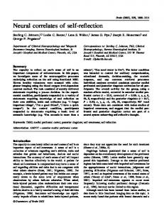
General Power Characteristics In Figure 2, general power characteristics of the task are depicted. The only statistical analysis we performed on these overall power characteristics was to confirm that there were no statistical differences (FDR corrected) between FM and WM trials before onset of the cue, as the two trial types are the same here. In Figure 2, it can be seen that there are no differences between the two trial types (FM–WM) from the onset of the green cue indicating the start of the trial (−500 msec relative to memory display) and cue onset (1250 msec relative to memory display) in parietal-occipital (Figure 2A), central-parietal (Figure 2B), or frontal-central (Figure 2C) electrodes. Indeed, for none of the electrode poolings, such a difference was found (all ps > .05, FDR corrected within this analysis). In both FM and WM trials, activity seemed Vandenbroucke et al.
5
Figure 2. General power characteristics of the task. For illustrative purposes, average power for the whole trial is depicted, but only data between the indication of the start of a trial (−500 msec) and the cue (1250 msec) were used for the correlational analyses. Reference for the timing of the trial is given below the graphs. (A) Example of average FM and WM power (top) and power difference between FM and WM trials (bottom) for PO7/PO8. (B) Example of average FM and WM power (top) and power difference between FM and WM trials (bottom) for CP1/CPz/CP2. (C) Example of average FM and WM power (top) and power difference between FM and WM trials (bottom) for FC5/ FC6. Gray dotted lines denote cutoffs for frequency bands. For illustrative purposes, average power for the whole trial is depicted, but only data between the indication of the start of a trial (−500 msec) and the cue (1250 msec) were used for the correlational analyses. Reference for the timing of the trial is given below.
to be most pronounced in the posterior electrodes (e.g., Figure 2B), with a clear theta enhancement after presentation of stimuli and cue and a decrease in alpha that persisted during the delay periods. This effect was similarly present at central electrodes (Figure 2C). After 1250 msec, a difference between FM and WM trials emerged that was manifested over posterior and central 6
Journal of Cognitive Neuroscience
electrodes: Theta enhancement was most pronounced for FM trials after (red coloring) presentation of the cue over posterior electrodes (Figure 2B), whereas theta enhancement was larger for WM trials (blue coloring) during this period over central electrodes (Figure 2C). At frontal electrodes (Figure 2D), there was a sustained decrease in alpha after presentation of the memory display Volume X, Number Y
for both FM and WM trials but no clear difference between FM and WM. Correlation between Capacity and Power To investigate whether FM and WM depend on different underlying oscillatory mechanisms, we correlated individual FM and WM capacities with time–frequency power before onset of the cue. Importantly, participants were not aware of the trial type they would receive before onset of the cue and thus could not prepare for the two conditions differently. Figure 2 shows that, in this task, there was no differential EEG signal in the two hemispheres before onset of the spatial retro-cue, thus justifying the pooling of electrodes over the left and right hemispheres. Furthermore, we confirmed that indeed no differences were present in power before onset of the retro-cue between FM and WM trials when averaged over participants. However, if different mechanisms support the formation of representations in FM and WM,
a divergence should be seen before onset of the retrocue in the correlation between individual FM/ WM capacity and time–frequency power. There were 35 time–frequency windows that showed a significant difference in correlation between FM and WM (FDR corrected at .05; Figure 3; Table 1 depicts all partial and full correlations for FM and WM). For P5/P6 and PO7/ PO8, there was a negative correlation between FM capacity and alpha power (8–15 Hz) at the initial preparation phase of the trial (−500 to −250 msec), whereas there was a slight positive correlation for WM capacity. To illustrate, the correlations for PO7/PO8 are depicted in Figure 4, top (FM: partial R = −.62, WM: partial R = .39, FM − WM difference: p < .001). For ease of interpretation, we plot the original variables next to the residualized data used in the partial correlation analyses (Figure 4B). This gives insight to the range of capacities and power between participants. A positive correlation between FM capacity and theta (4–7 Hz) was found during preparation (−250 to 0 msec at FC1/FCz/FC2) and presentation of the
Figure 3. Difference between FM and WM capacity–power correlations. Partial correlations between power and WM capacity were subtracted from partial correlations between power and FM capacity. We tested which electrode sites showed a difference between FM and WM in power dependency. Four different frequency bands (theta, alpha, beta, and gamma) were tested in seven 250-msec binned time windows (two before onset of the memory display, one during onset of the memory display, and four during the maintenance phase after offset of the memory display). White marks indicate electrode sites for which a significant difference was found between FM and WM correlations (FDR corrected at α = 0.05). Because WM correlations were subtracted from FM correlations, a positive Z value means that FM was more positively correlated with power during that time–frequency bin, whereas a negative Z value indicates that WM was more positively correlated with power. Note that only half of the topoplots are depicted because data were averaged over left and right hemispheres (see Methods). The middle electrodes were averaged together with their adjacent lateral electrodes. Data were spatially filtered before calculating power (see Methods).
Vandenbroucke et al.
7
Table 1. Partial and Full Correlations (Spearman’s Rho) for All Time–Frequency Windows That Significantly Differed between FM and WM
Electrode Pair
Frequency
Partial Correlations
Full Correlations
Time Bin
FM
FM
WM
WM
p Value Diff
P5/P6
Alpha
−500:−250 msec
−.518
.394
−.393
.154
.001
P07/P08
Alpha
−500:−250 msec
−.615
.385
−.523
.067