average firing rate of the neuronal populations responsible for the memory ... Understanding higher cognitive functions and their neural substrates is a key.
From THE DEPARTMENT OF WOMAN AND CHILD HEALTH Karolinska Institutet, Stockholm, Sweden
NEURAL MECHANISMS UNDERLYING WORKING MEMORY COMPUTATIONAL AND NEUROIMAGING STUDIES Julian Macoveanu
Stockholm 2006
All previously published papers were reproduced with permission from the publisher. Published and printed by Karolinska University Press Box 200, SE-171 77 Stockholm, Sweden © Julian Macoveanu, 2006 ISBN 91-7140-901-7
ABSTRACT The performance on various cognitive tasks, from language to selective attention and guidance of future actions depends on working memory (WM), the ability to hold and manipulate limited items of information for a period of up to a few seconds. During childhood development, WM capacity, the number of items one can maintain in WM, increases. However, the neural correlates of WM capacity, distractibility and maturational processes underlying WM development are still unclear. The present work addresses these issues by the integration of computational modeling, functional magnetic resonance imaging (fMRI) and behavioral methods. In the first study we used distracting visual stimuli in order to identify cellular mechanisms that account for the observed behavioral decrease in mnemonic accuracy as a function of the spatial distance to distractors. The computational model suggests that independently of the cellular and synaptic properties, increased neuronal firing rates accounted for higher mnemonic accuracy and resistance against distractors. In the second study we performed fMRI experiments on adults and children to monitor brain activity during a WM task. We isolated the delay-related activity and analyzed group differences and the distractor influence both behaviorally and in terms of changed brain activity. Accompanying the higher WM capacity and lower distractibility of adults, the fMRI study showed higher brain activity in middle frontal gyrus and intraparietal cortex in adults compared to children during the delay periods of WM tasks. In a subsequent study we addressed the cellular changes during WM development. The study combined a computational analysis with fMRI in order to establish putative maturational processes governing developmental changes in brain activity. We found that the increase in activity together with higher resistance against distractors could be explained by stronger connectivity between network areas. The final study addressed the limited storage capacity of previous WM models. Implementing structural and connectivity changes likely to occur during WM development in a biophysical WM model we have obtained multiple-item storage capacity similar to human WM performance. Furthermore, by using fMRI, we found that the informationactivity curve predicted by the model corresponds to that in the human posterior parietal cortex during performance of WM tasks. 1
In conclusion, in the context of neural networks dominated by reverberatory synaptic input, our studies demonstrate the correlation between a higher WM capacity, resistance to distractors, mnemonic accuracy, BOLD response and average firing rate of the neuronal populations responsible for the memory maintenance.
2
LIST OF PUBLICATIONS I. Julian Macoveanu, Torkel Klingberg and Jesper Tegnér Neuronal population firing rate predicts distance dependent distractor effects on mnemonic accuracy in a visuo-spatial working memory task II. Pernille J. Olesen, Julian Macoveanu, Jesper Tegnér and Torkel Klingberg Brain Activity Related to Working Memory and Distraction in Children and Adults
Cereb Cortex. 2006 Jun 26 III. Fredrik Edin, Julian Macoveanu, Pernille Olesen, Jesper Tegnér and Torkel Klingberg Stronger synaptic connectivity as a mechanism behind development of working memory-related brain activity during childhood IV. Julian Macoveanu, Torkel Klingberg and Jesper Tegnér A biophysical model of multiple-item working memory: a computational and neuroimaging study
Neuroscience. 2006;141(3):1611-1618
3
CONTENTS 1
INTRODUCTION
7
1.1 WORKING MEMORY – A KEY COMPONENT OF COGNITION 7 1.2 FUNCTIONAL PROPERTIES 7 1.3 ANATOMICAL CORRELATES 9 1.4 PHYSIOLOGICAL CORRELATES 10 1.5 WORKING MEMORY DEVELOPMENT 13 1.6 WORKING MEMORY AND DISTRACTION 14 1.7 EXPLORING THE NEURONAL CORRELATES USING COMPUTATIONAL MODELING 14 1.8 QUESTIONS ADDRESSED IN THE PRESENT WORK 15 1.8.1 EVALUATION OF DIFFERENT MECHANISMS BASED ON SYNAPTIC REVERBERATION – STUDY I 16 1.8.2 IMAGING DIFFERENCES BETWEEN CHILDREN AND ADULTS DURING VISUO-SPATIAL WM TASKS – STUDY II 16 1.8.3 CELLULAR MECHANISMS RESPONSIBLE FOR WM DEVELOPMENT DURING 17 CHILDHOOD – STUDY III 1.8.4 A BIOPHYSICAL MODEL OF MULTIPLE-ITEM WM – STUDY IV 18 2
METHODS
19
2.1 RESEARCH METHODS IN COGNITIVE NEUROSCIENCE 2.1.1 COMPUTATIONAL MODELS 2.1.2 FUNCTIONAL NEUROIMAGING 2.2 METHODOLOGICAL APPROACHES IN THE STUDIES 2.2.1 COMPUTATIONAL MODELS 2.2.2 THE FMRI STUDIES 2.2.3 THE BEHAVIORAL TASKS 2.2.4 COMPARISON BETWEEN SIMULATION AND EXPERIMENTAL DATA
19 19 22 24 24 27 27 28
3
30
RESULTS OF THE STUDIES
3.1 3.2
MNEMONIC ACCURACY, STUDY I. DIFFERENCES IN BRAIN ACTIVITY AND DISTRACTIBILITY BETWEEN CHILDREN AND ADULTS, STUDY II. 3.3 MATURATIONAL PROCESSES, STUDY III. 3.4 MULTIPLE-ITEMS WORKING MEMORY MODEL. STUDY IV.
30
4
33
DISCUSSION
4.1 SUMMARY 4.2 CURRENT COMPUTATIONAL CHALLENGES 4.3 FUTURE WORK 4.3.1 MODELING THE DOPAMINERGIC EFFECT ON WORKING MEMORY
4
31 31 32
33 35 36 36
4.3.2
LOCATION OF THE MEMORY STORAGE AND EXECUTIVE FUNCTIONS
36
5
ACKNOWLEDGEMENTS
38
6
REFERENCES
39
5
6
1 INTRODUCTION 1.1
WORKING MEMORY – A KEY COMPONENT OF COGNITION
Understanding higher cognitive functions and their neural substrates is a key objective of neuroscience today. Anatomical and imaging techniques have been used to map the neuronal circuits and today the major functional systems of the brain are starting to be uncovered. Integrating massive networks that link motor, perceptual and limbic regions, the prefrontal cortex is a key area for the control of information processing being the site where executive processes like planning and conducting future actions and finding solutions to novel problems take place. Another prominent function attributed to prefrontal cortex is the ability to hold information online for the guidance of goal-directed behavior as showed by lesion, brain imaging and electrophysiological studies (GoldmanRakic, 1987; Goldman-Rakic, 1995; Fuster, 1995). Thus, the prefrontal cortex is engaged in the execution of complex tasks when the essential cues for the behavioral response are not present but must be recalled from a short-term memory store. Referring to this capacity, Miller coined the term of working memory (WM) in 1960 (Miller et al., 1960). 1.2
FUNCTIONAL PROPERTIES
Psychological studies of patients with brain lesions causing specific losses in either forming new lasting memories or temporarily storing information have resulted in the distinction between short term memory (STM) and long term memory (LTM) (Atkinson and Shiffrin.R.M., 1968). It was proposed that perceptual information enters the LTM via the STM. During the 60's, the former view of short term memory as just a storage location of stimuli was challenged in a number of studies in favor of the WM concept. In 1974, Baddeley and Hitch published their classical model of the WM (Baddeley and Hitch G.J., 1974). Although other definitions also exists (Baddeley, 1992), modern models generally define WM as a limited capacity system allowing the online storage and manipulation of information central for cognitive performance, including selective attention, complex decision making, language and guiding future
7
actions (Norman, 1970; Baddeley, 1986). For instance, WM is used when remembering a phone number until it is dialed, remembering the directions to a certain location that somebody just gave you or keeping the total price of the groceries in a cart in mind, as each new item is added, so as not to exceed a predetermined amount. Baddeley and Hitch’s model originally consisted of three parts: the phonological loop, which stores and maintains auditory memory, the visuo-spatial sketchpad, which handles visual short term memories, and the central executive, which serves as a link between the two other systems. The phonological loop and the visuo-spatial sketchpad are regarded as two slave systems, whose management is performed by the central executive, which handles task management, attention and other functions, leading to a successful execution of action plans. In order to handle more versatile information, Baddeley (2000) recently introduced a fourth part in the model, the episodic buffer. Also controlled by the central executive, the episodic buffer is supposed to retrieve information from the episodic LTM. The buffer also integrates information from several modalities, thus it is the place where association of different short term memory traces occurs. The phonological loop located in the left hemisphere has been extensively investigated in behavioral studies (Baddeley, 2000). Auditory information first enters a phonological short term store of about 7 ± 2 items (Miller, 1956). Unless it is articulatorily rehearsed, the information in the phonological store decays within seconds. Information from other modalities can also enter the loop if that information is translated into an auditory code, which might occur e.g. during reading. Maybe due to the nature of the incoming stimuli, the loop is especially well suited for serial recall. Neuroimaging studies have supported the division of the phonological loop into a memory store located primarily in the supramarginal gyrus and a rehearsal system found in the frontal speech areas such as Broca's area (Paulesu et al., 1993; Smith and Jonides, 1999), where the activation of an area due to rehearsal could be analogous to that found during neuroimaging experiments. The visuo-spatial sketchpad consists of a passive memory store and an active rehearsal system, with functions largely analogous to those of the phonological loop. Compared to the phonological loop, the visuo-spatial sketchpad has an
8
improved capacity for complex stimuli but is more limited for serial information. The mechanisms of rehearsal are still unknown, but they could involve the sequential attention to components of the stimulus which are to be remembered (Baddeley, 2000). For visual tasks, neuroimaging have shown areas specifically activated during rehearsal, but since these are probably involved in selective attention as well, they cannot be entirely separated from the central executive in the same way as the auditory rehearsal areas (Paulesu et al., 1993; Smith and Jonides, 1999). Neuroimaging data has also led to a fractionation of the system into an object part, which stores information about the identity of objects, and a spatial part, which stores information about the spatial location of objects (D'Esposito et al., 1998). The spatial component, referred to as visuo-spatial WM, has been the exclusive focus of this thesis. The visuo-spatial WM capacity of healthy adults is today considered to be of approximately four items (Luck and Vogel, 1997; Cowan, 2001). 1.3
ANATOMICAL CORRELATES
Today there is substantial experimental evidence that prefrontal and posterior parietal cortical areas are closely associated with WM. In accordance with electrophysiological studies on non-human primates (Fuster and Alexander, 1971; Fuster, 1973; Goldman-Rakic, 1987; Funahashi et al., 1989; Fuster, 1997), which were first to find persistent neural activity in the prefrontal cortex, it has been suggested that prefrontal cortex is organized by the modality of the information stored, with ventral regions needed for non-spatial information and dorsal regions needed for visuo-spatial information (Wilson et al., 1993; Rao et al., 1997). These results led to the hypothesis that the ventral and dorsal streams, carrying ‘what’ and ‘where’ information respectively, extend into the lateral prefrontal cortex where information is temporarily stored. This hypothesis was questioned by a meta-analysis of human neuroimaging studies (D'Esposito et al., 1998). Instead, a hemispheric organization was suggested, with right hemispheric involvement during visuo-spatial WM tasks and left hemispheric involvement during non-spatial WM tasks. Also in contrast with the first division by modality of the prefrontal areas, another proposal supports an organization by process with ventrolateral regions mediating the actual storage
9
of information and dorsolateral regions having a role in the active manipulation of this stored information (Owen, 1997). Analogue with the electrophysiological studies on monkeys, functional neuroimaging experiments on humans have confirmed elevated activity in the prefrontal areas during WM tasks (Klingberg et al., 1997; Courtney et al., 1998; Ungerleider et al., 1998). However, some additional areas located in the posterior parietal cortex have also been found active during delay periods of WM tasks (Klingberg et al., 1997; Rowe et al., 2000) thus opening the possibility for a distributed neural system underlying the memory maintenance. Recent experimental studies have challenged the former view that posterior parietal regions only transmit visuospatial information to the prefrontal cortex (Todd and Marois, 2004; Vogel and Machizawa, 2004). These studies found that the activity of posterior parietal cortex correlates with the amount of visual information maintained ‘online’ during WM tasks and therefore proposed this cortical region as the neurophysiological correlate of visuo-spatial WM storage limitation. 1.4
PHYSIOLOGICAL CORRELATES
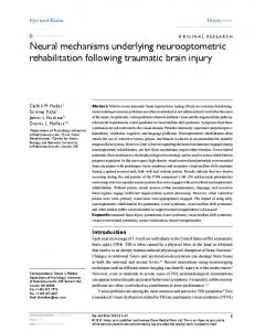
The neural basis of visuo-spatial WM was first addressed by early electrophysiological studies on monkeys. Monkeys were trained to perform WM tasks and the activity of their prefrontal and parietal cortex was recorded by direct measurement of single neuron activity using microelectrodes inserted into the cortex. WM-tasks can generally be divided into three phases: cue presentation, delay and response selection. Fuster et al. (1971) were among the first to identify persistent activity without external stimuli in prefrontal neurons during delayed response tasks. In later experiments performed by Funahashi et al. (1989) monkeys were trained to perform oculomotor delayedresponse tasks (ODR). During such tasks, the monkeys fixated a central spot on a computer screen during a brief peripheral cue presentation. The cue was followed by a delay period after which the monkeys responded by a saccadic eye movement to the cue location guided by the information stored in WM (Fig. 1A). ODR tasks offer the advantage of studying the neuronal response to multiple spatial locations in the visual field, a precise control over the onset
10
timings of the different task events, and a exact measurement of the response latency, trajectory, and amplitude of the response (Goldman-Rakic, 1995).
A
B
Figure 1. Oculomotor delayed-response (ODR) task (Chafee and Goldman-Rakic, 1998). A –. Diagrammatic representation of ODR trial events. The monkey's gazing location is indicated by ‘+’. The monkey fixates a central target (panel 1) and maintains fixation (panel 2) while a cue stimulus appears in the periphery (panel 3). Fixation of the central spot continues after cue offset for an additional 3-s delay period (panel 4). On offset of the fixation target, monkeys complete the trial by making a saccade toward the location where the cue appeared (panel 5). B – Activity recorded from a neuron in prefrontal area 8a. The neuron showed increased activity during cue and delay periods of the ODR only when the cue was presented at 225°.
11
The electrophysiological studies pioneered by Fuster and colleagues have revealed a population of neurons in monkey dorsolateral prefrontal cortex that is active during the entire delay period of WM tasks. The sustained activity of these neurons is thought to mediate the ‘on line’ storage and processing required by WM tasks. Using the ODR paradigm it was possible to show that prefrontal neurons have ‘memory fields’ characterized by elevated activity tuned to a specific location of the visual field (Funahashi et al., 1989). The neurons of a memory field are activated every time a cue is presented close to that particular visual angle (Fig. 1B). Interestingly, when the rate of firing of a cluster of neurons is increased the neurons with a spatially opposite memory field are inhibited, showing firing rates below baseline. The elevated sustained activity has been observed to last for delay periods exceeding 15 s (Fuster and Jervey, 1981), however because these neurons encode information that is ‘on line’ it is unlikely that they will remain active during longer periods. The elevated activity may decay in time due to feedback inhibition or it may be erased due to a strong inhibitory pulse that follows the enhancement of firing during the animal response phase (Funahashi et al., 1989). It has been shown that some of the prefrontal neurons are cue-selective and are activated phasically following cue presentation, some are delay-selective becoming active during the delay period, and some are response-selective becoming active in relation to the initiation of a memory-guided response. The activity of these neurons is both time locked to the events of the task and spatially tuned. However, most of the prefrontal neurons do not respond only during one phase of the delayed response tasks. Neurons can become active during two or all three phases (cue, delay and response) (Goldman-Rakic et al., 1990). On trials with erroneous saccadic responses the observed sustained activity during the delay period was found lower or totally absent. Similarly, if the sustained activity was disrupted by electrical stimulation or by distracting stimuli, the information about the cue location was lost (Funahashi et al., 1989; 1997).
12
Following these experimental findings Goldman-Rakic (1995) proposed a rather influential hypothesis for the neuronal mechanism underlying visuospatial WM. According to this model pyramidal cells with similar preferred visual angle reinforce each other through direct horizontal connections. Pyramidal cells with opposite preferred visual angles communicated via inhibitory interneurons. In this way a cluster of neurons activated by a cue inhibit the neurons with an opposite memory field. 1.5
WORKING MEMORY DEVELOPMENT
Cognitive processes greatly improve during childhood until their full capacity is reached in early adulthood. The maturation of prefrontal cortex plays a key role in understanding the increase of the higher cognitive functions. In particular, experimental studies have reported an increase in both WM capacity (Gathercole, 1999; Luna et al., 2004), and the ability to ignore distractors (Hale et al., 1997; Ridderinkhof et al., 1997). Performance of WM tasks seems to activate similar cortical areas in both children and adults. The increase in WM performance is paralleled by an increase in the activity of fronto-parietal areas (Luna et al., 2001; Klingberg et al., 2002; Kwon et al., 2002) and white matter maturation in the prefrontal lobe (Klingberg et al., 1999; Nagy et al., 2004). At a behavioral level, the significant increase in WM capacity can be attributed to e.g. increased controlled attention and improved memory encoding strategies as chunking and rehearsal. At the cellular level, the most important developmental changes are synaptic remodeling and myelination (Hubel and Wiesel, 1963; Yakovlev and Lecours, 1967; Huttenlocher, 1979; Rakic et al., 1986; Lamantia and Rakic, 1990). During the remodeling process the synapses are redistributed through processes like synaptic sprouting and synaptic pruning. The connectivity structure of the cortical networks is consequently modified. The process of myelination increases the velocity of propagation of the action potentials allowing a faster neuronal communication. However, it remains to be shown how these maturational changes at the cellular level influence intracortical interactions and brain activity.
13
1.6
WORKING MEMORY AND DISTRACTION
Miller (1996) revealed the key role played by the prefrontal cortex for an intact memory function and the ability to resist distraction. Following studies (de Fockert et al., 2001; Lavie et al., 2004) have disclosed even further the relationship between WM capacity and robustness against distractors. WM proved to play a fundamental role in the control of selective attention by maintaining the prioritization of relevant information. A high memory load was associated with a low resistance against distractors (de Fockert et al., 2001) and similarly subjects with relatively low WM capacity have difficulties in blocking out distracting information (Conway et al., 2001). Sakai (2002) proposed that memory representation is consolidated during the persistent delayed activity in prefrontal cortex and as a result gains resistance against distractors. This may be achieved by interactions between prefrontal cortex and posterior association areas. Interestingly, the distractor resistance seems to be influenced only by the prefrontal activity before the distractor presentation. The activity after distraction reflects the memory traces that survived distraction. Until present day, there is still very little knowledge about the neuronal mechanisms controlling the robustness against distractors. 1.7
EXPLORING THE NEURONAL CORRELATES USING COMPUTATIONAL MODELING
As tasks involving WM require a delay between a stimulus and a behavioral response, the recently received information must be first stored in a short-term buffer. Opposed to the long time storage of LTM, storing information in WM requires neuronal activity. As this sustained activity can be recorded and analyzed experimentally either through electrophysiological or functional imaging methods, the neural basis of this storage system is disclosed to computational modeling. This is further facilitated by an increased amount of biophysical data made available for the neurons and synapses of the prefrontal cortex. Today, there is solid evidence that persistent and stimulus selective elevated neural activity underlies the temporary storage of information in WM (Goldman-Rakic, 1995; Wang, 2001). Parallel with electrophysiological and functional imaging studies, several computational studies have undertaken the
14
challenge to explain the neuronal correlates of the observed persistent activity. Depending on the nature of the persistent activity, two major categories of models have emerged. Models that rely on intrinsic cellular mechanisms (Egorov et al., 2002; Loewenstein and Sompolinsky, 2003) and models based on synaptic reverberations in recurrent circuits (e.g. Amit, 1995; Amit and Brunel, 1997; Wang, 1999; Compte et al., 2000; Tegnér et al., 2002), the later being the most widely accepted hypothesis for the neuronal mechanisms of persistent activity. Recurrent WM models have therefore been studied extensively and they are also a major scope of the present thesis. 1.8
QUESTIONS ADDRESSED IN THE PRESENT WORK
Among the ultimate goals of cognitive neuroscience are the successful treatment of pathological conditions and improvement of cognitive functions, either through behavioral training or pharmacological therapy. Thus, understanding the neural basis of the cognitive processes is imperative. In particular, having a central role in cognitive processes, WM is today subject of extensive research. However, the underlying neuronal mechanisms are still elusive. This work incorporates computational modeling, behavioral data and fMRI in order to expose the neural correlates of visuo-spatial WM capacity, distractibility and maturational processes during development. Modeling the cortical activity underlying WM performance has been a key methodological approach in the present work. Using recurrent network models based on electrophysiological data with dynamics dominated by reverberatory excitation we have replicated WM tasks performed by recruited volunteers. Through a direct comparison between the collected behavioral and neuroimaging data with the simulation data we could both asses the validity of our WM model and make predictions about the cellular mechanisms underlying the limited WM capacity and developmental changes during development. Fig. 2 illustrates the methodological workflow.
15
Figure 2. The methodological workflow used in the present work.
1.8.1 Evaluation of different mechanisms based on synaptic reverberation – Study I Based on previous oculomotor delayed-response tasks used by GoldmanRakic and Funahasi (Funahashi et al., 1989; Goldman-Rakic, 1995) in Study I we have designed a visuo-spatial delayed-response task with distractors in order to identify the relationship between the cue–distractor distance and mnemonic accuracy. Our study combined behavioral experiments on human participants with computer simulations of a neural network model supported by reverberating synaptic input. The simulation experiment replicated the behavioral WM task. The network model was run in five different modes generating different prediction about the dynamics of the persistent activity state: weak or strong synaptic interactions, wide synaptic arborization, cellular bistability and reduced synaptic NMDA component. The predictions of the model simulations were contrasted with the behavioral results in order to reveal which mechanism endowed the best explanation of the behavioral data. 1.8.2 Imaging differences between children and adults during visuo-spatial WM tasks – Study II The frontal lobes, site for higher cognitive functions (Fuster, 2002), undergo a prolonged maturation process during development (Sowell et al., 1999). This may explain the capacity difference between adults and children for visuospatial WM, often associated with regions including superior frontal sulcus and dorsolateral prefrontal cortex. Furthermore, the increase in WM capacity during 16
development is tightly correlated with the ability to discern relevant information from irrelevant stimuli (Lavie et al., 2004). Consequently, sensitivity to distraction may be one of the reasons behind children’s lower WM capacity. In order to reveal more information about the neural basis of development, in Study II we evaluated the effect of distraction on WM in a group of 13-year-old children and adults. A delayed-response visuo-spatial WM task was performed by both groups while their brain activity was measured with functional magnetic resonance imaging (fMRI). The activity during the different phases of the WM task, cue presentation, delay, distraction and response selection was analyzed separately, allowing a direct comparison of the activity during the distraction and the activity during the delay period. 1.8.3 Cellular mechanisms responsible for WM development during childhood – Study III Several studies of visuo-spatial WM on humans have shown sustained delayactivity in both the prefrontal cortex and the posterior parietal cortex (Courtney et al., 1998; Rowe et al., 2000; Curtis et al., 2004). In order to evaluate hypotheses on how these two regions interact during WM processes and shed light on the maturation process of the WM during childhood, in Study III we integrated computational modeling and fMRI of the brain areas responsible for visuo-spatial WM. The single-region WM model from Study I was extended into a two-region network resembling prefrontal cortex and posterior parietal cortex. We implemented a reference adult network model which was altered according to different maturation processes. Synaptic remodeling was implemented by reshaping the cellular tuning curve, thus altering the specificity and selectivity for the stimuli location. Increasing the specificity of the neuronal response by sharpening the tuning curve may be a plausible mechanism for improved WM capacity analog with observations on the neural response of visual cortex stimulation (Hubel and Wiesel, 1963). A higher tuning curve will increase the activity contrast. Myelination is implemented by altering the inter-regional transmission delay. Five hypothetical cellular developmental mechanisms were studied: higher excitatory synaptic strength within a region, strengthening of connections between two regions, higher contrast of neuronal response, higher specificity
17
and increased conduction velocity. The hypotheses were assessed by comparing the induced change in the BOLD signal with the signal change from the fMRI experiments. 1.8.4 A biophysical model of multiple-item WM – Study IV The physiological correlate of multiple-item storage in WM has not yet been identified. In order to achieve multiple-item storage we extended the WMmodel used in Study I by implementing cellular mechanisms known to occur during the childhood development of WM. These mechanisms are responsible for both selective strengthening and weakening of inter-neuronal connections. The resulting structural and connectivity changes can be explained by a few hypothetical mechanisms: higher overall excitatory synaptic strength and higher contrast and specificity of the neuronal response. We made qualitative and quantitative evaluations of the generated network responses corresponding to developmental modifications. The model prediction about the relationship between the stored information and neural activity was tested against data collected during an fMRI experiment where three healthy participants performed a visuo-spatial WM task with a similar protocol as used for the simulations.
18
2 METHODS 2.1
RESEARCH METHODS IN COGNITIVE NEUROSCIENCE
With the aim of answering the questions at hand, cognitive neuroscience today focus on both experimental and theoretical investigations. The principal experimental fields are functional neuroimaging, electrophysiological studies on non-humans primates and behavioral studies of brain lesion patients. Based on the accumulated experimental data, mathematical / computational modeling has started to captured more and more attention because of it’s unique possibility to provide a detailed view of the neuronal processes. The present work addresses the neural correlates of visuo-spatial WM using both computational modeling and functional neuroimaging studies of the neuronal processes. 2.1.1 Computational models Computational models attempt to simulate observed natural processes in order to gain an understanding of these processes and to predict their behavior given a specific set of input parameters and initial conditions.
Modeling can be
successfully used in physics, chemistry or biology to simulate a particular system as long as the underlying mathematical description is known. Computational modeling or in silico experiments is a valuable compliment to in
vivo and in vitro studies offering the possibility to study the processes of a given system with high resolution and producing predictions that can be tested experimentally. Different parts of the system can also be explored separately and conclusions can be drawn on how different components interact. Computational modeling has become an important tool to study the interactions of many cellular events shaping the neuronal response. Today, the physiology of neurons is relatively well understood. The neurons output series of action potentials (electrical impulses) in response to stimulation. This is the key characteristic used when modeling their functionality. Depending on the level of abstraction neurons can be described by one or several differential equations.
19
2.1.1.1 Single cell models with different levels of abstraction Choosing a neuronal model implies some knowledge about the effects of the simplifying assumption made, i.e. how close the neuronal units need to be to their biological counterpart in order to preserve the key functionality. A first distinction between neuronal models can be made on the basis of time and its role in information processing. Neurons may convey the information either at spike frequency level i.e. the information is encoded by the firing rate of the neurons or at single action potential level, i.e. a unit of information is represented by one action potential.
Firing rate models Describing the neuronal activity by the firing rate is based on the observation that the spike frequency from one neuron is related to the magnitude of the input current, as revealed by early recordings (Adrian, 1932) . As individual spikes are of binary nature i.e. spike or no spike, the rate coding involves averaging the number of signals within a certain time frame. Thus, the output signals of the neuronal models can be seen as normalized spiking frequencies that are assumed to contain all information relevant to the postsynaptic targets. The simplest but quite influential model is the McCulloch-Pitts threshold neuron (McCulloch and Pitts, 1943). The unit sends a signal if the sum of its weighted incoming signals rises above a threshold value. This model has been effectively used in influential artificial neural networks like multi-layer perceptrons and Hopfield nets (Hopfield, 1982). A more powerful and biologically realistic neuronal model computes the output signals using a continuous activation function instead of the threshold function of the McCulloch-Pitts’s neurons. The continuous activation function, usually a sigmoid or hyperbolic tangent, makes them more suited for analog computation. As their output signals can take values between 0 and 1 the model is able to reproduce the intermediate frequencies of a biological neuron. Such neuronal units have been used in several studies of neuronal networks (Amari, 1977; Amit et al., 1994; Seung, 1996; Camperi and Wang, 1998).
20
Spiking models A problem with the firing rate models is that they need a long time-window for the averaging mechanism of the rate coding. Instead of using rate coding more realistic models use pulse coding i.e. the individual spikes are taken into consideration. Like real neurons, spiking neuronal models allow the incorporation
of
spatial-temporal
information.
The
spiking
neuron
is
characterized by a membrane potential and a threshold value. A commonly used model is the integrate-and-fire neuron (Lapicque, 1907). The neurons can be stimulated either through external current or synaptic input from the presynaptic neurons. The signals are integrated and a membrane potential is calculated. When the membrane potential reaches the threshold an action potential is generated. The form of an action potential is not described explicitly, thus it is fully characterized by its firing time. Networks of integrate-and-fire neurons have been successfully used for the modeling of self-sustaining stable states in recurrent neural networks (Amit and Brunel, 1997; Compte et al., 2000). The ‘gate’ model developed by Hodgkin and Huxley (1952) is used to describe both the voltage dependence and the time dependence of membrane conductances. The transmembrane proteins that allow the flow of ion currents across membranes are prone to variation of the electric potential. This variation can induce a conformational change of the ion channels causing the channel to move from open to closed states or vice versa. The gate model assumes a fistorder reaction between the states. The Hodgkin-Huxley model is the starting point for detailed neuron models, with realistic time course of the action potentials, numerous ion channels, different types of synapse and multi-compartment neurons which are able to account for the spatial geometry of a specific kind of neuron. Consequently, the model is able to reproduce electrophysiological measurements at a high degree of accuracy but it comes with the price of a high computational demand.
21
2.1.1.2 Network models: Modeling the visuo-spatial WM Although there are several hypotheses concerning the neuronal nature of the persistent activity, synaptic reverberation emerges as the dominant mechanism subserving visuo-spatial WM. Modeling the online maintenance of the mnemonic information is achieved by self-sustained attractor states of elevated activity in recurrent networks of neuronal units (Amari, 1977; Hopfield, 1982; Amit, 1995). In such models it proved to be a difficult task to control the rate of the attractor state. The reverberatory excitation that stabilizes the memories may become too strong and lead to a state of ‘runaway activity’. In contrast, a powerful inhibitory mechanism may suppress the state of persistent activity. Several cellular and synaptic mechanisms have been found to have a facilitating role for the generation and stability of persistent activity in recurrent network models. The most important are: strong reverberatory excitation, strong inhibitory feedback, and a dominant NMDA component of the recurrent connections (Compte, 2006). The dynamical stability of the attractor state and the synaptic properties have been examined by several studies based on different neuronal and synaptic instances of a recurrent neuronal network (Seung, 1996; Amit and Brunel, 1997; Fransen and Lansner, 1998; Compte et al., 2000; Seung et al., 2000; Tegnér et al., 2002; Koulakov et al., 2002; Goldman et al., 2003; Loewenstein and Sompolinsky, 2003; Wang et al., 2004). The most physiological models are based on recurrent networks of spiking neurons with synapses provided with gating kinetics that endow realistic postsynaptic time courses in accordance with experimentally measured cortical data. These models have led to specific physiological predictions concerning the neural dynamics, most of which are still to be observed in experimental studies. Thus, validating the proposed mathematical models using biophysical data is a central task for theoretical neuroscience today. 2.1.2 Functional neuroimaging Several techniques have been used to record the neural processes of the human brain. The methods contrast in their temporal and spatial resolution. Electrophysiological methods can provide near real-time temporal accuracy (10-100 ms) for the recorded neuronal activity by measuring either the electric
22
field change (electroencephalography or EEG) or magnetic field change (magneto-encephalography
or
MEG)
associated
with
the
neuronal
depolarization. However, because the measurement is performed outside the skull, especially in the case of EEG, the signal is distorted and ‘smoothed’ by the bone with loss of spatial resolution as result. In contrast, functional magnetic resonance imaging (fMRI) and positron emission tomography (PET) measure the neural activity indirectly through the associated increase in blood flow and energy consumption. These techniques give a relatively high spatial resolution (3 - 10 mm) but rather low temporal resolution (hundreds of milliseconds for fMRI, several seconds for PET), being limited by the slow hemodynamic changes that accompany neuronal depolarization. Measure of the cortical blood flows is also performed by optical imaging methods like near infrared spectroscopy (NIRS). However this technique is further limited by light scattering that occurs in the skull and by the inability to measure brain structures below the cortical surface. This thesis focuses on fMRI as this technique has been used in three of the included studies.
The physiological basis of BOLD fMRI The increased energy consumption that accompanies neural activity leads to an increased local blood flow (Duncan et al., 1987). The metabolic processes are however still unclear. It has been suggested that significant energy consumption
is
only
associated
with
excitatory
and
not
inhibitory
neurotransmitter release (Rees et al., 2000). However, other studies dispute this finding by observing energy consumptions during both excitatory and inhibitory synaptic activity (Glendenning et al., 1985; Mathiesen et al., 1998). The change in hemoglobin oxygenation associated with energy consumption and increased local blood flow lead to a local distortion in the magnetic field applied by the MR camera which modifies the magnetic properties of the surrounding water hydrogen nuclei. These changes are perceived by the MR camera and give rise to the blood-oxygenation level dependent (BOLD) contrast (Ogawa et al., 1990). The BOLD signal can therefore be used as an indirect measure of the synaptic activity (Jezzard et al., 2001). The properties of the BOLD signal were first studied in the visual cortex (Ernst and Hennig, 1994). Following a stimulus presentation there is an initial small
23
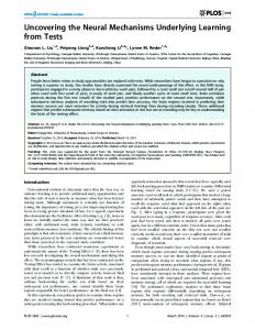
decrease in signal intensity followed by a progressive increase during a few seconds until a peak is reached. If the stimulus does not stop or lead to habituations the high signal level is maintained. When the stimulus stops the BOLD signal decreases over a few seconds until a level slightly below the initial level is reached. The signal intensity recovers then gradually up to the baseline. The time course of the BOLD signal is captured by the hemodynamic response
Signal change (%)
function (Fig. 2).
0
10
Time (s)
20
Figure 2. A standard hemodynamic response function
2.2
METHODOLOGICAL APPROACHES IN THE STUDIES
2.2.1 Computational models
2.2.1.1 Neurons The visuo-spatial WM models used in Study I, III and IV consisted of biologically realistic neuronal units following the Hodgkin-Huxley formalism for membrane conductance. The neurons had separate compartments for soma, proximate and distant dendrites integrating the most important ion channels. The neuronal input-output curves were calibrated using cortical-slice data (McCormick et al., 1985; Markram et al., 1997). We integrated two types of spiking neurons into networks: Excitatory (E) neurons of pyramidal type having both AMPA and NMDA mediated synaptic connections (Hestrin et al., 1990; Jahr and Stevens, 1990; Spruston et al., 1995) and Inhibitory (I) neurons of fast-spiking type with GABAA connectivity (Amari, 1977; Salin and Prince, 1996; Xiang et al., 2002). Details about membrane and synaptic equations and network parameters can be found in the ‘Methods’ sections of Study I,III, IV and in Tegnér et al. (2002).
24
Figure 3. A - Schematic representation of the model connectivity. For the Excitatory (E) cells the connectivity strength is a function of the difference between the preferred angles of the cells. The connectivity strength of the Inhibitory cells (I) is evenly distributed. B – Study I & IV, single-region model of the cortical circuitry represented by two populations of neurons, E and I. C – Study III, two-region model representing the prefrontal (PFC) and posterior parietal cortex (PPC).
2.2.1.2 Network The E and I populations had all-to-all connectivity both within and between the populations. The size of the E/I populations used were 128/32, 256/64 and 512/128 for Study III, IV and I respectively. The excitatory neuronal units in the 25
network were spatially distributed in a ring according to their preferred visual angle. The synaptic connection strength was highest for neurons with similar preferred visual angles and decreased with the difference of the visual angle, following a Gaussian distribution curve (Compte et al., 2000), Fig. 3A. Noise was introduced in the network by uncorrelated spike trains delivered randomly to all cells. The WM model used in Study I and IV represented a local cortical circuit (Fig. 3B). The WM model used in Study III integrated two regions representing prefrontal and posterior parietal cortical areas (Fig. 3B). The two regions, each composed of 128 E and 32 I neurons, were identical in parameter configuration and were connected only through their E cell projections. Local connections were instantaneous whereas the intra-regional connections were delayed according to a delta distribution. All external stimulating currents entered symmetrically in both regions.
2.2.1.3 Model simulations The network models maintained memories as states of persistent neural activity or memory bumps which were induced by transient excitatory currents (cues) distributed to a few adjacent neurons in the network. The stimulation protocol followed the behavioral tasks in terms of stimulation and delay time and in addition, in the case of Study IV, the number of presented cues. Distraction was achieved with a separate stimulus, identical to the cue but presented during the delay period at a different spatial location. Fig. 4 shows a typical network output.
Figure 4. Spatiotemporal firing pattern. The dots correspond to action potentials from E cells. The neurons are lined up on the Y-axis according to their preferred visual angle. The simulation time is on the X-axis.
26
2.2.2 The fMRI studies Healthy subjects were recruited for all brain imaging data acquisitions. All participants were scanned using a 1.5 Tesla Signa Excite General Electric MR scanner. Both anatomical images and functional data from the WM task performance were acquired. The participants performed the behavioral tasks while lying on the MR scanner bed and watching through a mirror a screen where the stimuli were back-projected. The behavioral tasks were composed of discrete events that could be analyzed separately. The recorded images were preprocessed and statistically analyzed using the general lineal model of fMRI time-series
(Friston
et
al.,
1995)
as
implemented
in
SPM2
(http://www.fil.ion.ucl.ac.uk/spm/). The brain activity was measured by means of the BOLD signal using contrasts between WM trials and control trials for study III and IV and additional contrasts for cue presentation, distractor and response selection for Study II. Main effect analyses were performed for each group and individual contrast images were analyzed for random effects. To determine whether any of the observed developmental differences (Study II and III) were also dependent on performance a correlation analyses between accuracy on distractor trials and event-related activity was performed. More details about scanning procedure, data acquisition, image processing and statistical analyses are available in the ‘Methods’ section of Study II, III and IV. 2.2.3 The behavioral tasks Voluntary participants were recruited for behavioral tasks that allowed us to measure their visuo-spatial WM performance and distractibility. The WM tasks of Study I and IV were specially designed in order to allow both the behavioral and computer simulation studies to share the same protocol Fig. 5A. The WM tasks used for Study II and III were adapted from a previous study (Rowe et al., 2000) Fig. 5B. Depending on the study, the participants were asked to remember the location of five (Study I) three (Study II and III) and one to six (Study IV) consecutively presented visual cues. The behavioral performance on the visuo-spatial WM tasks of Study I, II and III was evaluated by means of mnemonic accuracy, i.e. the distance between the coordinates of the cue
27
presentation and the coordinates of the response. The behavioral performance of WM task Study IV reflected the number of remembered cues out of the number of presented cues. In Study I, II and III besides the presented cues distracting stimuli were also presented, and the distraction effect was measured in terms of loss of accuracy of the remembered cues. The behavioral tasks for Study II, III and IV were performed inside the MR scanner. The programming of the tasks was performed in E-prime® (Psychology Software Tools, Inc., Pittsburgh, USA).
Figure 5. WM tasks performed inside the MR scanner. The participants fixated a central spot on a screen. Following a few seconds inter-trial interval (ITI) they were prompted by cues consisting of simultaneously presented dots. The participants remembered the location of all cues during a delay period of a few seconds and were then tested for the remembered cue locations. A – Study I and IV. During the response phase a dot appeared and the participants responded by pressing ‘yes’ and ‘no’ buttons if the dot appeared at the same location as a dot presented in the beginning of the trial. B – Study II and III. During a selection phase, a line crossing the position of one of the previously presented cues appeared. The response was given by clicking with a trackball on the intersection spot.
2.2.4 Comparison between simulation and experimental data
2.2.4.1 Simulation of the BOLD response In order to compare simulated neural activity with the BOLD signal measured during the fMRI experiments we evaluated a BOLD signal by convolving the total synaptic current as recorded during simulations with an experimentally measured canonical hemodynamic response function (Friston et al., 1998). As 28
the BOLD signal is thought to reflect the total synaptic activity in a brain area (Attwell and Iadecola, 2002), the total synaptic current was calculated by summating the absolute values of the NMDA, AMPA and GABA synaptic currents (Deco et al., 2004; Deco and Rolls, 2005).
2.2.4.2 Evaluation of the simulated behavioral performance The experimental mnemonic accuracy observed in Study I was compared to the memory drift in the network model, measured as the distance in degrees from the location of the cue presentation to the location where the memory bump is found after the delay period (Fig. 6A). The behavioral performance in Study IV was compared to the storage performance of the network model which was calculated as the number of memory bumps that were sustained during the entire simulation time (Fig. 6B)
Figure 6. Network simulations. A – Calculation of the accuracy drop-off of the memorized cue. The angle θA is the difference between the initial bump location and the post-delay location. B – Network model maintaining four memories during the entire delay period.
29
3 RESULTS OF THE STUDIES In the first study, which combined theoretical and behavioral methods, we tested a biophysical WM model’s prediction about mnemonic accuracy in the presence of distractors, against collected behavioral data. Study II addresses the neural basis of development using fMRI to quantify the differences in brain activity between children and adults. In Study III we combined modeling and fMRI data to evaluate several hypotheses about the cellular mechanisms of the developmental processes. In Study IV we used simulated developmental mechanisms to increase the storage capacity of a WM model until it matched behavioral data and then tested the information–activity relationship predicted by the model against collected fMRI data. 3.1
MNEMONIC ACCURACY, STUDY I.
Through the integration of computational modeling with behavioral experiments we have evaluated different suggested neural mechanisms for persistent neural activity. The accuracy of the memory location and its dependence on the distractor-target distance was found to be strikingly similar between the behavioral data and some of the tested network instances (Study I, Fig. 6A). In both experiments the accuracy decreased smoothly with closer distractor distances. By directly comparing the shape of the accuracy drop-off curve from simulation and behavioral results but also comparing the shape of the neuronal tuning curve with electrophysiological findings (Funahashi et al., 1989), we were able to rule out cellular mechanisms based on dominant AMPA receptor contribution to the total synaptic drive. Despite previous expectations, we could not single out one of the remaining tested network instances as one that fits best the experimental results. Interestingly, the firing rate of the attractor state that preserves the information about the cue location and not the underlying neural mechanisms seem to be essential to explain the behavioral data. High firing rates are associated with a lower sensitivity for distractors (Study I, Fig. 6C) and wider profile of the tuning curve (Study I, Fig. 5A), thus the most realistic distractor effects were found in network instances with strong NMDA
30
dominant recurrent connectivity with firing rates of the attractor neurons reaching the upper region of the physiological range (40-50Hz). 3.2
DIFFERENCES IN BRAIN ACTIVITY AND DISTRACTIBILITY BETWEEN CHILDREN AND ADULTS, STUDY II.
Using fMRI we measured the brain activity during a WM-task with distractors in a group of children and adults. The observed behavioral decrease in distractor influence in adults as well as the increase of the mnemonic accuracy could be associated with a different pattern of activation of the cortical areas responsible for WM function. Adults recruited the dorsolateral prefrontal cortex and intraparietal cortex to a greater extent than children, especially during the delay event of the WM task. The effect of distraction was however stronger in children than in adults which is reflected by a higher activity for the children in the superior frontal sulcus (Study II, Fig. 3). These results strongly point towards a key function that dorsolateral prefrontal cortex may have in explaining the improved WM capacity and distractor resistance of adults compared with children. The behavioral results show that both adults and children had significantly lower mnemonic accuracy during WM tasks with distraction. The group of children was more influenced by distractors but there were no significant difference in the reaction time of the groups. 3.3
MATURATIONAL PROCESSES, STUDY III.
The results of the fMRI experiment could clearly show a higher delay-related BOLD signal in the adult group compared with the children group in the areas responsible for WM function (Study III, Fig. 2). A similar developmental increase could be found on three of the five tested hypotheses: increased intraregional connectivity, increased inter-regional connectivity and increased tuning curve contrast. A narrower tuning curve caused a decrease in the BOLD signal and a increased conduction velocity had no effect, thus by themselves these mechanism are not able to explain the experimentally observed developmental change in brain activity in frontal and parietal lobes. If the correlation between the two areas is taken into account only the network instances with a stronger inter-regional connectivity are compatible with the experimental data.
31
Furthermore, a strong inter-regional connectivity showed a higher stability against distraction, consistent with experimental findings. Separate behavioral experiments effectuated in conjunction with Study III reveal a significant correlation between accuracy and WM capacity, which refers to the number of items a participant, could hold in mind at the same time. 3.4
MULTIPLE-ITEMS WORKING MEMORY MODEL. STUDY IV.
Modifying
the
connectivity
structure
according
to
the
implemented
developmental changes we obtained a network mode that exhibited similar performance as the average performance of the tested participants (Study IV, Fig. 4). The BOLD signal of the network mode proved to be highly correlated with the behavioral performance independent of size of the presentation set. Our computational study shows that the simulated structural and connectivity changes implemented in the WM model resulted in a significantly higher storage capacity and more robust information maintenance. Brain areas presenting a similar relationship between stored information and neural activity as predicted by the model were identified in the group of participants performing a vsWM task. This activity was localized to the posterior parietal cortex, an area that has previously been associated with the limited information stored in vsWM (Todd and Marois, 2004; Vogel and Machizawa, 2004).
32
4 DISCUSSION The studies incorporated in the present thesis add to previous theoretical as well as experimental studies exploring the neuronal correlates of visuo-spatial WM. Even if a lot of work remains to be done before the neuronal mechanisms underlying WM performance and development are completely elucidated, our studies bring a distinctive contribution to the field by the novel integrative methodological approach used. We have demonstrated how computational modeling can be integrated with both behavioral and neuroimaging experiments by a direct comparison between the theoretical and experimental data. Using the unique resolution provided by computational modeling we are now able to study many cellular and synaptic processes within the cortical network. We have successfully tested several hypothetical mechanisms and were able to rule out some of them. Based on our results we were also able to propose hypothesis that may be tested experimentally. Importantly, our results bring strong support for synaptic reverberation as the neuronal mechanism underlying visuo-spatial WM processes. Our experimental data (Study II) as well as previous data (Olesen et al., 2004) have shown a positive correlation between WM performance and measured BOLD response. Based on our theoretical data we suggest that the neurophysiological correlate of the observed increase in activity is an increased firing rate of the neurons responsible for the on line maintenance of information. This is further supported by our finding that higher neuronal activity during memory maintenance resulted in lower distractibility both behaviorally during the imaging experiments and in the computer simulations. In conclusion, to our knowledge our work is the first to demonstrate the correlation between the experimentally measured increase in delay related brain activity, elevated neuronal firing rates, mnemonic performance and resistance to distractors. 4.1
SUMMARY
The effects of visual distraction were visible in all tested groups. Even though the distractors did not significantly reduce the number of retained items the distractor influence was manifested as loss of accuracy of the remembered cue locations (Study I and II). Study I suggests that this effect is higher with 33
decreased distance between the visual distractor and the remembered cue location. Interestingly, none of the tested neuronal mechanisms were critical for this behavior per se (cellular bistability, connectivity footprint or the strength or type of the synaptic currents). Only the effective firing rate of the memory bump was found to be important for this. Elevated firing rates were associated with higher mnemonic accuracy thus less distractor influence. A neural correlate of the increased WM performance, either during childhood development (Study II) or during WM training (Olesen et al., 2004) has found to be an increased brain activity in WM specific cortical areas. Study II suggests that children may not maintain information as efficiently as adults since they do not show significant delay-related activity in the dorsolateral prefrontal cortex, a key area in the WM network. Furthermore, visual distractors affected activity in a region responsible for maintenance of information. Study III showed that similar developmental increase and correlation between fronto-parietal areas could be accounted for by mechanisms with a stronger inter-regional connectivity. The same mechanisms also showed a higher stability against distraction, consistent with our experimental findings. The mechanistic underpinning of the correlation between higher WM capacity and higher brain activity could be hypothesized to consist of both an increased firing rate of the neuronal population responsible for memory maintenance (Study I) and the recruitment of additional cortical areas. Study IV brings theoretical support to the former hypothesis showing that the capacity in WM could be increased in the model only by altering the connectivity structure without increasing the neuronal population size. The proposed changes, increased overall excitatory synaptic strength and higher contrast and specificity of the neuronal response can therefore be inferred as possible cellular modifications during childhood development but also as result of WM training. Study IV also demonstrates that a biophysical WM model is able to replicate with good accuracy the human performance of visuo-spatial WM tasks and identifies posterior parietal cortex as a likely location for the neuronal circuitry responsible for the information storage in WM.
34
4.2
CURRENT COMPUTATIONAL CHALLENGES
Although current WM network models based on recurrent connectivity are physiologically plausible, the models suffer from compatibility issues with several biological characteristics. First, mechanisms for spike-frequency adaptation, which are widely present in cortical neurons, prevent model neurons from reaching persistent activity. Second, persistent activity states can only exist if the synaptic weights of the model are fine-tuned (within a few percent accuracy), which is biologically unrealistic. Third, as neurons are recruited into persistent activity they reduce the variability of their spike trains compared to baseline firing. Experimentally, the trend is opposite. Furthermore, the baseline firing of the model cannot be as high as the biological counterpart without the appearance of spontaneous states of persistent activity. Previously, memory drift and the incapacity to maintain several states of persistent activity have also been counted to the limitations of present recurrent network models. However, in this work we have shown that memory drift is present to a similar extent even in behavioral tests (Study I) and that multiple-item storage can be achieved by the sole tuning of the inter-neuronal connectivity curves (Study IV). WM performance is not only dependent on the storage capacity of its underlying circuitry but also on the memory encoding strategies and other attention-base maintenance processes like rehearsal (Awh and Jonides, 2001). Simulating a realistic WM performance needs therefore the implementation of additional systems in the WM model, which in present form is only capable of passive retention of information without any attentional reinforcement of the maintained memories. Even if the proposed multiple-item WM model is capable of similar performance as measured in behavioral tasks, it is still unclear if the interaction with various cortical areas or the neuromodulatory effects of e.g. dopamine is necessary for the stabilization of the retained information.
35
4.3
FUTURE WORK
4.3.1 Modeling the dopaminergic effect on working memory Today there is substantial evidence that WM is modulated by the dopaminergic system (Ellis and Nathan, 2001). Brainstem dopamine afferents target the prefrontal cortex of primates (Lewis et al., 1988; Williams and Goldman-Rakic, 1993). The dopamine afferents appear to modulate the activity of prefrontal neurons by forming synapses on the same spines that are also target for glutamatergic excitatory input (Goldman-Rakic et al., 1989). Although the relative role of D1 and D2 receptors are yet to be clarified, increasing the dopamine levels in human subjects facilitates WM performance. In particular, D2 receptor agonist has been observed to improve performance in visuo-spatial delayed response tasks (Luciana 1992). It has been suggested that dopamine enhances the signal-to-noise ratio of mnemonic neural activity in prefrontal cortex (Servan-Schreiber et al., 1990; Sawaguchi et al., 1990). Elucidating the neural mechanisms of WM may imply a deeper understanding of how the dopaminergic system affects the neuronal circuits underlying WM performance. It is therefore highly relevant to study the dopaminergic modulation in a biophysical WM model either by studying the modulatory effect of dopamine on various synaptic currents or through the direct integration of dopamine receptors. 4.3.2 Location of the memory storage and executive functions Although there is consensus about the cortical areas involved in WM the actual role of these areas is subject for an ongoing debate. Functional neuroimaging studies have revealed that both prefrontal and intra-parietal cortical areas are activated during retention periods of WM tasks (Klingberg et al., 1997; Rowe et al., 2000). The cognitive processes attributed WM include both passive storage and active manipulation of the stored information. Electrophysiological studies on non-human primates have indicated the dorsolateral prefrontal cortex as the maintenance area of spatial information in WM (e.g. Fuster, 1973). The analogues functional area in the human brain is thought to be the superior frontal sulcus (Courtney et al., 1998) which is located more superiorly and
36
posteriorily and also show specific delay related activity during WM tasks (Courtney et al., 1998; Rowe et al., 2000; Klingberg et al., 2002). Based on the notion that the activation amplitude is dependent on the number of items held in WM, besides Study IV two other studies have recently succeeded to correlate intra-parietal activity with memory load thus attributing memory storage to this cortical area (Todd and Marois, 2004; Vogel and Machizawa, 2004). In both studies the activity increased with the number of remembered items until a peak at four items was reached. The prefrontal areas are thought to be the place of ‘top-down’ executive processes on these remembered items (Owen et al., 1996). More experimental work is needed to elucidate the anatomical localization of the passive and active components of WM.
37
5 ACKNOWLEDGEMENTS I would like to express my deepest gratitude to all people around me during the last four years, for their support, sharing of ideas and great time spent together in and outside the lab. In particular: My supervisors: Jesper Tegnér, first main supervisor and later co-supervisor, who introduced me to the exciting field of neuroscience, for excellent scientific discussions, for his patience, confidence and optimism. Torkel Klingberg, first co-supervisor and later main supervisor, who introduced me to neuroimaging, for his valuable guidance, sharp analytical view and constructive criticism. My co-workers: Fredrik Edin, Fabien Schneider, Pernille Olesen, Fiona McNab, Sissela Bergman and Gaëlle Leroux for great time together and valuable ideas and discussions. My colleagues and friends from Astrid Lindgren and MR centrum: Aurellien, Påvel, Carolina, Karin, Ulrika, Aurelija, Anders and None-Marie. In particular, Matteo and Christian for many ‘tough’ sessions at the KS gym, they were never easy on me. All my old friends who have been there all the time: David, Måns, Millan, Andreas, Anki, Krister, Jenny and Pontus. Maria, for her patience, encouragement, for believing in me and for her help during the final preparation of my doctoral degree. And most importantly, my family, for their precious advices, encouragement and support year after year through my whole education.
38
6 REFERENCES Adrian EDA (1932) The mechanism of nervous action: electrical studies of the neurone. Philadelphia: Univ. of Pennsylvania Press. Amari S (1977) Dynamics of pattern formation in lateral-inhibition type neural fields. Biol Cybern 27:77-87. Amit DJ (1995) The Hebbian paradigm reintegrated: local reverberations as internal representations. Behav Brain Sci 18:617-26. Amit DJ, Brunel N (1997) Model of global spontaneous activity and local structured activity during delay periods in the cerebral cortex. Cereb Cortex 7:237-252. Amit DJ, Brunel N, Tsodyks MV (1994) Correlations of cortical Hebbian reverberations: theory versus experiment. J Neurosci 14:6435-6445. Atkinson RL, Shiffrin.R.M. (1968) Human memory: A proposed system and its control processes. In K. W. Spence & J. T. Spence (Eds.), The psychology of learning and motivation: Advances inresearch and theory, Volume 2. New York: Academic Press. Attwell D, Iadecola C (2002) The neural basis of functional brain imaging signals. Trends Neurosci 25:621-625. Awh E, Jonides J (2001) Overlapping mechanisms of attention and spatial working memory. Trends Cogn Sci 5:119-126. Baddeley A (2000) The episodic buffer: a new component of working memory? Trends Cogn Sci 4:417-423. Baddeley A (1992) Working memory. Science 255:556-559. Baddeley A (1986) Working Memory. New York: Oxford University Press. Baddeley AD, Hitch G.J. (1974) Working memory. In: G. Bower, Editor. Recent advances in learning and motivation 8:47-90. Camperi M, Wang XJ (1998) A model of visuospatial working memory in prefrontal cortex: recurrent network and cellular bistability. J Comput Neurosci 5:383-405. Chafee MV, Goldman-Rakic PS (1998) Matching patterns of activity in primate prefrontal area 8a and parietal area 7ip neurons during a spatial working memory task. J Neurophysiol 79:2919-2940. Compte A (2006) Computational and in vitro studies of persistent activity: edging towards cellular and synaptic mechanisms of working memory. Neuroscience 139:135-151. Compte A, Brunel N, Goldman-Rakic PS, Wang XJ (2000) Synaptic mechanisms and network dynamics underlying spatial working memory in a cortical network model. Cereb Cortex 10:910-923. Conway AR, Cowan N, Bunting MF (2001) The cocktail party phenomenon revisited: the importance of working memory capacity. Psychon Bull Rev 8:331-335.
39
Courtney SM, Petit L, Maisog JM, Ungerleider LG, Haxby JV (1998) An area specialized for spatial working memory in human frontal cortex. Science 279:13471351. Cowan N (2001) The magical number 4 in short-term memory: a reconsideration of mental storage capacity. Behav Brain Sci 24:87-114. Curtis CE, Rao VY, D'Esposito M (2004) Maintenance of spatial and motor codes during oculomotor delayed response tasks. J Neurosci 24:3944-3952. D'Esposito M, Aguirre GK, Zarahn E, Ballard D, Shin RK, Lease J (1998) Functional MRI studies of spatial and nonspatial working memory. Brain Res Cogn Brain Res 7:1-13. de Fockert JW, Rees G, Frith CD, Lavie N (2001) The role of working memory in visual selective attention. Science 291:1803-1806. Deco G, Rolls ET (2005) Attention, short-term memory, and action selection: A unifying theory. Prog Neurobiol 76:236-256. Deco G, Rolls ET, Horwitz B (2004) "What" and "where" in visual working memory: a computational neurodynamical perspective for integrating FMRI and single-neuron data. J Cogn Neurosci 16:683-701. Duncan GE, Stumpf WE, Pilgrim C (1987) Cerebral metabolic mapping at the cellular level with dry-mount autoradiography of [3H]2-deoxyglucose. Brain Res 401:43-49. Egorov AV, Hamam BN, Fransen E, Hasselmo ME, Alonso AA (2002) Graded persistent activity in entorhinal cortex neurons. Nature 420:173-178. Ellis KA, Nathan PJ (2001) The pharmacology of human working memory. Int J Neuropsychopharmacol 4:299-313. Ernst T, Hennig J (1994) Observation of a fast response in functional MR. Magn Reson Med 32:146-149. Fransen E, Lansner A (1998) A model of cortical associative memory based on a horizontal network of connected columns. Network 9:235-264. Friston KJ, Fletcher P, Josephs O, Holmes A, Rugg MD, Turner R (1998) Event-related fMRI: characterizing differential responses. Neuroimage 7:30-40. Friston KJ, Holmes AP, Poline JB, Grasby PJ, Williams SC, Frackowiak RS, Turner R (1995) Analysis of fMRI time-series revisited. Neuroimage 2:45-53. Funahashi S, Bruce CJ, Goldman-Rakic PS (1989) Mnemonic coding of visual space in the monkey's dorsolateral prefrontal cortex. J Neurophysiol 61:331-349. Fuster JM (1973) Unit activity in prefrontal cortex during delayed-response performance: neuronal correlates of transient memory. J Neurophysiol 36:61-78. Fuster JM (2002) Frontal lobe and cognitive development. J Neurocytol 31:373-385. Fuster JM (1997) The Prefrontal Cortex: Anatomy, Physiology, and Neuropsychology of the Frontal lobe. New York: Lippincott-Raven. Fuster JM (1995) Memory in the cerebral cortex. Cambrige, Mass.: MIT press. Fuster JM, Alexander GE (1971) Neuron activity related to short-term memory. Science 173:652-654.
40
Fuster JM, Jervey JP (1981) Inferotemporal neurons distinguish and retain behaviorally relevant features of visual stimuli. Science 212:952-955. Gathercole SE (1999) Cognitive approaches to the development of short-term memory. Trends Cogn Sci 3:410-419. Glendenning KK, Hutson KA, Nudo RJ, Masterton RB (1985) Acoustic chiasm II: Anatomical basis of binaurality in lateral superior olive of cat. J Comp Neurol 232:261-285. Goldman MS, Levine JH, Major G, Tank DW, Seung HS (2003) Robust persistent neural activity in a model integrator with multiple hysteretic dendrites per neuron. Cereb Cortex 13:1185-1195. Goldman-Rakic PS (1995) Cellular basis of working memory. Neuron 14:477-485. Goldman-Rakic PS (1987) Circuitry of primate prefrontal cortex and regulation of behavior by representational memory. In: Handbook of physiology pp 373-417. Bethesda, MD: American Physiology Society. Goldman-Rakic PS, Funahashi S, Bruce CJ (1990) Neocortical memory circuits. Cold Spring Harb Symp Quant Biol 55:1025-1038. Goldman-Rakic PS, Leranth C, Williams SM, Mons N, Geffard M (1989) Dopamine synaptic complex with pyramidal neurons in primate cerebral cortex. Proc Natl Acad Sci U S A 86:9015-9019. Hale S, Bronik MD, Fry AF (1997) Verbal and spatial working memory in school-age children: developmental differences in susceptibility to interference. Dev Psychol 33:364-371. Hestrin S, Sah P, Nicoll RA (1990) Mechanisms generating the time course of dual component excitatory synaptic currents recorded in hippocampal slices. Neuron 5:247-253. Hodgkin A, Huxley A (1952) Currents carried by sodium and potassium ions through the membrane of the giant axon of Loligo. J Physiol 116:449-472. Hopfield JJ (1982) Neural networks and physical systems with emergent collective computational abilities. Proc Natl Acad Sci U S A 79:2554-2558. Hubel DH, Wiesel TN (1963) Receptive fields of cells in striate cortex of very young visually inexperienced kittens. J Neurophysiol 26:994-1002. Huttenlocher PR (1979) Synaptic density in human frontal cortex - developmental changes and effects of aging. Brain Res 163:195-205. Jahr CE, Stevens CF (1990) Voltage dependence of NMDA-activated macroscopic conductances predicted by single-channel kinetics. J Neurosci 10:3178-3182. Jezzard P, Matthews PJ, Smith SM (2001) Functional Magnetic Resonance Imaging: An Introduction to Methods. Oxford University Press, USA. Klingberg T, Forssberg H, Westerberg H (2002) Increased brain activity in frontal and parietal cortex underlies the development of visuospatial working memory capacity during childhood. J Cogn Neurosci 14:1-10. Klingberg T, O'Sullivan BT, Roland PE (1997) Bilateral activation of fronto-parietal networks by incrementing demand in a working memory task. Cereb Cortex 7:465471.
41
Klingberg T, Vaidya CJ, Gabrieli JD, Moseley ME, Hedehus M (1999) Myelination and organization of the frontal white matter in children: a diffusion tensor MRI study. Neuroreport 10:2817-2821. Koulakov AA, Raghavachari S, Kepecs A, Lisman JE (2002) Model for a robust neural integrator. Nat Neurosci 5:775-782. Kwon H, Reiss AL, Menon V (2002) Neural basis of protracted developmental changes in visuo-spatial working memory. Proc Natl Acad Sci U S A 99:13336-13341. Lamantia AS, Rakic P (1990) Cytological and quantitative characteristics of four cerebral commissures in the rhesus monkey. J Comp Neurol 291:520-537. Lapicque L (1907) Recherches quantitatives sur l'excitation electrique des nerfs traitée comme une polarisation. J Physiol Pathol Gen 9:620-635. Lavie N, Hirst A, de Fockert JW, Viding E (2004) Load theory of selective attention and cognitive control. J Exp Psychol Gen 133:339-354. Lewis DA, Foote SL, Goldstein M, Morrison JH (1988) The dopaminergic innervation of monkey prefrontal cortex: a tyrosine hydroxylase immunohistochemical study. Brain Res 449:225-243. Loewenstein Y, Sompolinsky H (2003) Temporal integration by calcium dynamics in a model neuron. Nat Neurosci 6:961-967. Luck SJ, Vogel EK (1997) The capacity of visual working memory for features and conjunctions. Nature 390:279-281. Luna B, Garver KE, Urban TA, Lazar NA, Sweeney JA (2004) Maturation of cognitive processes from late childhood to adulthood. Child Dev 75:1357-1372. Luna B, Thulborn KR, Munoz DP, Merriam EP, Garver KE, Minshew NJ, Keshavan MS, Genovese CR, Eddy WF, Sweeney JA (2001) Maturation of widely distributed brain function subserves cognitive development. Neuroimage 13:786-793. Markram H, Lubke J, Frotscher M, Roth A, Sakmann B (1997) Physiology and anatomy of synaptic connections between thick tufted pyramidal neurones in the developing rat neocortex. J Physiol 500 ( Pt 2):409-440. Mathiesen C, Caesar K, Akgoren N, Lauritzen M (1998) Modification of activitydependent increases of cerebral blood flow by excitatory synaptic activity and spikes in rat cerebellar cortex. J Physiol 512 ( Pt 2):555-566. McCormick DA, Connors BW, Lighthall JW, Prince DA (1985) Comparative electrophysiology of pyramidal and sparsely spiny stellate neurons of the neocortex. J Neurophysiol 54:782-806. McCulloch WS, Pitts W (1943) A logical calculus of the ideas immanent in nervous activity. Bull Math Biol 5:115-133. Miller EK, Erickson CA, Desimone R (1996) Neural mechanisms of visual working memory in prefrontal cortex of the macaque. J Neurosci 16:5154-5167. Miller GA (1956) The magical number seven plus or minus two: some limits on our capacity for processing information. Psychol Rev 63:81-97. Miller GA, Galanter E, Pribram KH (1960) Plans and the structure of behavior. New York: Holt, Rinehart & Winston.
42
Nagy Z, Westerberg H, Klingberg T (2004) Maturation of white matter is associated with the development of cognitive functions during childhood. J Cogn Neurosci 16:1227-1233. Norman DA (1970) Models of human memory. New York: Academic Press. Ogawa S, Lee TM, Kay AR, Tank DW (1990) Brain magnetic resonance imaging with contrast dependent on blood oxygenation. Proc Natl Acad Sci U S A 87:9868-9872. Olesen PJ, Westerberg H, Klingberg T (2004) Increased prefrontal and parietal activity after training of working memory. Nat Neurosci 7:75-79. Owen AM (1997) The functional organization of working memory processes within human lateral frontal cortex: the contribution of functional neuroimaging. Eur J Neurosci 9:1329-1339. Owen AM, Evans AC, Petrides M (1996) Evidence for a two-stage model of spatial working memory processing within the lateral frontal cortex: a positron emission tomography study. Cereb Cortex 6:31-38. Paulesu E, Frith CD, Frackowiak RS (1993) The neural correlates of the verbal component of working memory. Nature 362:342-345. Rakic P, Bourgeois JP, Eckenhoff MF, Zecevic N, Goldman-Rakic PS (1986) Concurrent overproduction of synapses in diverse regions of the primate cerebral cortex. Science 232:232-235. Rao SC, Rainer G, Miller EK (1997) Integration of what and where in the primate prefrontal cortex. Science 276:821-824. Rees G, Friston K, Koch C (2000) A direct quantitative relationship between the functional properties of human and macaque V5. Nat Neurosci 3:716-723. Ridderinkhof KR, van der Molen MW, Band GP, Bashore TR (1997) Sources of interference from irrelevant information: a developmental study. J Exp Child Psychol 65:315-341. Rowe JB, Toni I, Josephs O, Frackowiak RS, Passingham RE (2000) The prefrontal cortex: response selection or maintenance within working memory? Science 288:1656-1660. Sakai K, Rowe JB, Passingham RE (2002) Active maintenance in prefrontal area 46 creates distractor-resistant memory. Nat Neurosci 5:479-484. Salin PA, Prince DA (1996) Electrophysiological mapping of GABAA receptormediated inhibition in adult rat somatosensory cortex. J Neurophysiol 75:15891600. Sawaguchi T, Matsumura M, Kubota K (1990) Catecholaminergic effects on neuronal activity related to a delayed response task in monkey prefrontal cortex. J Neurophysiol 63:1385-1400. Servan-Schreiber D, Printz H, Cohen JD (1990) A network model of catecholamine effects: gain, signal-to-noise ratio, and behavior. Science 249:892-895. Seung HS (1996) How the brain keeps the eyes still. Proc Natl Acad Sci U S A 93:13339-13344. Seung HS, Lee DD, Reis BY, Tank DW (2000) Stability of the memory of eye position in a recurrent network of conductance-based model neurons. Neuron 26:259-271.
43
Smith EE, Jonides J (1999) Storage and executive processes in the frontal lobes. Science 283:1657-1661. Sowell ER, Thompson PM, Holmes CJ, Jernigan TL, Toga AW (1999) In vivo evidence for post-adolescent brain maturation in frontal and striatal regions. Nat Neurosci 2:859-861. Spruston N, Jonas P, Sakmann B (1995) Dendritic glutamate receptor channels in rat hippocampal CA3 and CA1 pyramidal neurons. J Physiol 482 ( Pt 2):325-352. Tegnér J, Compte A, Wang XJ (2002) The dynamical stability of reverberatory neural circuits. Biol Cybern 87:471-481. Todd JJ, Marois R (2004) Capacity limit of visual short-term memory in human posterior parietal cortex. Nature 428:751-754. Ungerleider LG, Courtney SM, Haxby JV (1998) A neural system for human visual working memory. Proc Natl Acad Sci U S A 95:883-890. Vogel EK, Machizawa MG (2004) Neural activity predicts individual differences in visual working memory capacity. Nature 428:748-751. Wang XJ (1999) Synaptic basis of cortical persistent activity: the importance of NMDA receptors to working memory. J Neurosci 19:9587-9603. Wang XJ (2001) Synaptic reverberation underlying mnemonic persistent activity. Trends Neurosci 24:455-463. Wang XJ, Tegnér J, Constantinidis C, Goldman-Rakic PS (2004) Division of labor among distinct subtypes of inhibitory neurons in a cortical microcircuit of working memory. Proc Natl Acad Sci U S A 101:1368-1373. Williams SM, Goldman-Rakic PS (1993) Characterization of the dopaminergic innervation of the primate frontal cortex using a dopamine-specific antibody. Cereb Cortex 3:199-222. Wilson FA, Scalaidhe SP, Goldman-Rakic PS (1993) Dissociation of object and spatial processing domains in primate prefrontal cortex. Science 260:1955-1958. Xiang Z, Huguenard JR, Prince DA (2002) Synaptic inhibition of pyramidal cells evoked by different interneuronal subtypes in layer v of rat visual cortex. J Neurophysiol 88:740-750. Yakovlev PI, Lecours AR (1967) The myelogenetic cycles of regional maturation of the brain. In A. Minkowsky (Eds). Regional Development of the Brain in Early Life. Blackwell Scientific Publications, Oxford.
44