PRESENCE 2005
Neural processing of Spatial Information: What we know about Place cells and what they can tell us about Presence 1
Jorge Brotons 1,2, Shane O’Mara 2 and Maria V. Sanchez-Vives 1 Instituto de Neurociencias de Alicante, Universidad Miguel Hernández-CSIC, San Juan de Alicante, Spain. 2 Institute of Neuroscience, Trinity College, Dublin, Ireland.
[email protected];
[email protected];
[email protected]
Abstract Brain processing of spatial information is a very prolific area of research in neuroscience. Since the discovery of place cells (PCs)[1] researchers have tried to explain how these neurons integrate and process spatial and non-spatial information. Place cells (PCs) are pyramidal neurons located in the hippocampus and parahippocampal region which fire with higher frequency when the animal is in a discrete area of space. Recently, PCs have been found in the human brain. The processing of spatial information and the creation of cognitive maps of the space is the result of the integration of multisensory external and internal information and the brain’s own activity. In this article we review some of the most relevant properties of PCs and how this knowledge can be extended to the understanding of human processing of spatial information and to the generation of spatial presence. Keywords--- Hippocampus, Place cells, Subiculum, Spatial Processing, Spatial Presence.
1. Introduction. Spatial navigation is a fundamental form of interaction with the environment. Animals and humans must move about in their environments in search for food, shelter or mate, actions which are basic for the survival of the individual and the species. The brain in different species has evolved in an effort to make individuals capable of navigating their environments in an efficient manner. The understanding of the brain mechanisms underlying the generation of internal maps of the external world, the storage (or memory) of these maps, and the use of them in the form of navigation strategies is the field of study of a large number of researchers in the neuroscience community. On the other hand, the study of navigation in real and virtual environments (VE) has been a broad field of study, including a diverse range of topics from model city design to the generation of VEs that successfully result in spatial presence and that are optimal for the transfer of spatial information between virtual and real worlds. In this article we review data (including our own) on the neural basis of spatial navigation, mostly centered on hippocampal and parahippocampal neurons called “place cells” that are specialized in responding to spatial position.
The functional properties of these neurons embody many aspects of human navigation that are well known from a behavioral point of view. It is our purpose to demonstrate that the understanding of the neuronal basis of spatial processing is relevant to the understanding and successful generation of spatial presence. Furthermore, we will suggest that the similar activation of brain structures during navigation in virtual compared to real worlds can be in itself an objective measurement of presence. In other words if place cells activation occurs in the same way in a virtual environment (VE) as it does in a physical environment then this is one level of evidence, a very important one, that presence is occurring within that VE. In Section 2 we review general mechanisms and strategies of navigation and the underlying brain structures that control them. We go on to center our attention on the best known structure that codes for spatial information (Section 3), the hippocampus and parahippocampal region. Its anatomical structure is briefly described, as well as the properties of one of the most prominent electrophysiological signatures of this region, the “theta rhythm”. This rhythm is important because it synchronises activity within the hippocampal formation and it affects the firing of place cells. For this reason it has been repeatedly implicated in integrative functions related to the navigation tasks –i.e. sensory-motor integration-, and therefore it is worth mentioning. Once the general framework for investigating place cells has been described, we go on to explain their specific functional properties, with an emphasis on the factors that determine their spatial firing fields (location, visual or other sensory cues, behavioral relevance of the area, etc) and the involvement of other areas of the brain in other relevant aspects of navigation, such as place significance or reward. These functional properties that are studied at the cellular level are supposed to support many of the well known features of navigation and their understanding results in the knowledge of the elements that could induce spatial presence. Based on that knowledge, in Section 4 we review relevant aspects of place cells and we suggest how this information could be useful to the understanding on how the brain processes spatial information in VR. To expand on how this could be relevant to presence research, we suggest some empirical experiments and predictions based on observations made in place cells.
59
PRESENCE 2005 2. Spatial navigation in animals and humans.
2.1. Allocentric and egocentric navigation
Species varying from migratory birds to humans need to utilize different information to generate knowledge of environments to navigate successfully. O’Keefe and Dostrovsky [1] suggested that the hippocampus was the central brain structure implicated in spatial navigation and the neuronal substrate in which a “cognitive map” of the external environment is created. A “cognitive map” is an internal representation of an environment that allows subjects to choose the best way to get to an objective by making calculations based on the relations between different environmental landmarks. Other strategies could be used by humans and animals in an effort to navigate such as egocentric navigation (see below), and these route or ‘taxon’-based strategies depend on non-hippocampal brain systems. Birds with hippocampal lesions can navigate during migration using a compass strategy, following a fixed direction, but they get lost in their local area because they are not capable of generating a cognitive map of the area [2]. Classic studies of migratory birds shed light on the strategies of these expert navigators to make use of different types of available information to orient themselves throughout long distances in their migratory flights or in their short trips in search for food. [3] showed that if naïve migratory birds in their first flight were captured and transported in a perpendicular direction to that which they were directed they would miss the final destiny by the amount of kilometers they were transported. These birds were flying towards a fixed goal using a compass strategy [4]. On the other hand if the same procedure was implemented in experienced birds, these would correct the distance they were transported, reaching successfully the final goal. Experienced birds use a more elaborate approach to navigation involving knowledge of the environment. Therefore, cognitive mapping would depend on experience and learning, ruling out the possibility of instinctive knowledge of migratory routes. While using a compass strategy birds can use three different sources of information, the sun, geomagnetism and the stars [5]. Experiments which have manipulated the internal (circadian) clock of birds have demonstrated that they use the sun to orientate themselves with respect to their internal clock [6]. Animals use also geomagnetism to orientate themselves and by applying magnets in the head of the animals, they can be redirected towards a specific direction if the skies are overcast. More recently, some studies have demonstrated that pigeons, while flying to their nests, can also use highways and their exits as cues using compass adjustment during the middle part of the fly and a cognitive map when approaching the loft area [7]. No evidence has been found in the human brain of magnetic sensors contributing to spatial orientation. However, there are recent advances in the understanding of the cellular networks underlying human navigation by means of single neuron recordings in implanted patients [8] and fMRI studies [9] in virtual environments.
Two basic navigational strategies allow animals and humans to navigate successfully:
60
1) Allocentric navigation enables humans and animals to generate a internal representational system based on the global coordinates of the environment. Thus, a topographical representation of the environment is generated by using multiple relevant landmarks of their surroundings. These external cues are used to establish a complex representation which would include the distance between them and to the subject´s own relative position. This facilitates a precise navigation to specific goals even if those are not visible. For example it has been proved that rats are able of swimming to a hidden platform which allows them to escape from a pool. To achieve this kind of successful navigation rats had to use multiple environmental cues available in the experimental room which allow them to generate a map-like representation of the environment [10]. Subsequently, studies were performed on experimental groups that were trained to navigate in the watermaze searching for the hidden platform using external cues, while having different degrees of damage in the parahippocampal region [11]. Those studies revealed that rats had a strong degree of impairment to find the hidden platform but not in the visible platform version of the task. The results of these two experiments would suggest that these different structures of the parahipocampal complex and the hippocampus are necessary for the allocentric navigation strategy [12] supporting the original suggestion [1]. Similar evidence has been found in humans, whose hippocampus and parahipocampal region appeared activated in fMRI studies in which subjects navigated in a virtual environment [13, 14]. The activation of these structures followed a different pattern depending on the type of navigation, wayfinding or route following [9]. 2) The egocentric navigation implies using other available information such as internal cues, motor input, vestibular and directional information. All these sources of information allow the subject to calculate its present and its future position by summing all different movements and turns, also called path integration. The hippocampus [15] and other areas such as the parietal cortex seem to be involved [15-17]. The parietal cortex and other structures are involved in path integration while humans navigate in a virtual reality environment using a route strategy [18]. The egocentric strategy would be the dominant while navigating in situations in which there is no allocentric information available, for example while navigating in the darkness.
3. Neuronal substrates of spatial navigation. So far we have briefly described some of the neuronal bases of spatial navigation and the two basic strategies that are used during navigation. To better understand how the
PRESENCE 2005 brain integrates spatial information it is necessary to briefly describe the anatomy of the hippocampus, its physiology, and the functionality of hippocampal place cells.
Figure 1. Anatomy of the rat hippocampus
3.1. The anatomy of the hippocampus.
freely for food in a small arena. They described a group of cells whose firing increased whenever the animal was in a discrete location of the environment and this location was called the “firing field” (FF) of that particular neuron. The firing of these neurons seemed to be independent of other variables such as view, direction or speed of movement; location or position was the best predictor of their firing [28]. Subsequent research has supported the original finding and PCs were seen as the first objective measurable neuronal basis of an advanced or higher-order cognitive process. The study of PCs has generated a broad body of investigation and research but initially they were only recorded in rodents, and proved difficult to detect in primates [29, 30] until recently [31]. It was questionable if this same mechanism would be also present in the human brain. Lately, recordings from subcortical implanted electrodes in epileptic patients revealed that cells in the human hippocampus fire strongly in specific locations while the subject navigated a virtual environment [8], thus proving the existence of PCs in humans. Furthermore, it is evident that human hippocampal formation is strongly activated virtual navigation and exploration using brain imaging [9, 32].
A distinction between the hippocampus proper and the hippocampal region must be made. The hippocampus proper consists of two interlocked cell layers with the shape of a C consisting of the dentate gyrus and the cornus ammonis comprising areas CA1 and CA3, the two main subfields. The parahippocampal region comprises the entorhinal cortex, the periallocortical area of the perirhinal area, the subicular complex, presubiculum, parasubiculum and subiculum [19].
3.2. Electrophysiology of the hippocampus. It has been suggested that the major electrophysiological activity involved in sensory and motor integration is hippocampal theta rhythm [for a review see 20]. Theta activity is characterized by a regular sinusoidal activity between 4-8 Hz. Its changes in amplitude and frequency are directly related to sensory inputs reflecting changes in any sensory pathway and also changes in motor behaviour [20-22]. The fact that hippocampal PCs firing is related to the theta rhythm [23] strengthens the idea that this sensory and motor integration process conveys at least some of the essential information required for spatial navigation. Theta rhythm has been detected in humans while navigating a virtual maze [24], being related to the difficulty of the maze [25]. This rhythm appears to be dissociated from other components of the task, being associated with navigation [26]. Nevertheless, some authors find association of theta rhythm just with the motor act of exploring, but find no correlations between any theta characteristics and the cognitive demand of the tasks [27].
3.3. Place cells
Figure 2. Simplifed anatomical afferent and efferent connections between the hippocampus and brain areas that are relevant to spatial processing, including some of their attributed functions.
O´Keefe and Dostrovsky [1] recorded single neurons in the hippocampus from chronically implanted rats foraging
61
PRESENCE 2005 Standard methods for studying the spatial selectivity of hippocampal formation neurons require freely-moving rats to traverse mazes or open-fields (sometimes foraging for food); neuronal activity is recorded and correlated with the rats’ moment-to-moment position, from which colourcoded contour maps are generated (representing normalized/averaged spike firing density at all points occupied by the rat; see Figs. 3,4). Different parameters have been studied to better understand how PCs code spatial information among which we can highlight stability, directionality of PCs firing, sensory information and cue control of PCs firing.
Figure 3. Recording from PCs. Left, animal tracking signal and spike firing. Right, firing density according to position or firing field of a particular neuron, located in the lower right corner. 3.3.1. Place cell stability. Stability of place cells, or the opposite, plasticity of place cells are relevant to the understanding of remapping of space when we enter a virtual world. How stable are the maps coded by PCs? PCs tend to fire in a stable manner if no spatial or other manipulation is implemented in the environment. Thompson and Best [33] reported a neuron whose firing field was stable over 153 days of recording, using the same recording arena. Hill [34] suggested that PCs firing fields (FF) are generated as quickly as the animal explores the environment. Subsequent studies have demonstrated that PCs learn to code salient cues in the environment. Thus, hippocampal PCs can generate a progressive differential representation of two different arenas [35]. Although firing patterns were similar in both arenas at the beginning this spatial representation, they diverged after repetitive exposure. This new representation was stable one month later for each of the environments. Therefore, although FFs can be stable for long periods of time they also reflect spatial and neuronal plasticity. Indeed, blockade of NMDA receptors (involved in synaptic plasticity) impaired PCs firing stability in new environments [36]. However, other authors [37, 38] postulated that PCs firing depends not only on a learning process but it is relatively hard-wired in the hippocampus during brain development, a view that is challenged by data showing the great plasticity of place cells under appropriate circumstances. 3.3.2. Directionality of place cells. Although it is clear that PCs fire in relation to the animals’ location [39] it was not clear if PCs also coded for the direction of the
62
movement. It has been reported that the firing frequency of PCs was higher when the animal was running in an inward direction in a radial arm maze [40]. Later research suggested that PCs directional firing was related to the physical characteristics of the maze and to the task´s demands. Thus, directional firing of PCs was higher in the radial arm maze and also in an open field arena whenever the animal had to move in a linear track to retrieve a reward [41]. Taube et al [42, 43] described a type of cell whose firing coded for head direction (HDC) firing only whenever the animal head it is oriented to a specific direction. These type of cells are found in different structures of the parahipocampal complex as well as in other subcortical structures [44]. The firing of these neurons conveys information about where the animal’s head is pointing. They seem to use environmental cues to calibrate their directional firing and they depend on vestibular input without which their firing disappears. A group of cells were found in the presubiculum with firing codes for location and direction [45].This type of cells could synthesize spatial information and direction information being the bridge between both systems. 3.3.3. Place cells and goal navigation. An efficient navigational system must be able to integrate the significance of a place in the cognitive map for efficient spatial navigation. It is not enough to know where you are but to know where you want to go [29]. O’Keefe and Nadel (1978) suggested in their model that PCs do not code for goals or hedonics aspects of navigation. On the other hand some authors have suggested that place cells have to do with the meaning of a place [46]. Speakman and O’Keefe [47] found that goal location changes did not affect the location of FFs in a radial arm maze, although prefrontal lesions do impair performance on this goal navigation task [48] suggesting that goal-related information might be located in prefrontal cortex.. The fact that FFs of place cells are stable while the forage for pellets of food thrown to random locations in the arena would suggest that they are not coding for the goal aspect of navigation [29]. However, when animals were trained to escape a watermaze using in a hidden platform, a strong concentration of PCs near the escape platform was found, suggesting that areas of space of behavioural significance could be over represented by the hippocampus [49]. Subiculum and nucleus accumbens’ cells firing predicted reward administration and also coded for spatial location [50]. Similarly, PC firing changes due to task demands and that those changes correlated with efficient performance [51]. Gemmell and O’Mara [48] have suggested that the prefrontal cortex might be the central structure involved in goal coding during navigation. Likewise [52] found cells in the prefrontal cortex of the rat which they suggested coded for goal and it has been proved that prefrontal cells are able of differentiate between high and low frequency rewarded arms in the radial arm maze [53]. The amygdala is another structure involved in place preference learning [49, 54]. In humans, while hippocampal PCs code for location, neurons in the
PRESENCE 2005 parahippocampal region as well as throughout the frontal and temporal lobes were found to respond to the subject’s navigational goals and to conjunctions of place, goal and view [8]. We could summarize that hippocampal place cells are susceptible to changes in navigational tasks adapting the to new demands in relation to reward location changes. Also, hippocampal place cells could over-represent relevant areas of space. These changes could be due to integration of place significance in other areas of the brain such as the nucleus accumbens, amygdala and prefrontal cortex, areas which are all strongly interconnected with the hippocampus [55]. 3.3.4. What does affect the firing of PCs? Environmental cues help animals and humans to make navigational decisions, to locate themselves and to calculate different trajectories to reach relevant goals [29]. How does multisensory information affect position coding? [56] introduced different manipulations of the recording arena to study the different effects on PCs. The recordings were carried out in a cylindrical arena with a cue card attached at the wall acted as a distal cue. Rotation of this visual cue produced a rotation of the FFs keeping the same angular relation as in the original configuration and removal of this cue card produced FF to rotate to unpredictable positions. However, manipulations of the cue size did not affect FFs. Placing a small barrier over the location of a previously recorded FFs was enough to make the FFs disappear. Doubling the size of the area and walls height produced that some cells expanded their FFs in relation to the new size although most cells generated new FFs, producing what has been called remapping [57, 58]. In the same way if the arena shape was changed from a cylinder to square, cells also remapped. The removal of existing cues has different effects depending on the proximity of the cues [59]. It was found that removal of a cue proximal to the FF reduced the size of the FF, while removal of a distal cue would produce an enlargement of FF size. In [60] a visual cue was manipulated either when the animal was present or before he was placed in the recording arena. PCs did not rotate their FF if the cue was moved in their presence but if the cue was rotated while away then FF would also rotated. Rats learned to rely on egocentric information when the visual cue was not reliable. [61] placed objects centrally in the arena. They found that this configuration did not exert any control on the FF. On the contrary if these objects were placed against the walls of the arena then they were able of exerting control on FF. [62] rotated in different directions proximal and distal cues producing that some of the cells rotated with the distal cues and other with the proximal cues. Recordings in animals deprived of visual and auditory information revealed that the PCs of these animals were stable in despite of the lack of sensory input [63] [64-66]. It is then clear that a mechanism other than allocentric information is being used by the animals. PCs would be using some sort of path integration or egocentric information to keep their firing stable [38]. PCs could strongly respond to features of the environment such as barriers [35, 67, 68]. The recorded
PCs while animals foraged for food in an open field in which a high barrier was located. “Barrier cells” would fire around this and their FF would move with the barrier if this was moved. The barrier would exert similar control if the animal was located in second new environment.
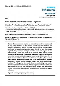
Figure 4. Firing fields of differing subicular place cells under different lighting conditions. Subicular firing fields are typically large. Firing fields (FFs) are optimally seen with a colour scale. A. Neuron with FF that remaps with a change in light condition and remains in the next location. B. Neuron whose firing field remaps from the top left corner to the left low corner in the dark and remaps again when back to light.
Previous research found that the geometry of the arena exert a quite strong control on PCs firing [67, 69]. Sharp [70] hypothesized that hippocampal place cells not only code spatial information but contextual spatial information. Place cells would be then modulated by geometric and non geometric changes in the environment. This would explain that subtle changes in context might generate extreme changes in the establishing firing field of a place cell and that geometric changes sometimes would not affect FFs that strongly. This hypothesis predicts that if place cells represent a unique spatial context then all place cells should remap under the different manipulations the experimenter could develop. There was, however, a high heterogeneity in the remapping of all different cells, leading to the conclusion that contextual information does not affect place cells in a whole block but in a fragmented way. Similarly the fact that hippocampal PCs display different maps in different environments [71] could be seen as evidence that the hippocampus is coding spatial and non spatial aspects of the environmental context. On the contrary, subicular and entorhinal cortical PCs tend to represent different environments in similar ways [70]. However, in our laboratory we recorded place cells in a square arena of 50 cm x 50 cm and a 60 cm height wall. The animals were first trained to forage for food in the light and in the dark. We found that PCs in the subiculum do
63
PRESENCE 2005 indeed show a large heterogeneity regarding their stability under different light conditions, as illustrated in Figure 4. In it we show two different types of subiculum PCs whose FFs remapped and remained (A) and remapped and returned to the original location (B) when studied in a light-dark-light protocol. Those neurons that remap under different light conditions would integrate visual information in their spatial coding. We also observed a third population with FFs that did not change with the light. It is clear that multiple factors are being coded by the hippocampus and the parahippocampal region. There is a clear influence of visual information on PCs firing, influence that is not enough to disrupt firing of PCs under multiple circumstances. The fact that some PCs can keep their FF in the darkness or after being blind is strong evidence that the animals are using other information to keep their representation. Also, 3D objects are able to produce an effect on PC firing if these are located distal from the centre of the arena. PCs adapt in different ways when the size of the arena is manipulated but this adaptation seems to be different in different brain areas. It has been well described that the hippocampus would code more than spatial information while other areas of the region would be less sensitive to these aspects. Therefore it is of great interest to investigate how different areas of the parahippocampal region and the hippocampus code different aspects of the “where” experience as well other elements of the context. Recent research has probed that rats can learn to navigate in a VR environment [72] and this opens a new door to use VR as a valuable tool in the quest for the understanding of spatial processing.
4. Place cells and Presence research. Presence research and research on spatial processing are strongly interrelated. On one side, the understanding of the factors that most influence our sense of location in space and that induce the creation of internal cognitive maps of the space can be exploited to induce presence. Reciprocally, the use of virtual environments is one of the fundamental tools to comprehend spatial processing. We have reviewed in this article the neuroscience literature devoted to spatial coding, concentrating mostly on hippocampal and parahippocampal place cells which comprise the best defined neuronal populations that participate in an internal representation of the external world. What can we learn from how spatial information is processed in the brain that can be useful in the field of presence research? We follow the operational definition of presence that it is successful substitution of real by virtual sensory data, where success is indicated by participants acting and responding to virtual sensory data in a VE as if it were real world sensory data, and where response is multilevel [73]. From that point of view, and since PCs code for particular locations in the space, we propose that if the firing of PCs during virtual navigation corresponds to the firing of these PCs in the equivalent real space, this would provide one component of a measure of presence based on
64
brain activity. It has been shown that indeed PCs in humans respond to particular locations within VEs [8]. However, a systematic use of this tool to measure presence is so far unattainable since it is only rarely, in pre-surgical brain patients with deep implanted electrodes, that such kind of single unit recordings can be obtained in humans. Otherwise, it would be appealing to test if presence correlates with the appropriate firing of place cells in VEs under a variety of experimental conditions (differences in visual realism, frame rate, etc), or to measure to what extent the pattern of PCs activation was transferable from a real to a virtual representation of the same space and vice versa. Although the difficulties to carry out these experiments are obvious, in theory they could provide a tool to better understand brain processing of spatial information both in real and VE. This theoretical consideration will still be valid if we consider that other methods of measuring brain activity such as brain imaging (fMRI) have already been used to detect the activation of neural structures during virtual navigation [9]. The limitations in this case are determined by the spatial resolution of the techniques (no single PCs can be detected). Another limitation is that the subject must navigate while remains motionless, since fMRI cannot be performed so far in moving around subjects. Therefore, it does not provide the means to compare human brain activity under real and virtual navigation. However, with the fast transformation that brain recording techniques have experienced in the last decades, it is reasonable to think that all these limitations will only lessen over time. So far, as we have presented in this review, most of the studies on the neural mechanisms underlying spatial navigation in real environments have been studied in animal models. Recently, the first really effective VE for rats has been described [72]. In it, a group of animals were trained to navigate to specific locations in order to obtain a series of rewards. A second group of rats were trained in the equivalent real environment without finding any sort of behavioral difference between them both [72]. We could take this result as an evidence of spatial presence in the VE. The obvious next step that has not been yet taken is to record from PCs in these animals in the equivalent real and VE and to try to correlate the stability of the PCs firing fields with the successful transfer of information between both experimental conditions. Accoding to our hypothesis and operational definition of presence, the similar firing of PCs in both environments would underlie a similar processing of the spatial information and would reveal presence in the VE. The fact that hippocampal cells are very sensitive to spatial contextual changes could be used to measure how different a VE is perceived in relationship to its correspondent real environment. It also provides the means to experiment on the impact that different streams of sensory information have on the brain processing of space, exploiting the possibility of disrupting sensory modalities in VEs that always appear together in real environments. Thus, in a Ves, visual, vestibular, somatosensory, auditory or propioceptive information could be dissociated, providing an excellent tool for the evaluation of their individual role on spatial processing.
PRESENCE 2005 It is a fact that has been described by different authors, that VEs are useful for acquiring spatial knowledge [74], although these findings are not exempt from controversy [75]: differences in the fidelity of the environments or the training methods can yield different behavioral results. We know that when cells “learn” to fire in order to code for a new space, this pattern of firing can be maintained for at least a month [35]. This variable transfer of spatial knowledge between virtual and real environments [75] could be due to the efficiency of the VE to generate a stable, cognitive map of space, that remains functional when the subject is moved to operate in the equivalent environment in the real world. The success of this transfer could therefore reflect the activation of the same network of PCs both in the virtual and the real environments. For this reason, the transfer success could be taken as a surrogate of the stability of the map coded in the PCs and, furthermore, as a measure of spatial presence during virtual navigation. At the same time that spatial mapping in place cells can be very stable, PCs are plastic and one observation that reveals this plasticity is the fact that areas of the space that are relevant from a behavioral point of view, have been reported to have larger representation in the hippocampal map [50]. This means that if a particular area of the space goes on to increase its relevance for the subject, the number of neurons that code for that particular area of space increases. Based on this observation it seems reasonable to predict that those VEs with higher behavioral significance for the subject are going to induce higher spatial presence. Or, what is the same, that a relatively crude VE could induce high spatial presence if what is represented is behaviorally relevant for the subject.
Conclusions Place cells in the hippocampus and parahippocampal formation create an internal cognitive map of the external space that integrates information about location, multisensory inputs and internal information (propioceptive, vestibular, etc). Chronic recordings of PCs in animal experiments and eventually in humans have yielded valuable information about the functional properties of these neurons that we have reviewed in this study. We believe that this information is relevant for presence research since these neurons constitute the roots of spatial presence, without understimating the involvement of other areas of the brain (parietal, frontal cortex) in the process. In this paper we suggest that if place cells activation operates in the same way in a VE as it does in its equivalent physical environment then this is one level of evidence that presence is occurring within that VE. We propose that this similar activation of PCs in virtual and real spaces should have its behavioral correlation in a succesful transfer of spatial information across both environments.
Acknowledgements This work has been sponsored by PRESENCIA (European Union FET IST-2001-37927) and MCYT (BFI2002-03643) to MVSV.
References [1]
[2]
[3]
[4]
[5] [6]
[7]
[8]
[9]
[10]
[11]
[12]
[13]
[14]
[15]
[16]
O'Keefe, J. & Dostrovsky, J. The hippocampus as a spatial map. Preliminary evidence from unit activity in the freely-moving rat. Brain Res 34, 171-5. 1971 Bingman, V. P. & Yates, G. Hippocampal lesions impair navigational learning in experienced homing pigeons. Behav Neurosci 106, 229-32. 1992 Watson, J. & Lashley, K. Homing and related activities of birds. Carnegie Institute Publication 211, 1-104. 1915 Griffin, D. Bird navigation. in Recent studies oin avian biology, ed. ed., A. W. (University of Illinois Press, Urbana, Illinois, pp. 154-197. 1955 Keeton, W. T. The mystery of pigeon homing. Sci Am 231, 96-7. 1974 Bingman, V. P. & Jones, T. J. Sun compass-based spatial learning impaired in homing pigeons with hippocampal lesions. J Neurosci 14, 6687-94. 1994 Lipp, H. P., Vyssotski, A. L., Wolfer, D. P., Renaudineau, S., Savini, M., Troster, G. & Dell'Omo, G. Pigeon homing along highways and exits. Curr Biol 14, 1239-49. 2004 Ekstrom, A. D., Kahana, M. J., Caplan, J. B., Fields, T. A., Isham, E. A., Newman, E. L. & Fried, I. Cellular networks underlying human spatial navigation. Nature 425, 184-8. 2003 Hartley, T., Maguire, E. A., Spiers, H. J. & Burgess, N. The well-worn route and the path less traveled: distinct neural bases of route following and wayfinding in humans. Neuron 37, 877-88. 2003 Morris, R. G., Garrud, P., Rawlins, J. N. & O'Keefe, J. Place navigation impaired in rats with hippocampal lesions. Nature 297, 681-3. 1982 Schenk, F. & Morris, R. G. Dissociation between components of spatial memory in rats after recovery from the effects of retrohippocampal lesions. Exp Brain Res 58, 11-28. 1985 Morris, R. G., Schenk, F., Tweedie, F. & Jarrard, L. E. Ibotenate Lesions of Hippocampus and/or Subiculum: Dissociating Components of Allocentric Spatial Learning. Eur J Neurosci 2, 1016-1028. 1990 Aguirre, G. K., Detre, J. A., Alsop, D. C. & D'Esposito, M. The parahippocampus subserves topographical learning in man. Cereb Cortex 6, 823-9. 1996 Jordan, K., Schadow, J., Wuestenberg, T., Heinze, H. J. & Jancke, L. Different cortical activations for subjects using allocentric or egocentric strategies in a virtual navigation task. Neuroreport 15, 135-40. 2004 Whishaw, I. Q., Hines, D. J. & Wallace, D. G. Dead reckoning (path integration) requires the hippocampal formation: evidence from spontaneous exploration and spatial learning tasks in light (allothetic) and dark (idiothetic) tests. Behav Brain Res 127, 49-69. 2001 Save, E., Paz-Villagran, V., Alexinsky, T. & Poucet, B. Functional interaction between the associative parietal
65
PRESENCE 2005
[17]
[18]
[19]
[20]
[21]
[22]
[23]
[24]
[25]
[26]
[27]
[28]
[29]
[30]
[31]
[32]
[33]
66
cortex and hippocampal place cell firing in the rat. Eur J Neurosci 21, 522-30. 2005 Commins, S., Gemmell, C., Anderson, M., Gigg, J. & O'Mara, S. M. Disorientation combined with bilateral parietal cortex lesions causes path integration deficits in the water maze. Behav Brain Res 104, 197-200. 1999 Wolbers, T., Weiller, C. & Buchel, C. Neural foundations of emerging route knowledge in complex spatial environments. Brain Res Cogn Brain Res 21, 401-11. 2004 Witter, M. P., Groenewegen, H. J., Lopes da Silva, F. H. & Lohman, A. H. Functional organization of the extrinsic and intrinsic circuitry of the parahippocampal region. Prog Neurobiol 33, 161-253. 1989 Bland, B. H. & Oddie, S. D. Theta band oscillation and synchrony in the hippocampal formation and associated structures: the case for its role in sensorimotor integration. Behav Brain Res 127, 119-36. 2001 Whishaw, I. Q. & Vanderwolf, C. H. Hippocampal EEG and behavior: changes in amplitude and frequency of RSA (theta rhythm) associated with spontaneous and learned movement patterns in rats and cats. Behav Biol 8, 461-84. 1973 Kramis, R., Vanderwolf, C. H. & Bland, B. H. Two types of hippocampal rhythmical slow activity in both the rabbit and the rat: relations to behavior and effects of atropine, diethyl ether, urethane, and pentobarbital. Exp Neurol 49, 58-85. 1975 O'Keefe, J. & Recce, M. L. Phase relationship between hippocampal place units and the EEG theta rhythm. Hippocampus 3, 317-30. 1993 Bischof, W. F. & Boulanger, P. Spatial navigation in virtual reality environments: an EEG analysis. Cyberpsychol Behav 6, 487-95. 2003 Kahana, M. J., Sekuler, R., Caplan, J. B., Kirschen, M. & Madsen, J. R. Human theta oscillations exhibit task dependence during virtual maze navigation. Nature 399, 781-4. 1999 de Araujo, D. B., Baffa, O. & Wakai, R. T. Theta oscillations and human navigation: a magnetoencephalography study. J Cogn Neurosci 14, 70-8. 2002 Kelemen, E., Moron, I. & Fenton, A. A. Is the hippocampal theta rhythm related to cognition in a nonlocomotor place recognition task? Hippocampus 15, 472-9. 2005 Poucet, B., Lenck-Santini, P. P., Paz-Villagran, V. & Save, E. Place cells, neocortex and spatial navigation: a short review. J Physiol Paris 97, 537-46. 2003 O'Mara, S. M. Spatially selective firing properties of hippocampal formation neurons in rodents and primates. Prog Neurobiol 45, 253-74. 1995 O'Mara, S. M., Rolls, E. T., Berthoz, A. & Kesner, R. P. Neurons responding to whole-body motion in the primate hippocampus. J Neurosci 14, 6511-23. 1994 Ludvig, N., Tang, H. M., Gohil, B. C. & Botero, J. M. Detecting location-specific neuronal firing rate increases in the hippocampus of freely-moving monkeys. Brain Res 1014, 97-109. 2004 Roche, R., Mangaoang, M., Commins, S. & O'Mara, S. Hippocampal Contributions To Neurocognitive Mapping In Humans: A New Model. Hippocampus in press. 2005 Thompson, L. T. & Best, P. J. Long-term stability of the place-field activity of single units recorded from the dorsal hippocampus of freely behaving rats. Brain Res 509, 299-308. 1990
[34] [35]
[36]
[37]
[38]
[39]
[40]
[41]
[42]
[43]
[44]
[45]
[46]
[47]
[48]
[49]
[50]
Hill, A. J. First occurrence of hippocampal spatial firing in a new environment. Exp Neurol 62, 282-97. 1978 Lever, C., Wills, T., Cacucci, F., Burgess, N. & O'Keefe, J. Long-term plasticity in hippocampal placecell representation of environmental geometry. Nature 416, 90-4. 2002 Kentros, C., Hargreaves, E., Hawkins, R. D., Kandel, E. R., Shapiro, M. & Muller, R. V. Abolition of long-term stability of new hippocampal place cell maps by NMDA receptor blockade. Science 280, 2121-6. 1998 McNaughton, B. L., Barnes, C. A., Meltzer, J. & Sutherland, R. J. Hippocampal granule cells are necessary for normal spatial learning but not for spatially-selective pyramidal cell discharge. Exp Brain Res 76, 485-96. 1989 McNaughton, B. L., Barnes, C. A., Gerrard, J. L., Gothard, K., Jung, M. W., Knierim, J. J., Kudrimoti, H., Qin, Y., Skaggs, W. E., Suster, M. & Weaver, K. L. Deciphering the hippocampal polyglot: the hippocampus as a path integration system. J Exp Biol 199 ( Pt 1), 173-85. 1996 McNaughton, B. L., Barnes, C. A. & O'Keefe, J. The contributions of position, direction, and velocity to single unit activity in the hippocampus of freely-moving rats. Exp Brain Res 52, 41-9. 1983 Markus, E. J., Qin, Y. L., Leonard, B., Skaggs, W. E., McNaughton, B. L. & Barnes, C. A. Interactions between location and task affect the spatial and directional firing of hippocampal neurons. J Neurosci 15, 7079-94. 1995 Muller, R. U., Bostock, E., Taube, J. S. & Kubie, J. L. On the directional firing properties of hippocampal place cells. J Neurosci 14, 7235-51. 1994 Taube, J. S., Muller, R. U. & Ranck, J. B., Jr. Headdirection cells recorded from the postsubiculum in freely moving rats. II. Effects of environmental manipulations. J Neurosci 10, 436-47. 1990 Taube, J. S. Head direction cells recorded in the anterior thalamic nuclei of freely moving rats. J Neurosci 15, 70-86. 1995 Goodridge, J. P., Dudchenko, P. A., Worboys, K. A., Golob, E. J. & Taube, J. S. Cue control and head direction cells. Behav Neurosci 112, 749-61. 1998 Cacucci, F., Lever, C., Wills, T. J., Burgess, N. & O'Keefe, J. Theta-modulated place-by-direction cells in the hippocampal formation in the rat. J Neurosci 24, 8265-77. 2004 Breese, C. R., Hampson, R. E. & Deadwyler, S. A. Hippocampal place cells: stereotypy and plasticity. J Neurosci 9, 1097-111. 1989 Speakman, A. & O'Keefe, J. Hippocampal Complex Spike Cells do not Change Their Place Fields if the Goal is Moved Within a Cue Controlled Environment. Eur J Neurosci 2, 544-555. 1990 Gemmell, C. & O'Mara, S. M. Medial prefrontal cortex lesions cause deficits in a variable-goal location task but not in object exploration. Behav Neurosci 113, 465-74. 1999 Hollup, S. A., Molden, S., Donnett, J. G., Moser, M. B. & Moser, E. I. Accumulation of hippocampal place fields at the goal location in an annular watermaze task. J Neurosci 21, 1635-44. 2001 Martin, P. D. & Ono, T. Effects of reward anticipation, reward presentation, and spatial parameters on the firing of single neurons recorded in the subiculum and nucleus accumbens of freely moving rats. Behav Brain Res 116, 23-38. 2000
PRESENCE 2005 [51]
[52]
[53]
[54]
[55]
[56]
[57]
[58]
[59]
[60]
[61]
[62]
[63]
[64]
[65]
[66]
[67]
[68]
[69]
Kobayashi, T., Nishijo, H., Fukuda, M., Bures, J. & Ono, T. Task-dependent representations in rat hippocampal place neurons. J Neurophysiol 78, 597613. 1997 Hok, V., Save, E., Lenck-Santini, P. P. & Poucet, B. Coding for spatial goals in the prelimbic/infralimbic area of the rat frontal cortex. Proc Natl Acad Sci U S A 102, 4602-7. 2005 Pratt, W. E. & Mizumori, S. J. Neurons in rat medial prefrontal cortex show anticipatory rate changes to predictable differential rewards in a spatial memory task. Behav Brain Res 123, 165-83. 2001 White, N. M. & McDonald, R. J. Acquisition of a spatial conditioned place preference is impaired by amygdala lesions and improved by fornix lesions. Behav Brain Res 55, 269-81. 1993 O'Mara, S. M., Commins, S., Anderson, M. & Gigg, J. The subiculum: a review of form, physiology and function. Prog Neurobiol 64, 129-55. 2001 Muller, R. U. & Kubie, J. L. The effects of changes in the environment on the spatial firing of hippocampal complex-spike cells. J Neurosci 7, 1951-68. 1987 Sharp, P. E. Subicular place cells expand or contract their spatial firing pattern to fit the size of the environment in an open field but not in the presence of barriers: comparison with hippocampal place cells. Behav Neurosci 113, 643-62. 1999 Sharp, P. E. Subicular cells generate similar spatial firing patterns in two geometrically and visually distinctive environments: comparison with hippocampal place cells. Behav Brain Res 85, 71-92. 1997 Hetherington, P. A. & Shapiro, M. L. Hippocampal place fields are altered by the removal of single visual cues in a distance-dependent manner. Behav Neurosci 111, 20-34. 1997 Jeffery, K. J. & O'Keefe, J. M. Learned interaction of visual and idiothetic cues in the control of place field orientation. Exp Brain Res 127, 151-61. 1999 Cressant, A., Muller, R. U. & Poucet, B. Failure of centrally placed objects to control the firing fields of hippocampal place cells. J Neurosci 17, 2531-42. 1997 Shapiro, M. L., Tanila, H. & Eichenbaum, H. Cues that hippocampal place cells encode: dynamic and hierarchical representation of local and distal stimuli. Hippocampus 7, 624-42. 1997 Hill, A. J. & Best, P. J. Effects of deafness and blindness on the spatial correlates of hippocampal unit activity in the rat. Exp Neurol 74, 204-17. 1981 Quirk, G. J., Muller, R. U. & Kubie, J. L. The firing of hippocampal place cells in the dark depends on the rat's recent experience. J Neurosci 10, 2008-17. 1990 Save, E., Cressant, A., Thinus-Blanc, C. & Poucet, B. Spatial firing of hippocampal place cells in blind rats. J Neurosci 18, 1818-26. 1998 Paz-Villagran, V., Lenck-Santini, P. P., Save, E. & Poucet, B. Properties of place cell firing after damage to the visual cortex. Eur J Neurosci 16, 771-6. 2002 O'Keefe, J. & Burgess, N. Geometric determinants of the place fields of hippocampal neurons. Nature 381, 425-8. 1996 Rivard, B., Li, Y., Lenck-Santini, P. P., Poucet, B. & Muller, R. U. Representation of objects in space by two classes of hippocampal pyramidal cells. J Gen Physiol 124, 9-25. 2004 Anderson, M. I. & Jeffery, K. J. Heterogeneous modulation of place cell firing by changes in context. J Neurosci 23, 8827-35. 2003
[70]
[71]
[72]
[73]
[74]
[75]
Sharp, P. E. Complimentary roles for hippocampal versus subicular/entorhinal place cells in coding place, context, and events. Hippocampus 9, 432-43. 1999 Deadwyler, S. A. & Hampson, R. E. Differential but complementary mnemonic functions of the hippocampus and subiculum. Neuron 42, 465-76. 2004 Holscher, C., Schnee, A., Dahmen, H., Setia, L. & Mallot, H. A. Rats are able to navigate in virtual environments. J Exp Biol 208, 561-9. 2005 Sanchez-Vives, M. V. & Slater, M. From presence to consciousness through virtual reality. Nat Rev Neurosci 6, 332-9. 2005 Bliss, J., Tidwell, P. & Guest, M. The effectiviness of virtual reality for adminestering spatial navigation training to firefighters. Presence-Teleoperators and Virtual Environments 6, 73-86. 1997 Darken, R. & Banker, W. in Proceedings of the IEEE Virtual Reality Annual International Symposium. ed. Press, C. S., Los Alamitos, CA). 1998
67