DOI: 10.1093/brain/awg082
Brain (2003), 126, 928±945
Neural resources for processing language and environmental sounds Evidence from aphasia
AysËe Põnar Saygõn,1 Frederic Dick,1 Stephen W. Wilson,2 Nina F. Dronkers3 and Elizabeth Bates1 1Department of Cognitive Science, University of California, San Diego, 2Neuroscience Interdepartmental Program, University of California, Los Angeles, and 3VA Northern California Health Care System, Martinez, USA
Summary
Although aphasia is often characterized as a selective impairment in language function, left hemisphere lesions may cause impairments in semantic processing of auditory information, not only in verbal but also in nonverbal domains. We assessed the `online' relationship between verbal and nonverbal auditory processing by examining the ability of 30 left hemisphere-damaged aphasic patients to match environmental sounds and linguistic phrases to corresponding pictures. The verbal and nonverbal task components were matched carefully through a norming study; 21 age-matched controls and ®ve right hemisphere-damaged patients were also tested to provide further reference points. We found that, while the aphasic groups were impaired relative to normal controls, they were impaired to the same extent in both domains, with accuracy and reaction time for verbal and nonverbal trials revealing unusually high correlations (r = 0.74 for accuracy, r = 0.95 for reaction
Correspondence to: AysËe Põnar Saygõn, Department of Cognitive Science, University of California, San Diego, 9500 Gilman Drive 0515, La Jolla, CA 92093-0515, USA E-mail:
[email protected]
time). Severely aphasic patients tended to perform worse in both domains, but lesion size did not correlate with performance. Lesion overlay analysis indicated that damage to posterior regions in the left middle and superior temporal gyri and to the inferior parietal lobe was a predictor of de®cits in processing for both speech and environmental sounds. The lesion mapping and further statistical assessments reliably revealed a posterior superior temporal region (Wernicke's area, traditionally considered a language-speci®c region) as being differentially more important for processing nonverbal sounds compared with verbal sounds. These results suggest that, in most cases, processing of meaningful verbal and nonverbal auditory information break down together in stroke and that subsequent recovery of function applies to both domains. This suggests that language shares neural resources with those used for processing information in other domains.
Keywords: aphasia; auditory agnosia; environmental sounds; Wernicke's area; lesion mapping Abbreviations: AQ = aphasia quotient; ERD = event-related desynchronization; ERP = event-related potentials; fMRI = functional MRI; IPL = inferior parietal lobule; LHD = left hemisphere-damaged; pMTG = posterior middle temporal gyrus; pSTG = posterior superior temporal gyrus; RHD = right hemisphere-damaged; ROI = region of interest; RT = reaction time; VLSM = Voxel-based lesion-symptom mapping; UCSD = University of California San Diego; WAB = Western Aphasia Battery
Introduction
The relationship between language impairments and de®cits in other cognitive and sensorimotor domains has been of interest since the early days of neurology. That aphasia itself may be symptomatic of a more general sensorimotor or cognitive disturbance is an idea with ample historical roots (Head, 1926; Goldstein, 1948). Indeed Jackson (1878), who observed a high incidence of nonverbal impairments in aphasic patients, believed that they suffer from a more Brain 126(4) ã Guarantors of Brain 2003; all rights reserved
general disturbance (sometimes referred to as asymbolia) and may be `lame in thinking'. Many studies have provided evidence for a range of nonverbal impairments in aphasic patients (see for example Gainotti and Lemmo, 1976; Ammon, 1979; Chertkow et al., 1997). It was noted early on (Jackson, 1878; Head, 1926) as well as more recently (De Renzi et al., 1968; Varney, 1978; Duffy and Duffy, 1981; Wang and Goodglass, 1992), that
Language and environmental sounds de®cits in comprehension and production of gesture and pantomime are strongly associated with receptive and expressive language disorders. Impairments in nonverbal domains in aphasic patients have been demonstrated in such tasks as associating pictures with corresponding objects (De Renzi et al., 1968), colours with pictures (De Renzi et al., 1972), gestures with objects (De Renzi et al., 1968; Varney, 1978) and sounds with pictures (Spinnler and Vignolo, 1966; Faglioni et al., 1969; Varney, 1980). Unfortunately, only a few studies have attempted to test aphasics systematically on nonverbal tasks that are comparable to those that show de®ciencies in the language domain (Spinnler and Vignolo, 1966; Varney, 1980; Bates et al., 2001). Of particular interest here is auditory agnosiaÐa rare neurological disorder characterized by a relatively isolated de®cit in auditory comprehension despite normal hearing. When the disorder affects only verbal material, it is often called word deafness; when the de®cit is in recognizing environmental sounds, it is often termed nonverbal auditory agnosia. Much of the literature on auditory agnosia consists of case studies. The associated lesions are not particularly consistent and have included unilateral right (Spreen et al., 1965; Haguenauer et al., 1979; Vignolo, 1982; Eustache et al., 1990; Fujii et al., 1990), unilateral left (Vignolo, 1982; Haguenauer et al., 1979; Eustache et al., 1990; Pasquier et al., 1991; Clarke et al., 2000) and bilateral cortical lesions (Albert et al., 1972; Haguenauer et al., 1979; Miceli, 1982; Rosati et al., 1982; Vignolo, 1982; Lechevalier et al., 1984; Motomura et al., 1986; Mendez and Geehan, 1988; Buchtel and Stewart, 1989; Lambert et al., 1989; Engelien et al., 1995; Kaga et al., 2000). Subcortical lesions can also cause this de®cit (Kazui et al., 1990). Auditory agnosia restricted to nonverbal material is a rather rare phenomenon, previously associated with bilateral (Spreen et al., 1965; Albert et al., 1972; Kazui et al., 1990) or right hemisphere (Fujii et al., 1990) lesions. Based on these studies, two forms of auditory agnosia have been proposed: (i) perceptual-discriminative, with patients failing to identify whether two consecutive sounds are identical; and (ii) associative-semantic, with patients being impaired at audio-visual matching or naming. Bilateral lesions appear to be implicated in severe discriminative disorders (Albert et al., 1972; Rosati et al., 1982; Vignolo, 1982; Lechevalier et al., 1984; Motomura et al., 1986; Mendez and Geehan, 1988; Buchtel and Stewart, 1989; Kazui et al., 1990; Taniwaki et al., 2000). Unilateral right hemisphere lesions can lead to normal association with impaired discrimination (Vignolo, 1982; Eustache et al., 1990), de®cient association with normal discrimination (Spreen et al., 1965) or de®cient association and de®cient discrimination (Fujii et al., 1990). Unilateral left hemisphere lesions have been reported to cause de®cient association and normal discrimination (Vignolo, 1982); however, in many cases discrimination has not been tested. A clear picture does not emerge from these ®ndings, due in part to the heterogeneity of the tests used. Thorough reviews of the relevant case
929
study literature are provided by Clarke et al. (1996, 2000) and Grif®ths et al. (1999). Environmental sounds share quite a few perceptual and informational features with language (Gygi, 2001), thus making them useful in exploring possible links between aphasia and (associative) auditory agnosia, and also more broadly between verbal and nonverbal auditory processing. Functional neuroimaging studies of human auditory processing have begun to reveal areas in the temporal lobes that are more activated for certain types of sounds than others. However, it is not yet clear whether these effects re¯ect divisions based on the type (e.g. music versus speech), semantic content, or spatial and temporal complexity of the sound stimuli used (Belin et al., 2000; Binder et al., 2000; Zatorre and Belin, 2001). Functional activation related to environmental sounds has been reported in only a few studies (Humphries et al., 2001; Lewis et al., 2001; Maeder et al., 2001; Adams and Janata, 2002; Dick et al., 2002b). Although contrasts with linguistic sounds were not always carried out or discussed in these studies, environmental sounds were observed to activate some middle and superior temporal areas in the left hemisphere that have been associated with language-related activation in earlier studies (e.g. Wise et al., 1991; DeÂmonet et al., 1992). These results are consistent with the idea that language shares some neural mechanisms with certain nonverbal processes. In an eventrelated potentials (ERP) study, Van Petten and Rheinfelder (1995) explored this hypothesis and found that target words were similarly modulated when preceded by contexts consisting of environmental sounds or sentences, suggesting verbal and nonverbal information may in¯uence a common semantic or associative space. Again using ERP, Cycowicz and Friedman (1998) showed that both types of stimuli elicit brain activity with similar characteristics as a function of familiarity and frequency. Looking at event-related desynchronization (ERD), Lebrun et al. (1998, 2001) observed leftlateralization for the semantic, but not perceptual processing of environmental sounds. Identifying differences and similarities in the brain mechanisms for processing these different types of auditory input is likely to be a fruitful line of research. Experimental studies of environmental sound processing in groups of patients with brain lesions have also provided insights. In a series of papers, Vignolo, Spinnler and Faglioni reported disturbances of environmental sound recognition due to unilateral hemispheric damage (Spinnler and Vignolo, 1966; Faglioni et al., 1969; Vignolo, 1982). They observed that right hemisphere-damaged (RHD) patients performed signi®cantly worse than controls on perceptual tests involving environmental sounds while left hemisphere-damaged (LHD) patients performed signi®cantly worse on associative tests. Intrigued by the ®nding that left hemisphere lesions can cause associative auditory agnosia, Varney (1980) used environmental sounds in order to examine verbal and nonverbal comprehension de®cits in aphasic patients, an undertaking similar to the present study. He found that defects in
930
A. P. Saygin et al.
environmental sound recognition were seen only in subjects with impaired verbal comprehension and that all aphasic patients with intact verbal comprehension performed well on sound recognition. There were, however, aphasic patients who were impaired in verbal comprehension, but not in sound recognition. Verbal comprehension was always as impaired as sound recognition, whereas sound recognition performance could be better than verbal comprehension. Interestingly, a similar relationship has been reported between pantomime recognition and reading comprehension (Varney, 1978). More recently, Schnider et al. (1994) observed that both LHD and RHD patients performed signi®cantly worse than a group of normal controls on an environmental sound recognition test. They found no signi®cant differences in the performance of the two patient groups; however, the pattern of errors appeared to differ over groups: LHD patients made more semantically-based errors, while RHD patients and control subjects made almost exclusively acoustic errors. For all patients, accuracy in recognizing environmental sounds correlated with language comprehension as measured by the Western Aphasia Battery (WAB) (Kertesz, 1979). Lesion-behaviour correlations showed that LHD patients with impaired environmental sound recognition tended to have damage to the posterior superior temporal gyrus (pSTG) and the inferior parietal lobe. Clarke et al. (1996, 2000) also tested patients with brain damage on different aspects of sound recognition. Here, patients who were de®cient in the sound recognition task exhibited much variability. In these studies, however, language comprehension was not tested in relation to sound processing; the purpose of some of the experiments was to contrast sound identi®cation and localization, topics that have recently been the focus of much research (e.g. Belin and Zatorre, 2000; Rauschecker and Tian, 2000). There are several reasons why the above literature tends to provide fragmentary and incomplete answers when addressing the relationship between verbal and nonverbal auditory processes. Many previous studies of environmental sound processing by aphasics did not attempt to make a direct comparison between verbal and nonverbal auditory processing in the same patients. Those that did compare performance between domains used different tasks or tests in the two domains and did not control for factors such as stimulus frequency and identi®ability, or the relationship between the auditory and visual stimuli (Varney, 1980; Schnider et al., 1994). Such factors are known to have effects on performance in verbal and nonverbal tasks. In addition, all previous studies used four or ®ve picture displays in sound to picture matching tasks, entailing a lengthy visual processing component to the task. Thus, in analysing performance in sound processing, these studies were limited to analysing accuracy data onlyÐ losing potentially important information about the time course of processing. The work we report allows us to address directly the relation between verbal and nonverbal auditory comprehension in chronic aphasic patients, using an `online' (timed) recognition paradigm, with verbal and nonverbal stimuli that are matched
for several factors. First, we brie¯y describe a norming study on a large set of environmental sound recordings. This study allowed us to test the sound stimuli for recognizability as well as to extract linguistic labels to be used in the verbal trials of the main experiment. Then we report on an online task with aphasic patients and age-matched controls in which stimuli in both domains are matched for identi®ability, frequency, and semantic relationship to the visual target. In line with earlier studies by Vignolo, Faglioni and Spinnler (Spinnler and Vignolo, 1966; Faglioni et al., 1969), we also address the effect of semantic competition on both domains across our patient groups in order to observe whether processing in the two domains is similarly modulated by higher-level semantic or conceptual constraints.
Methods Environmental sounds norming study Participants
Participants were 31 undergraduate and graduate students at University of California San Diego (UCSD), aged 18± 31 years with normal vision and hearing. All received class credit for their participation. Prior to the experiment, participants completed a handedness assessment questionnaire and a language history questionnaire. Subjects gave informed consent to participate in the study, which was approved by the UCSD Human Research Protections Program.
Materials
The sound stimuli were taken from digital sound effect libraries including Digifex and BBCâ. The sampling rate of the sounds was 44.1 kHz, with 16-bit quantization.
Procedure
Following a procedure used by Ballas (1993), we asked subjects to listen to sounds and to press a button as soon as they believed they had identi®ed the source of each sound. After the sound ended, subjects gave a verbal description, having been instructed to provide both a noun and a verb (e.g. dog barking, engine running). Subjects completed a practice block of eight trials and an experimental block of 236 trials. Verbal responses were coded by two independent raters. Accuracy was computed using the raters' codes, and response time was computed only for correct trials. More detail about the procedure and results is available in Saygin (2001) and Saygin et al. (2002).
Aphasia study Participants
Patients were voluntary participants recruited from Veterans' Administration Medical Centers from San Diego, CA, USA,
Language and environmental sounds
931
Table 1 Characteristics of aphasic and RHD patients Initials
Age
Patient group
AQ
Site
Lesion site
B.E. B.K. C.H. C.W. D.C. D.D. D.F. E.B. E.C. E.R. F.N. F.Y. G.G. H.K. H.M. J.A. J.B. J.C. J.D. J.G. J.H. J.Q. J.S. J.W. K.W. L.L. L.R. M.B. P.B. P.P. R.K. R.S. RS V.H. W.G.
24 55 66 72 63 56 46 32 43 81 58 77 50 62 72 59 66 81 72 63 62 76 51 72 64 76 56 50 75 50 52 74 55 71 82
Broca's Anomic Anomic RHD Broca's Broca's Broca's Broca's Anomic RHD RHD Wernicke's Anomic Wernicke's* Broca's Anomic Broca's Anomic Anomic Anomic Anomic Broca's Broca's Anomic Anomic Anomic Anomic Broca's Anomic Wernicke's RHD Wernicke's RHD Wernicke's Wernicke's
71.6 84.4 92.2 ± 74.8 18.9 49.6 68.3 91.7 ± ± 64.1 90.3 47.6 26.7 79.9 13.8 91.1 89.8 80.8 92.4 11.2 48.8 90.9 98.0 78.9 79.2 31.0 98.0 78.0 ± 33.3 ± 78.6 51.5
SD M SD M SD M M M M SD SD M SD M M M SD SD M M SD SD SD SD SD M SD SD SD SD SD M M SD M
Frontal, temporal, parietal, insula, basal ganglia Basal ganglia, insula Basal ganglia Right hemisphere (not included in group lesion analyses) Frontal, insula, basal ganglia Temporal, parietal, frontal, insula Temporal, parietal, frontal, insula Frontal, parietal, insula Frontal, temporal, parietal, insula Right hemisphere (not included in group lesion analyses) Right hemisphere parietal (not included in group lesion analysis) Inferior parietal, small region on superior temporal Small, posterior to temporal lobe Frontal, medial temporal, insula, subcortical Frontal, temporal, parietal N/A MCA-territory, acute scan shows expanding frontal lesion N/AÐacute scan shows no lesion boundaries Frontal, anterior temporal Basal ganglia Frontal, tip of anterior temporal Frontal, temporal, parietal, insula Frontal, temporal, parietal Temporal, parietal Frontal Excluded from all analysesÐpossibility of multiple infarcts Frontal, temporal, parietal Frontal, insular and subcortical extension, parietal Medial frontal Frontal, temporal, parietal, insula Right hemisphere (not included in group lesion analyses) Temporal Right hemisphere temporal, parietal (not included in group lesion analyses) Frontal, anterior temporal Temporal, parietal
*Criteria for classi®cation for Wernicke's aphasia based on WAB subscores are as follows: ¯uency = 5±10; comprehension = 0±6.9; repetition = 0±7.9; naming = 0±9. Criteria for transcortical sensory aphasia are identical except for repetition (8±10). Since repetition is not a component of the task here, we found it appropriate to analyse this subject's data in the Wernicke's aphasia group. M = Martinez, CA, USA; SD = San Diego, CA, USA. Lesion summaries are based on CT or MRI scans or medical records.
or Martinez, CA, USA, and were paid $25.00 for their participation. Thirty LHD patients with varying types and severity of aphasia and ®ve RHD patients with no measurable aphasia participated in the experiment. CT or MRI scans and the medical records of all patients were evaluated by a neurologist; only patients with unilateral lesions due to a single cerebrovascular accident were included. Exclusionary criteria included diagnosed or suspected hearing dif®culties, dementia, head trauma, tumours or multiple infarcts. Aphasic patients were classi®ed using the WAB (Kertesz, 1979) as anomic (n = 14), Broca's (n = 10) or Wernicke's aphasics (n = 6). Details are provided in Table 1. Age-matched controls were 21 adults aged 53±78 years, with no history of audiological, neurological or psychiatric disorders; all had normal or corrected-to-normal vision, and were tested for hearing impairment with a standard question-
naire and/or with an audiometer. All were paid $25.00 for their participation. Informed consent was obtained from all subjects in accordance with guidelines of the UCSD Human Research Protections Program. Data from two control subjects were excluded (one talked to the experimenter throughout the testing session and one reported low-frequency hearing loss afterwards). Data from one patient (L.L., anomic) were excluded due to a possibility of multiple infarcts.
Experimental design and materials
A 2-within- 3 1-between-subjects design was used, with domain (verbal versus nonverbal) and semantic competition (visual target related to distracter versus visual target unrelated to distracter) as within-subject factors, and patient
932
A. P. Saygin et al.
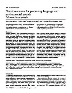
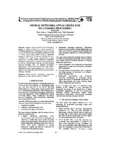
group (control, RHD, Broca's, Wernicke's, anomic in the main analysis; LHD, RHD, control in a supplementary analysis) as the between-subjects factor. Stimuli were black-and-white line drawings, nonverbal sounds and speech sounds. Visual stimuli were 10.6 cm 3 10.6 cm digitized drawings culled from extensively normed picture databases. Naming norms for these pictures have been reported elsewhere (Bates et al., 2003). Forty-®ve nonverbal sound stimuli were selected from the set normed in the preliminary study explained above. Selection criteria included identi®ability (moderate to high), inter-rater reliability for identi®ability, imageability (identi®ability/availability of picture) and recognition time. Selected sounds included animal cries (n = 10; e.g. cow mooing, bird chirping), human sounds (n = 6; e.g. sneezing, laughing), vehicle noises (n = 5; e.g. train, car, tractor noises), tool/machinery sounds (n = 4; e.g. drill, lawnmower noises), alarms/bells (n = 5; e.g. telephone ringing, bells tolling), water sounds (n = 6; e.g. dripping, pouring), sports (n = 4; e.g. bowling, golf) and music (n = 5; e.g. piano, violin). A full list of sounds used, as well as norming results on these sounds, are reported in Saygin et al. (2002). Speech stimuli were phrases based on the most common labels provided by the subjects in the preliminary experiment. Grammatical complexity was kept constant by putting together commonly reported nouns and verbs in `noun phrase + verbing (+ object)' constructions. Examples of phrases used were `cow mooing', `water boiling' and `someone eating an apple'. All phrases were read by a 38-year-old male speaker of American English and were digitally recorded at a sampling rate of 44.1 kHz with 16-bit quantization. Three line drawings were matched to each sound pair: a target, a related distracter and an unrelated distracter. For example, for the sound of a cow `mooing' or its verbal description, the target drawing was `cow', the semantically related distracter was `sheep', and the unrelated distracter was `violin' (see Fig. 1). In order to ensure that the semantically related and unrelated distracters were appropriately assigned, we made use of the semantic relatedness measure latent semantic analysis (Landauer et al., 1998). The average latent semantic analysis index for semantically related pairs was 0.36; for unrelated pairs it was 0.04. Over the course of the experiment, each picture appeared eight times in a fully counterbalanced fashion: picture type (target/distracter) 3 domain (verbal/nonverbal) 3 distracter type (related/unrelated to the target). Each of the 45 sound `types' (e.g. `cow') was also crossed with domain (verbal/ nonverbal) and distracter type (related/unrelated). A full list of items used is reported in Saygin et al. (2002).
Procedure
The experiment was run on Apple Macintosh PowerBook 3400c computers using the PsyScope experimental driver (Cohen et al., 1993). Participants sat in front of a VGA monitor, Yamaha YST-M7 speakers were placed on each side, and a standard PsyScope button box was used to collect
responses. The experimenter read a set of instructions to each participant and asked him or her to complete a practice session of six trials. The experimental block consisted of 180 experimenteradvanced trials. In each trial, subjects were presented with a two-picture display on the screen. After 1000 ms, the sound stimulus (either verbal or nonverbal) was presented through the speakers. This delay allowed subjects enough time to process the visual stimuli, thus mitigating visual processing contributions to reaction time data. Subjects pushed the button under the picture they believed matched the sound. Reaction time and accuracy were recorded for each trial. Subjects were continuously monitored for attention to the task, and were asked at intervals whether they needed a break. The nature of errors was noted, as were any comments made during or after the experiment. Special care was taken to note whether or not the subject was immediately aware of the error (as indicated by an overt verbal or physical response). Motivational feedback (e.g. `you are doing great so far') was provided as often as considered necessary to keep participants engaged in the task (for aphasic patients, this was approximately once every 20 trials); however, this feedback did not relate any information about the subject's accuracy in a particular trial.
Lesion analysis
As noted above, head CT or MRI images were obtained for all of the patients. For 20 of our LHD patients, computerized lesion reconstructions to be used in lesion overlay analyses were available. For another six patients, we had MRI or CT scans showing lesion boundaries, which were used in some analyses but not for the lesion overlays. Only acute scans were available for the remaining three LHD subjects; chronic scans showing distinct lesion boundaries could not be obtained. Lesion reconstructions were available only for two of the RHD patients who participated in this study, so we did not include this group in our lesion analyses. Lesion reconstructions were based on CT or MRI scans at least 3 weeks post-onset and were hand-drawn onto 11 axial slice templates based on the atlas of DeArmond et al. (1976), They were then entered into a Macintosh computer via electronic bitpad using software developed at the VA Medical Center in Martinez, California (Frey et al., 1987). All reconstructions were completed by a board-certi®ed neurologist, experienced in neuroradiology, but blind to the behavioural de®cits of the patients. Individual variations in gyral patterns and any differences in imaging angles were compensated for by using subcortical structures as landmarks. To determine common areas of infarction in patients who exhibit similar behavioural pro®les, we overlapped their lesions using the voxel-based lesion symptom mapping (VLSM) software developed by our group (Wilson et al., 2002).
Language and environmental sounds
933
Fig. 1 Summary of the experimental design. Domain (verbal/nonverbal) and distracter type (related to target/unrelated to target) were within-subject factors, and subject group was the between-subjects factor. The target `cow' appeared four times, twice with verbal sound stimuli (the phrase `cow mooing'), twice with non-verbal stimuli (the sound of a cow mooing), twice with `sheep' as the distracter (related condition), and twice with `violin' as the distracter (unrelated condition). All these trial types with the target `cow' are depicted in the pictures. Forty-®ve pictures and sounds were used as targets and related and unrelated foils, giving rise to 45 triplets such as `cow±sheep±violin'. A total of 180 trials was administered. Twenty quasi-random orders of the list were rotated among the subjects.
Statistical analysis
Performance across groups was compared using repeated measures analysis of variance (ANOVA). Regression and correlation analyses were performed to examine the relationships between performance in the two domains. We also conducted outlier analyses to identify any dissociations in performance. All analyses were performed using JMP and StatView statistical packages (Sall et al., 2001).
Results
Here we examine differences in accuracy and reaction time between patient and control groups, the correlation in performance across verbal and nonverbal domains and the relationship between lesion site and processing de®cits.
Is nonverbal processing spared in aphasic patients?
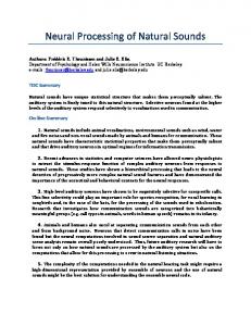
We examined accuracy and reaction time (RT) for the aphasic (LHD) and RHD subjects, and their age-matched controls. LHD subjects were grouped according to aphasia subtype (as determined by the WAB) into Broca's, Wernicke's and anomic groups. RTs were analysed only for correct responses. We analysed RT data in several different ways (e.g. patients' RT measured as the difference from the normal controls' RT, or converted into standardized scores) with no change in the pattern of results. Therefore, we report results for the simple case of RTs measured from the onset of sound. As depicted in Fig. 2, groups differed in their overall accuracy [F(4,48) = 8.533, P < 0.0001]; planned comparisons showed that control, anomic and RHD groups did not differ signi®cantly from each other (all making very few errors),
934
A. P. Saygin et al.
Fig. 2 Accuracy depicted across verbal and nonverbal domains for all subject groups. Groups differed in their overall accuracy (P < 0.0001) with control = RHD = anomic > Broca's > Wernicke's (all comparisons corrected P < 0.01).
whereas Broca's aphasics and Wernicke's aphasics were less accurate than all other groups and, furthermore, differed signi®cantly from each other, with Broca's more accurate than Wernicke's (P < 0.01 for all signi®cant differences, with correction for multiple comparisons). Distracter type had an effect on accuracy, such that subjects were less accurate when the distracter picture was semantically related to the target picture [F(1,48) = 62.920, P < 0.0001]. The effect of distracter type was also modulated by group [F(4,48) = 5.612, P = 0.0009]. Patient groups were more adversely affected when the distracter was related to the target (see Fig. 3). This interaction appears to be driven mainly by the Broca's and Wernicke's aphasics. When both of these severely affected groups were excluded from the analyses, the distracter type by group interaction was no longer signi®cant [F(2,34) = 1.458, P = 0.25]; conversely, ANOVA comparisons between either Broca's or Wernicke's patients and normal controls revealed signi®cant distracter type by group interactions [F(1,27) = 10.730, P = 0.0029 and F(1,23) = 57.816, P < 0.0001, respectively]. There was no main effect of domain; accuracy in verbal and nonverbal conditions did not differ signi®cantly [F(1,48) = 2.895, P = 0.095]. Domain did not interact with distracter type [F < 1], nor was there an interaction of group by domain [F(4,48) = 1.333, P = 0.27] or a three-way interaction of group, domain and distracter type [F(4,48) = 1.397, P = 0.25]. The fact that the group by domain interaction did not reach signi®cance is especially notable, as we might expect aphasic groups to commit more errors in verbal trials compared both with normals and with patients with RHD. In fact, the (nonsigni®cant) numerical results were in the opposite direction (verbal accuracy > nonverbal) for all groups except for Wernicke's aphasics, the most impaired group. To determine whether the anticipated interaction would hold if we restricted our attention only to these patients, the ANOVA was repeated for Wernicke's and controls only. In this case, the group by domain interaction reached signi®cance [F(1,23) = 4.442, P = 0.046]. Comparable re-analysis
Fig. 3 Accuracy depicted across related and unrelated distracter conditions for all subject groups. There was a main effect of distracter type (P < 0.0001). There was also an interaction of distracter type with group (P < 0.01), driven mainly by the Broca's and Wernicke's aphasics.
comparing each of the other patient groups with normals did not reveal any evidence for a group by domain interaction (see Fig. 2). RT was analysed for the accurate trials only. We found signi®cant differences in RT over patient group, as plotted in Fig. 4 [F(4,48) = 9.891, P < 0.0001]. Pairwise comparisons showed the following ordering of RT (from slowest to fastest, P < 0.0001 for all differences): Wernicke's = Broca's > anomic = RHD > control patients. As with accuracy, there were signi®cant effects of distracter type on RT [F(1,48) = 254.849, P < 0.0001], where RTs to semantically related target and distracter pairs were higher than those to unrelated ones. The distracter type interaction with patient group just reached signi®cance [F(4,48) = 2.605, P = 0.047]. Here, control subjects were slightly less affected in their response latencies by the distracters compared with all groups except RHD (Ps < 0.05); none of the other groups differed from one another. There was no main effect of domain on reaction times [F < 1], but contrasts were carried out to examine whether there was a differential effect of domain across patient groups. Comparing each patient group with controls revealed that anomic patients [F(1,30) = 4.485, P = 0.042] and, to a lesser extent, the RHD patients [F(1,22) = 4.118, P = 0.055] tended to respond slower relative to controls on the nonverbal material. For anomics, this is the opposite to what might be predicted in a traditional account of aphasia. There were no signi®cant interactions between controls and Broca's [F(1,27) = 0.242, P = 0.63] or Wernicke's [F(1,23) = 2.771, P = 0.11] patients. In summary, our analyses did not reveal a sparing of nonverbal processing in aphasic patients; in particular, LHD patients performed poorly in the nonverbal domain at levels comparable to their performance in the verbal domain.
Analyses over hemisphere of lesion
Although the analyses reported above examine the effects of lesion side on performance in the experiment, we also report
Language and environmental sounds results of analyses in which aphasic subjects were considered as a single LHD group. This is mainly to enable comparison with previous studies that used side of lesion as the grouping variable across subjects and did not form groups based on aphasia type. For accuracy, there were main effects of group (LHD, RHD, controls) [F(2,50) = 4.625, P < 0.014] and of distracter type [F(1,50) = 18.864, P < 0.0001]. Controls and RHD patients performed better than LHD patients. The group by distracter type interaction reached signi®cance, with the LHD group making more errors when related distracters were presented [F(2,50) = 5.872, P = 0.0051]. Once again, the group by domain interaction was not signi®cant [F < 1]. The RT data closely parallel the accuracy data and previous analyses: The main effects of group [F(2,50) = 12.563,
Fig. 4 Reaction time for correct responses depicted across verbal and nonverbal domains for all subject groups. Groups differed in their response latencies (P < 0.0001) with control < RHD = anomic < Broca's = Wernicke's (all comparisons corrected P < 0.01)
935
P < 0.0001] and distracter type [F(1,50) = 160.925, P < 0.0001] are signi®cant. The LHD group was the slowest; the RHD group was faster than the LHD group, but slower than the control subjects. The group by distracter type interaction reached signi®cance, with the LHD group more adversely affected by related distracters [F(2,50) = 5.307, P = 0.0081]. The group by domain interaction was again not signi®cant [F(2,50) = 1.311, P = 0.28]. To summarize, hemisphere of lesion did not signi®cantly affect the relative impairment on the verbal and nonverbal conditions in this experiment. There was, however, a reliable effect of semantically related distracters: LHD patients found them harder to process than RHD and control subjects.
Associations between task performance across domains and outlier analyses
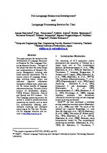
Within the LHD group, accuracy in verbal and nonverbal domains was very tightly correlated (r = 0.74, P < 0.0001), with reaction time data demonstrating an even closer relationship, approaching an identity function (r = 0.95, P < 0.0001). Impairments in verbal and nonverbal domains go hand in hand in our data. Fig. 5A and B show correlation scatter plots and linear ®ts for accuracy and RT in LHD subjects over the two domains. We also assessed the relationship between patients' WABderived aphasia quotient (AQ), a measure of overall aphasia severity, and performance in our task. Note that AQ is a taskexternal measure of language impairment. Overall, accuracy was correlated with AQ (r = 0.526, P = 0.0033); when split by domain, both verbal and nonverbal performance were correlated with severity of aphasia (verbal: r = 0.539,
Fig. 5 Correlation of performance in the verbal and nonverbal domains within the aphasic group for (A) accuracy and (B) reaction time. Linear ®ts and density ellipses using a con®dence interval of 95% are shown. Correlations are signi®cant (P < 0.0001 for both) and high (r = 0.74 and 0.95, respectively). Data points outside the ellipses are outliers based in Mahalanobis distances. + denotes patient R.S. and * denotes patient J.W; the two patients who show signs of possible dissociations between the two domains.
936
A. P. Saygin et al.
P = 0.0025; nonverbal: r = 0.440, P = 0.017). RT measures also correlated with AQ (r = 0.619, P = 0.0003); when split by domain, both the verbal performance (r = 0.689, P < 0.0001) and the nonverbal performance (r = 0.546, P = 0.0022) showed signi®cant relationships to aphasia severity. Very similar results have been reported by Schnider et al. (1994). In order to explore the outliers in the dataset, we calculated density ellipses using a con®dence interval of 95% using the outlier analysis tool of the JMP statistical software package. These ellipses are based on Mahalanobis distances and, assuming a bivariate normal distribution, show where a given percentage of the data is expected to lie. The Mahalanobis distance takes into account the correlation structure of the data as well as the individual scales (Appelbaum et al., 1999; Sall et al., 2001). We used a 95% con®dence ellipse for both of our measures. These outlier analyses report only the aphasic (LHD) population; we also carried out the analysis including RHD subjects and found very similar results. For accuracy, three subjects remained outside the ellipse and were identi®ed as outliers, (as shown in Fig. 5A). For RT, we identi®ed only one outlierÐas can be seen in Fig. 5B. In order to compare these results with what would be expected by chance, we carried out a small-scale randomization test. The verbal and nonverbal accuracy scores were shuf¯ed 50 times and outlier analysis was performed each time. The mean number of outliers obtained was 2.9 (range 2±4). This demonstrates that the procedure identi®es roughly the same number of outliers regardless of the correlation structure of the data; thus, no special signi®cance should be attached to the number of patients identi®ed. Rather, the advantage of outlier analysis is that it provides a quantitative method of identifying patients who may potentially exhibit dissociations. The actual process of identifying genuine dissociations is more qualitative. Based on Fig. 5A, we saw that patient J.W. (*) showed a striking dissociation with 100% accuracy on verbal (better than healthy controls) and 87.7% accuracy on nonverbal trials. Patient R.S. (+) showed some dissociation with 92.2% accuracy on nonverbal and 84.4% accuracy on verbal trials. Patient W.G. was an outlier by virtue of the fact that he was severely impaired with 81% accuracy in both domains. RT analyses pinpointed J.W. as the sole outlier, shown in Fig. 5B. R.S. and W.G. are both severe Wernicke's aphasics, with large lesions involving temporal and parietal regions. In order to further investigate R.S. as a patient exhibiting a potential dissociation, we re-tested him on the same task after a sixmonth delay. His performance was better, with 95% accuracy on nonverbal and 90.2% accuracy on verbal trials. At the time of re-testing, he had made more gains in the nonverbal domain and, with these scores, he would no longer be an outlier with respect to the rest of the sample. However, a relatively low score in the verbal domain remains in his pro®le; we conclude that his dissociation should be noted but interpreted with care.
For J.W. on the other hand, who showed a striking dissociation in a rather unexpected direction (worse nonverbal processing in an aphasic patient) which was also re¯ected in his RT scores, follow-up testing revealed that the dissociation was persistent and reliable. J.W. has an unusual neurological pro®le: despite a large temporoparietal lesion, he presents with a very mild aphasia (anomic) with almost completely intact verbal auditory comprehension. We carried out several additional tests on this patient after a nine-month delay and veri®ed that he has severe auditory agnosia for nonverbal sounds (Saygin and Moineau, 2002).
Lesion location analyses
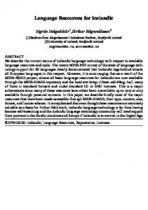
We performed a lesion analysis to investigate further the neural correlates of auditory comprehension. First, we overlapped the computer-reconstructed lesions of the patients who exhibited behavioural pro®les of interest (e.g. poor performance in nonverbal sounds) to determine if they shared a common area of infarction. Next, we used these shared areas of injury as regions of interest (ROIs) to determine statistical differences between groups of patients whose lesions either spared or involved these particular areas. For the lesion overlays, the 20 LHD patients for whom we had lesion reconstructions were grouped together based on their performance in the task, regardless of aphasia classi®cation. We used accuracy and RT values, respectively; both converted into z-scores with respect to normal controls as a measure of patients' degree of impairment. The VLSM software (Wilson et al., 2002) was used to assess the degree of spatial overlap in lesions shared by patients with similar behavioural de®cits. Patients who performed >2 SDs below the normal controls were considered de®cient and their lesions overlapped to determine if a common area of infarction could be found. For accuracy, overlays are provided in the top panel of Fig. 6, broken down by stimulus domain. Here, we show the results on three axial slices that pass through the middle temporal (slice 1), superior temporal (slice 2) and inferior parietal regions (slice 3). Based on the criteria used here, eight patients were de®cient in nonverbal sounds and 10 were de®cient in verbal sounds. As can be seen, the overlays are very similar across these two domains. Consistent with Schnider et al. (1994) and recent neuroimaging studies of environmental sound processing (e.g. Adams and Janata, 2002), the areas of maximal overlap for patients impaired in the nonverbal domain are centred in the posterior superior temporal gyrus (pSTG, in slice 2) extending into some middle temporal (in slice 1) and inferior parietal (in slice 3) regions. The implicated areas for verbal sound processing are strikingly similar, though a slightly smaller fraction of patients overlap on these regions, i.e. not all patients with poor verbal comprehension have damage to the areas of maximal overlap. For reaction time, overlays for patients who were identi®ed as de®cient based on slow response latencies appear to be
Language and environmental sounds
Fig. 6 Lesion overlays for LHD patients who performed poorly in the nonverbal and verbal domains based on accuracy and reaction time. The overlays consist of lesions from patients who performed >2 SDs below the age-matched controls in the corresponding measure in each condition. Lesions are depicted on three axial slices that go through middle temporal, superior temporal and inferior parietal lobes. The colour maps indicate the percentage of patients whose lesions involve that particular region.
almost identical for the two domains (verbal versus nonverbal). The lower panel of Fig. 6 shows that the same pSTG region identi®ed for accuracy is once again the focal point for patients with slow RT (Fig. 6). The pattern of results looks very similar to that revealed by the overlays based on accuracy, although for RT, there seems to be a stronger overlap in the insula. The analyses above selected patients based on behavioural de®cits and identi®ed the common areas of infarction.
937
Another possible method is to examine the behavioural pro®les associated with the ROIs previously discussed. This type of analysis enables us to see how general the localization results are and allows us to assess quantitatively which areas are differentially implicated in verbal versus nonverbal processing. For the analyses below, we identi®ed as ROIs the maximal areas of infarction for each slice in the lesion overlays. Then we divided the patients into groups consisting of those who had a lesion in that ROI and those whose lesions spared that area. This permitted us to compare and contrast performance in relation to the ROIs and the two domains quantitatively. Only patients whose lesion reconstructions (or scans, for the six patients for whom we did not have digital reconstructions) clearly involved or spared the ROIs were included in the groups. Patients were ®rst divided into two groups consisting of those who had a lesion in the region with the darkest colour in Fig. 6, slice 2 (pSTG) and those whose lesions did not involve this region [n(lesioned) = 11, n(intact) = 14]. For accuracy, there was a signi®cant main effect of pSTG lesion [F(1,23) = 5.714, P = 0.025]; patients who had lesions in this location had signi®cantly lower accuracy scores than patients who did not have a lesion here [mean(lesioned) = 92.8%, mean(intact) = 97.2%]. There was also an interaction of pSTG lesion with domain [F(1,23) = 4.349, P = 0.048], mainly driven by the nonverbal errors (see Fig. 7). The difference between patients with pSTG lesions versus those without was larger in the nonverbal domain than in the verbal domain. Furthermore, the difference between verbal and nonverbal domains was larger in the pSTG-lesioned patients than the difference between domains in those without lesions to this area. Additionally, pSTG-lesioned patients were signi®cantly slower than the patients whose lesions spared this region [mean(lesioned) = 2223 ms, mean(intact) = 1649 ms, F(1,23) = 5.338, P = 0.030]. However, there was no interaction of domain and pSTG lesion for RT [F(1,23) = 1.529, P = 0.23], consistent with the great similarity of the overlays for the two domains in Fig. 6. For the posterior middle temple gyrus (pMTG) region that is identi®ed as possibly important for sound processing in slice 1, a similar analysis with eight lesioned patients and 16 non-lesioned patients revealed a main effect of pMTG on accuracy [F(1,22) = 7.582, P = 0.012]. Patients with lesions here were signi®cantly less accurate than patients whose lesions spared this region [mean(lesioned) = 91.6%, mean(intact) = 96.9%]. However, this region did not make a differential contribution to the two domains: the interaction of pMTG lesion and domain was not signi®cant [F < 1]. In a separate analysis on reaction times, patients with pMTG lesions were signi®cantly slower than those without lesions here [mean(lesioned) = 2450 ms, mean(intact) = 1613 ms, F(1,22) = 11.767, P = 0.0024], but again the pMTG lesion and domain interaction did not reach signi®cance [F(1,23) = 2.214, P = 0.15], as was also the case for RT for the pSTG.
938
A. P. Saygin et al.
Fig. 7 Summary of statistics on the regions of interest based on Fig. 6: pSTG, pMTG, IPL and insula. There is a signi®cant main effect of pSTG and pMTG on accuracy. We also found signi®cant interactions indicating the involvement of the pSTG and IPL in nonverbal de®cits above and beyond verbal de®cits. As can be seen, there is no clear implication to the portion of the insula that we examined on accuracy in either of the domains.
For the inferior parietal lobule (IPL) region in slice 3, we carried out the analogous analyses [n(lesioned) = 11, n(intact) = 13]. Here, although there was no main effect of group on accuracy [F(1,22) = 1.827, P = 0.19] or RT [F(1,22) = 2.680, P = 0.12], the IPL lesion by domain interaction was signi®cant for accuracy [F(1,22) = 6.695, P = 0.017], largely due to the fact that those patients whose lesions included this region were signi®cantly less accurate for the nonverbal trials [mean(lesioned,verbal) = 95.2%, mean(lesioned, nonverbal) = 92.1%]. These latter ®ndings suggest that the IPL region may be especially important for processing nonverbal sounds; however, the absence of a main effect demands caution in drawing strong conclusions. Recall that on slice 2, there was some evidence from the RT data for insula involvement. However when we examined all the LHD patients [n(lesioned) = 14, n(intact) = 7], damage involving this portion of the insula was not signi®cantly associated with accuracy or RT in either domain [all Fs < 1]. Although we observed extremely high correlations between performance in the two domains, for exploratory purposes we also performed lesion overlay analysis for patients who performed relatively better in one domain compared with the other (Fig. 8). First, we identi®ed patients whose accuracy in one domain was >1 SD different from their performance in the other domain. We then constructed an overlay of lesions for those patients who performed more poorly in the nonverbal domain (n = 10) and those who performed more poorly in the verbal domain (n = 5). As can be seen, the patients who were relatively more impaired in the nonverbal domain have lesions along the middle and posterior portions of the superior temporal gyrus and in the IPL. Notice that these are the same areas already identi®ed as being important in Fig. 6 and showed some signi®cant quantitative effects after lesion-location-based group analy-
ses. We see now that these regions (especially the pSTG) are implicated even when the patients whose de®cits are comparable in the verbal and nonverbal domains are excluded from this highly correlated dataset. This lends further support to the importance of these regions for environmental sound processing. On the other hand, once the patients whose de®cits also equally involve the nonverbal domain are excluded, the lesion overlay for the patients with verbal de®cits becomes less focal and has a visibly more anterior and medial focus moving towards the anterior insula, basal ganglia and caudate nucleus. Turning to results for reaction time, we regrouped the patients into those whose performance was slower in the nonverbal than the verbal domain (n = 10) and compared them with those whose verbal performance was slower in the verbal compared with the nonverbal domain (n = 11). Here a similar anterior focus for relatively slow response times in the verbal domain can be seen, whereas the focus for relatively slow response times in the nonverbal domain does not change. However, unlike the posterior foci analysed above, this anterior area which is implicated in patients who perform relatively poorly in the verbal domain is not signi®cantly associated with our behavioural measures. When analogous statistics are computed between groups of patients with (n = 10) and without (n = 11) lesions to the region of maximal overlap for worse performance in the verbal domain in Fig. 8, no differences can be found between these patients in accuracy or RT. Nor are there any interactions or tendencies towards selective involvement in one domain versus the other [all Fs < 1]. Note that this common lesion location in patients with poorer performance in the verbal domain may re¯ect the participation of aphasic patients with haemorrhagic stroke who tend to have more subcortical involvement.
Language and environmental sounds
939
Fig. 9 Lesion overlay on slice 2 depicting patients who are `spared' in the nonverbal domain along with an overlay depicting patients who are impaired. Note that the latter overlay is the same as slice 2 of the top panel in Fig. 6, replicated for easy contrast.
data, we cannot identify a speci®c region that is differentially and speci®cally implicated for verbal processing. We performed one more lesion overlay to determine whether the pSTG region that is signi®cantly implicated in nonverbal processing in prior analyses is essential for performing well in this domain. Four patients in our sample were 100% accurate on nonverbal trials, thus performing better than normal controls. Fig. 9 depicts the lesion overlays on slice 2 for these patients (on a blue colour scale to emphasize that this is a map of sparing) along with an overlay for patients who were de®cient in the nonverbal domain (2 SDs below the controls; same as the overlay in Fig. 6). As can be seen, all of the patients who performed well in the nonverbal domain have lesions that spare the pSTG region (as well as the pMTG region, data not shown). Thus, based on our sample, it appears that the pSTG region may be crucial for normal nonverbal auditory processing.
Discussion Aphasic patients do not have spared nonverbal processing Fig. 8 Lesion overlays for patients whose performance in one domain was worse compared with the other domain. For accuracy, the overlays consist of lesions from patients whose accuracy zscores were >1 SD apart between the two domains. For reaction time, the scores were too tightly correlated and very few patients had scores that were 1 SD apart. The overlays for this measure were thus computed based on positive or negative z-score differences. Patients whose lesions are overlapped in the top panel for the RT overlays were thus relatively slower in responding to nonverbal trials than they were to verbal trials. Conversely, lesion overlays in the bottom panel depicts patients who were slower in responding to the verbal trials.
Thus, the results for the relative impairment overlays corroborate prior results that point to the pSTG as a critical region for nonverbal sound processing. This area is also important for verbal comprehension. However, despite obtaining a different pattern of lesions for patients who are more de®cient in the verbal domain, based on the present
The data revealed no clear evidence of an advantage for nonverbal auditory processing in these aphasic patients. We did not ®nd a consistent interaction of stimulus domain (verbal versus nonverbal) by patient group in the direction of spared performance on the nonverbal domain by the clinically language-impaired subjects. We did ®nd differences between patient groups that were reliable and systematic: Broca's and Wernicke's aphasics performed similarly in the task, with the latter group faring slightly worse, while anomic and RHD patients performed similarly to each other. All patient groups were impaired relative to normal control subjects. However, impairment in the verbal condition tended to go hand-in-hand with impairment in the nonverbal condition for all patient groups. In the single instance where processing of language stimuli was less accurate than nonverbal stimuli (in latter patients), the result was not mirrored in the RT data. In fact, the latter measure showed that anomic and RHD patients had longer reaction times for nonverbal than verbal stimuli,
940
A. P. Saygin et al.
thus implying that our nonverbal stimuli were even more challenging for these groups than were the verbal stimuli. In short, there was little evidence for a speci®c de®cit in language processing in our group of patients.
Impairments in the two domains go hand-inhand
That our aphasic patients are not selectively de®cient in linguistic processes is an interesting result, but the lack of a statistically signi®cant difference between verbal and nonverbal processing does not necessarily imply a similarity or contiguity in processing. However, additional results and analyses strengthen our contention that these two domains may draw on some of the same processing resources. Similar to ®ndings by Schnider et al. (1994) and Varney (1980), but unlike ®ndings of Clarke et al. (1996), we observed correlated patterns between behavioural de®cits of our patients across the two domains. First, aphasia severity was correlated strongly not only with performance on the verbal condition of our task, but almost equally as well with performance on the nonverbal condition. This is consistent with results reported by Schnider et al. (1994). That a signi®cant amount of the variance in our nonverbal task was predicted by a separate measure of aphasic patients' language competence is suggestive of an association between processing of verbal and nonverbal auditory information. Secondly, within the LHD group, high cross-domain correlations over both RT and accuracy (Fig. 5) demonstrate that the severity of language de®cit goes hand-in-hand with the severity of the de®cit in environmental sound recognition. A potential alternative explanation for the associations we show here may be that subjects are engaging in verbal/subvocal mediation in the processing of environmental sounds. However, there is some evidence against this explanation. First, both younger controls (Saygin, 2001) and the elderly control subjects reported here were signi®cantly faster in processing the environmental sounds stimuli than the verbal stimuli (see Fig. 4). If subjects were using verbal mediation for both tasks, then we could expect reaction times for environmental sounds to be at least equal to, if not longer than, those for verbal material. Secondly, we made an explicit test of this sub-vocal rehearsal hypothesis (Dick et al., 2002a). We asked subjects to perform the nonverbal portion of the task with and without sub-vocal naming of the sounds. The results were clear; while using the verbal mediation strategy, subjects responded an average of 20% more slowly than when using no linguistic mediation. Given the pattern of reaction times we have obtained in these experiments, it seems unlikely that sub-vocalization or naming is the root of the close relationships observed here. Another hypothesis to entertain is that the behavioural correlations we see in the patients are not due to a systematic neural relationship between the processing of nonverbal and verbal sounds, but are simply due to lesion size. It could be
that patients with larger lesions perform poorly in both domains because they are likely to have damage to both verbal and nonverbal processing systems that may actually have separate neural substrates. Similarly, patients with smaller lesions would be less likely to have damage to either system and hence have relatively spared processing in both domains. To explore this possibility, we examined the effect of lesion size on performance in the verbal and non-verbal domains. For the 20 patients with reconstructed lesions, we computed lesion volume (in cm3) on standardized space. While we had a range of lesion volumes in this group (ranging from 6.4 cm3 to 162.6 cm3 with mean volume of 66.7 cm3), the correlation of lesion size with overall accuracy (r = 0.04) or RT (r = 0.21) did not approach signi®cance (Fs < 1), nor were there any signi®cant correlations of lesion size within the verbal or the nonverbal domains (Fs < 1). This suggests that it is unlikely that lesion size alone could suf®ce to explain the high degree of correlation we observed on performance across the two domains. Furthermore, if lesion size were a crucial factor, it might be expected that similarly high correlations would be observed between any two behavioural measures. To investigate this possibility, we examined correlations between different WAB subscale scores in a larger set of 97 LHD patients (including most of the LHD patients included in this study). Signi®cant correlations with effect sizes comparable to the verbal/nonverbal correlations reported above (r = 0.75 for accuracy, r = 0.95 for RT) are only found within the respective verbal and nonverbal domains on the WAB. Table 2 summarizes some examples of within- and acrossdomain correlations for three verbal subscales and three nonverbal subscales. The three verbal scales reported here are `auditory word comprehension' (AudComp; a word-topicture matching task), `object naming' (ObjName; a cued picture-naming task) and `¯uency' (a performance rating of speech output, incorporating factors such as phrase length, word-®nding, and grammatical complexity). The nonverbal subscales are `Raven's coloured progressive matrices' (Raven; assesses visuospatial perception and processing), `block design' (Block; is a subtest derived from a performance IQ test), and `calculation' (Calc.; tests arithmetical ability utilizing one or two digit numbers controlling for any comprehension or reading de®cits). While correlations between pairs of verbal measures are generally high, as are correlations between pairs of nonverbal measures, correlations between verbal and nonverbal measures tend to be lowÐsometimes even non-signi®cant. In other words, the correlation between performance on environmental sounds and their matched linguistic descriptors `looks like' a correlation between two closely related language tasks on the WAB. In summary, these analyses indicate that higher correlations in patient datasets are not merely due to covariates such as lesion size, but are likely caused by further perceptual, neural and cognitive commonalities between behaviourally
Language and environmental sounds
941
Table 2 Within- and between-domain correlations among aphasic patients' WAB subscale scores WAB subscale
AudComp ObjName Fluency Raven Block Calc.
Verbal
Nonverbal
AudComp
ObjName
Fluency
Raven
Block
Calc.
1
0.87** 1
0.63** 0.77** 1
0.32** 0.26** 0.21 (n.s.) 1
0.22* 0.17 (n.s.) 0.19 (n.s.) 0.77** 1
0.37** 0.29** 0.27* 0.78** 0.81** 1
Verbal measures depicted here are auditory word comprehension (AudComp), object naming (ObjName) and ¯uency of speech production. The nonverbal measures are Raven's progressive matrices (Raven), block design (Block) and calculation (Calc.) tests. **Correlation signi®cant at P < 0.01 level; *Correlation signi®cant at P < 0.05 level; n.s = correlation not signi®cant.
correlated processes. These commonalities in turn may be direct (e.g. some common brain areas are involved in the processes of interest) or indirect (e.g. the processes have some more basic components in common), or a combination. It is not possible from such data alone to determine whether the associations between correlated measures are direct or indirect, but it is possible to say that for the present data, the high correlations are indicative of shared neural resources and processes.
Hemisphere of lesion and distracter effects
Like some previous studies (e.g. Spinnler and Vignolo, 1966; Faglioni et al., 1969), but unlike Schnider et al. (1994), we observed signi®cant differences between LHD and RHD groups in our task. The RHD group (all non-aphasic) performed overall at a very similar level to the mildest aphasic group (anomics), but faster and more accurately than either the Broca's or the Wernicke's patients. However, LHD patients were signi®cantly more affected by the semantic distracter manipulation. On closer inspection, this effect is seen to be driven by the two more severely language impaired LHD groups (Broca's and Wernicke's; see Fig. 3). Interestingly, this distracter effect did not interact with domain. Thus, not only did performance levels go hand-inhand in the verbal and nonverbal domains, but the two domains also display similar effects of semantic distance. Furthermore, while there is some evidence that de®cits in semantic processing follow posterior lesions of the left hemisphere (Cappa et al., 1981; Hart and Gordon, 1990; Chertkow et al., 1997), a differential impairment in dealing with semantic competition was not seen in this study even on the subset of our patients with posterior lesions (see Saygin, 2001). That a semantic manipulation affected performance more in the aphasic subjects but did not differentially affect language processing is an outcome that agrees with several prior studies (e.g. De Renzi et al., 1972; Duffy and Duffy, 1981). Such results may support the more general hypothesis advanced by earlier pioneers in neurology (e.g. Jackson, 1878; Head, 1926) that aphasia is correlated with or is itself a more general symbolic or conceptual de®cit rather than being
restricted only to the linguistic domain. However, these hemispheric ®ndings should be interpreted with some caution because of the disparity in sample sizes in the present study.
On dissociations
In an earlier study, Varney (1980) reported de®cits in nonverbal comprehension only in patients who also exhibited de®cits in verbal comprehension. However, he did ®nd dissociations in the opposite direction, i.e. aphasic patients who were impaired in verbal comprehension but not in sound recognition. In contrast, Clarke et al. (1996) did ®nd one patient who was de®cient in the nonverbal auditory domain but had no diagnosed verbal comprehension de®cits. Clarke et al. (1996, 2000) also report on subjects with impaired language comprehension who performed well on nonverbal sounds, implying that there could be dissociations of verbal and nonverbal comprehension in aphasic subjects, in both directions. In our experiment, both task and items were closely matched across domains, response latencies as well as accuracy were recorded, and outliers were analysed quantitatively taking correlations at the group level into account. Under these conditions, we saw that de®cits in the two domains largely went hand-in-hand. Three outliers for accuracy (patients J.W., R.S., W.G.) and one for RT (patient J.W.) were identi®ed. Subsequent testing con®rmed that J.W. has a persistent and reliable nonverbal auditory agnosia. Patient R.S. exhibits some evidence for a weaker dissociation in the opposite direction. W.G. was classi®ed as an outlier based on very low scores in both domains and thus does not represent a theoretically interesting dissociation. R.S. and J.W. both have extensive lesions that are largely overlapping and thus we cannot make any localization inference based simply on the dissociations we identi®ed. We believe that the differences between our results and others' are due primarily to task differences, to differences in experimental design and, perhaps, partially to the random distribution of patients studied. Note that previous dissociations suggested by Varney (1980) and Clarke et al. (1996) were both based on a classi®cation of impaired performance
942
A. P. Saygin et al. In summary, our data indicate that, while performance in the verbal and nonverbal domains is highly correlated, it is possible to identify not only patients who perform worse in the verbal domain (i.e. the expected result based on an aphasic sample), but we can also identify reliably patients who perform worse in the nonverbal domainÐan unexpected and rarely reported outcome. However, we did not observe any systematic pattern in the lesion locations or behavioural pro®les of the few patients who exhibited dissociations. It is possible that these dissociations are due to variation between individuals' pre-morbid brain organization for these functions, as well as non-uniform post-stroke recovery patterns across patients and across domains.
Fig. 10 Histogram depicting the distribution of z-score differences between subjects' accuracy scores in the two domains. Con®rming previous ®ndings by others and as expected given the clinical diagnosis of the population, we found aphasic patients whose performance in the verbal domain was worse than their performance in the nonverbal domain. However, we also identi®ed several patients who exhibited the opposite pattern despite having diagnosed language disorders. Note that the histogram's outlier scale depicts the three patients identi®ed earlier in Fig. 5A, patients J.W. (*), R.S. (+) and W.G. (´).
as performing below the level of the worst control subject. While we advocate quantifying dissociations in neuropsychology using more quantitative methods such as outlier analyses, we can compare our results to these prior studies by using the same criteria. According to these criteria, nine patients in the current study performed worse than the poorest performing control subject in the verbal domain and eight patients did so in the nonverbal domain. Furthermore, with these criteria, ®ve of our patients would be considered to show dissociations: three impaired in verbal processing but not in nonverbal processing (M.B., P.P., C.H.) and two de®cient in the nonverbal domain but not in the verbal domain (J.W., F.Y.). Note that according to this analysis, R.S.'s performance was de®cient in both domains. While Clarke et al. (1996, 2000) did not focus upon comparing performance in verbal and nonverbal domains and did not test or report language processing in much detail, Varney's study featured verbal versus nonverbal processing as an experimental condition (Varney, 1980). Varney also examined processing in the two domains without stipulating performance cut-offs by using standardized score differences in verbal and nonverbal tests, and found that de®cits in nonverbal recognition were consistently associated with de®cits in verbal recognition of equal or larger severity. We examined the distribution of standardized score differences in our sample in a similar fashion (see Fig. 10) and saw that, while the distribution is skewed in the direction Varney observed, several patients in our sample had worse impairments in the nonverbal domain. Once again, the difference between Varney's results (Varney, 1980) and ours may well be due to the fact that we used the same controlled online task across both conditions.
Localization
The strong behavioural correlations elicited by our verbal and nonverbal stimuli suggest that these two domains may utilize common brain regions and/or processes. In an effort to understand where in the brain those directly or indirectly shared resources may reside, we used lesion±symptom correlation analyses. We performed lesion overlays on patients who exhibited speci®c behavioural de®cits and identi®ed some posterior regions in the middle temporal gyrus, superior temporal gyrus and IPL that were associated with impairments in our task. These regions most likely correspond to Brodmann's areas 41 and 42, posterior portions of areas 21 and 22, the superior portion of area 37, and inferior portions of areas 39 and 40. There was some indication that the insula may also be involved. The overlay maps were similar for both the verbal and the nonverbal domains. The regions of maximum overlap were more clearly de®ned for the nonverbal domain, whereas for the verbal condition the foci were less strong. We then reanalysed the behavioural data based on these regions to see how general the results were and to quantify any domain differences that might arise. We found that the pMTG and pSTG regions contributed signi®cantly to performance in both domains, while the IPL region showed a tendency linked primarily to non-verbal processing. In addition, the pSTG region was signi®cantly implicated as important for nonverbal processing above and beyond its importance for the verbal domain. Patients who performed well in the nonverbal domain all had lesions sparing the posterior areas we identi®ed with lesion overlays, speci®cally the pSTG focus. We did not identify any areas for which verbal processing had quanti®ably more impact compared with nonverbal processing. Interestingly, the left-hemisphere regions we identi®ed using overlays and further veri®ed with quantitative analyses are typically considered to be language-speci®c areas of the human brain. Indeed the pSTG region we identi®ed in the current study (the posterior portion of Brodmann's area 22) corresponds to the original Wernicke's area held since the early days of neurology to be crucial for language comprehension. We now see that Wernicke's area and the surround-
Language and environmental sounds ing mid-temporal and parietal regions are implicated strongly in environmental sound processing as well. What is rather surprising is the ®nding that Wernicke's area itself, while it is signi®cantly associated with de®cits in both domains, is identi®ed to be signi®cantly more associated with de®cits in the nonverbal domain above and beyond the de®cits in the verbal domain. Our claim is not that Wernicke's area is selectively involved in environmental sound processing; but it is reliably and signi®cantly implicated in our data in relation to de®cits in the processing of familiar environmental sounds. The ®nding that environmental sound processing relies on the same areas that are known to be important for linguistic comprehension is dif®cult to reconcile with very strong views on domain-speci®c brain regions for language. However, it is consistent with recent research on the superior temporal region. Mammalian temporal lobes contain multiple auditory areas that respond to different types and complexities of auditory stimuli (Rauschecker and Tian, 2000). Human imaging studies are revealing that different kinds of auditory stimuli are processed in various regions of the brain (e.g. Belin et al., 2000). In fact, a recent functional MRI (fMRI) study by Binder et al. (2000) concludes that the human superior temporal region consists primarily of auditory sensory cortex. Considering the fact that speech sounds and environmental sounds are both complex auditory signals that have rich semantic associations, they could indeed be expected to share neural representations and resources. Indeed, language-related areas in the left hemisphere have recently also been implicated in the processing of environmental sounds in other lesion studies (Schnider et al., 1994) and fMRI studies (e.g. Lewis et al., 2001; Adams and Janata, 2002; Dick et al., 2002b). Note that we did not ®nd a speci®c region that was clearly more important for performance in the verbal domain in this task. Instead, we saw that verbal de®cits are associated with similar lesion locations to nonverbal de®cits but less uniformly and less focally. Again, this ®nding is perhaps not surprising given that language is a complex phenomenon, relying perhaps upon more diverse and diffuse neural and cognitive resources. Thus following brain damage, there may be more ways for language processes to break down compared with environmental sound processing.
Conclusion
Although aphasia is often characterized as a selective impairment to language, we found that patients typically have nonverbal auditory comprehension de®cits as well. In a carefully normed and controlled task involving the matching of environmental sounds and corresponding phrases to pictures, Broca's and Wernicke's aphasics were the most impaired, while anomic and right hemisphere-damaged patients showed less severe de®cits. Interestingly, we found no sparing of nonverbal processing in the aphasic patients; instead, impairments in verbal and nonverbal domains went
943
hand-in-hand. Lesion analysis revealed that the patients with pMTG, pSTG and IPL lesions were especially impaired, suggesting a role for these regions in the processing of not only verbal but also nonverbal sounds. Furthermore, we identi®ed the posterior part of the left superior temporal gyrus (corresponding to Wernicke's area) to be important for nonverbal sound processing above and beyond verbal sound processing. Our results and others suggest that aphasia is not a circumscribed linguistic de®cit and that language may share neural resources utilized for the processing of meaningful information across cognitive domains. Further investigation will expand on the current results, exploring for instance whether they re¯ect general impairments in auditory comprehension, de®cits in associating auditory and visual information, or problems in accessing from memory the semantic associations of auditory input. It would also be ideal to test more patients with right hemisphere damage to gain more insight on hemispheric differences as well as to study patients with left-hemisphere damage without a diagnosis of aphasia to examine verbal and nonverbal auditory processing in a sample without a priori language disorders. We are currently carrying out related fMRI experiments with normal controls in order to shed more light on the nature of the interactions between verbal and nonverbal processes in the human brain (Dick et al., 2002b).
Author note
The experimental stimuli and norms can be made available to researchers who wish to study or contrast verbal and nonverbal auditory processing in different subject populations (Saygin et al., 2002). For more information on running this test on new populations, please contact the corresponding author.
Acknowledgements
We wish to thank Marta Kutas, Carl Ludy, Suzanne Moineau and Marty Sereno for helpful comments and/or assistance with testing. This research was supported by NIH/NIDCD 2RO1 DC00216 to E.B. References Adams RB, Janata P. A comparison of neural circuits underlying auditory and visual object categorization. Neuroimage 2002; 16: 361±77. Albert ML, Sparks R, Von Stockert T, Sax D. A case study of auditory agnosia: linguistic and non-linguistic processing. Cortex 1972; 8: 427±43. Ammon KH. Common dimensions of visual and auditory agnosia and an explanation of the auditory recognition de®cit in aphasia. Int J Neurosci 1979; 9: 11±5. Appelbaum M, Bates E, Pizzamiglio L, Marangolo P. Quantifying dissociations in aphasia. Brain Lang 1999; 69: 313±6.
944
A. P. Saygin et al.
Ballas JA. Common factors in the identi®cation of an assortment of brief everyday sounds. J Exp Psychol Hum Percept Perform 1993; 19: 250±67. Bates E, Marangolo P, Pizzamiglio L, Dick F. Linguistic and nonlinguistic priming in aphasia. Brain Lang 2001; 75: 1±8. Bates E, D'Amico S, Jacobsen T, Szekely A, Andonova E, Devescovi A, et al. Timed picture-naming in seven languages. Psych Bull Rev. In press 2003. Belin P, Zatorre RJ. `What', `where' and `how' in auditory cortex. Nat Neurosci 2000; 3: 965±6. Belin P, Zatorre RJ, Lafaille P, Ahad P, Pike B. Voice-selective areas in human auditory cortex. Nature 2000; 403: 309±12. Binder JR, Frost JA, Hammeke TA, Bellgowan PS, Springer JA, Kaufman JN, et al. Human temporal lobe activation by speech and nonspeech sounds. Cereb Cortex 2000; 10: 512±28. Buchtel HA, Stewart JD. Auditory agnosia: apperceptive or associative disorder? Brain Lang 1989; 37: 12±25. Cappa S, Cavallotti G, Vignolo LA. Phonemic and lexical errors in ¯uent aphasia: correlation with lesion site. Neuropsychologia 1981; 19: 171±7. Chertkow H, Bub D, Deaudon C, Whitehead V. On the status of object concepts in aphasia. Brain Lang 1997; 58: 203±32. Clarke S, Bellmann A, De Ribaupierre F, Assal G. Non-verbal auditory recognition in normal subjects and brain-damaged patients: evidence for parallel processing. Neuropsychologia 1996; 34: 587± 603. Clarke S, Bellmann A, Meuli RA, Assal G, Steck AJ. Auditory agnosia and auditory spatial de®cits following left hemispheric lesions: evidence for distinct processing pathways. Neuropsychologia 2000; 38: 797±807. Cohen JD, MacWhinney B, Flatt M, Provost J. PsyScope: An interactive graphic system for designing and controlling experiments in the psychology laboratory using Macintosh computers. Behav Res Methods Instrum Comput 1993; 25: 257±71. Cycowicz YM, Friedman D. Effect of sound familiarity on the event-related potentials elicited by novel environmental sounds. Brain Cogn 1998; 36: 30±51. DeRenzi E, Pieczuro A, Vignolo LA. Ideational apraxia: a quantitative study. Neuropsychologia 1968; 6: 41±52. DeRenzi E, Faglioni P, Scotti G, Spinnler H. Impairment in associating colour to form, concomitant with aphasia. Brain 1972; 95: 293±304. DeArmond SJ, Fusco MM, Dewey MM. Structure of the human brain: a photographic atlas. 2nd ed. New York: Oxford University Press; 1976. DeÂmonet JF, Chollet F, Ramsay S, Cardebat D, Nespoulous JL, Wise R, et al. The anatomy of phonological and semantic processing in normal subjects. Brain 1992; 115: 1753±68. Dick F, Bussiere J, Saygin, AP. The effects of linguistic mediation on the identi®cation of environmental sounds. Center Res Lang Newsletter 2002a; 14: 3±9.
Dick F, Galati G, Pitzalis S, Hagberg G, Bentrovato S, D'Amico S, et al. Bold fMRI response evoked by processing of environmental sounds and their linguistic equivalents [abstract]. Soc Neurosci Abstr 2002b; 28: Program No. 583.5. Available from: http:// web.sfn.org/Content/Publications/AnnualMeeting Duffy RJ, Duffy JR. Three studies of de®cits in pantomimic expression and pantomimic recognition in aphasia. J Speech Hear Res 1981; 24: 70±84. Engelien A, Silbersweig D, Stern E, Huber W, DoÈring W, Frith C, et al. The functional anatomy of recovery from auditory agnosia. A PET study of sound categorization in a neurological patient and normal controls. Brain 1995; 118: 1395±409. Eustache F, Lechevalier B, Viader F, Lambert J. Identi®cation and discrimination disorders in auditory perception: a report on two cases. Neuropsychologia 1990; 28: 257±70. Faglioni P, Spinnler H, Vignolo LA. Contrasting behavior of right and left hemisphere-damaged patients on a discriminative and a semantic task of auditory recognition. Cortex 1969; 5: 366±89. Frey RT, Woods DL, Knight RT, Scabini D, Clayworth C. De®ning functional areas with averaged CT scans [abstract]. Soc Neurosci Abstr 1987; 13: 1266. Fujii T, Fukatsu R, Watabe S, Ohnuma A, Teramura K, Kimura I, et al. Auditory sound agnosia without aphasia following a right temporal lobe lesion. Cortex 1990; 26: 263±8. Gainotti G, Lemmo MS. Comprehension of symbolic gestures in aphasia. Brain Lang 1976; 3: 451±60. Goldstein K. Language and language disturbances. New York: Grune and Stratton; 1948. Grif®ths TD, Rees A, Green GGR. Disorders of human complex sound processing. Neurocase 1999; 5: 365±78. Gygi B. Factors in the identi®cation of environmental sounds. Psychology and cognitive science. Bloomington (IN): Indiana University; 2001. Haguenauer JP, Schott B, Michel F, Dubreuil C, Romanet P. [Three case histories of cortical and sub-cortical auditory lesions. Audiological and tomodensimetrical confrontations]. [French]. Ann Otolaryngol Chir Cervicofac 1979; 96: 185±96. Hart J Jr, Gordon B. Delineation of single-word semantic comprehension de®cits in aphasia, with anatomical correlation. Ann Neurol 1990; 27: 226±31. Head H. Aphasia and kindred disorders of speech. London: Cambridge University Press; 1926. Humphries C, Buchsbaum B, Hickok G. Perception of speech, music and sounds: an fMRI study [abstract]. Soc Neurosci Abstr 2001; 27: Program No. 949.9. Available from: http://web.sfn.org/ Content/Publications/AnnualMeeting Jackson H. On af¯ictions of speech from disease of the brain. Brain 1878; 1: 304±30. Kaga K, Shindo M, Tanaka Y, Haebara H. Neuropathology of auditory agnosia following bilateral temporal lobe lesions: a case study. Acta Otolaryngol 2000; 120: 259±62.
Language and environmental sounds
945
Kazui S, Naritomi H, Sawada T, Inoue N, Okuda J. Subcortical auditory agnosia. Brain Lang 1990; 38: 476±87.
aphasia. Department of Cognitive Science. La Jolla (CA): UCSD; 2001.
Kertesz A. Aphasia and associated disorders: taxonomy, localization, and recovery. New York: Grune & Stratton; 1979.
Saygin AP, Moineau S. Auditory agnosia with preserved verbal comprehension after unilateral left hemisphere lesion involving Wernicke's area [abstract]. Soc Neurosci Abstr 2002; 28: Program No. 673.7. Available from: http://web.sfn.org/Content/Publications/ AnnualMeeting.
Lambert J, Eustache F, Lechevalier B, Rossa Y, Viader F. Auditory agnosia with relative sparing of speech perception. Cortex 1989; 25: 71±82. Landauer TK, Foltz PW, Laham D. An introduction to latent semantic analysis. Discourse Process 1998; 25: 259±84. Lebrun N, Clochon P, EteÂvenon P, Baron JC, Eustache F. Effect of environmental sound familiarity on dynamic neural activation/ inhibition patterns: an ERD mapping study. Neuroimage 1998; 8: 79±92. Lebrun N, Clochon P, Etevenon P, Lambert J, Baron JC, Eustache F. An ERD mapping study of the neurocognitive processes involved in the perceptual and semantic analysis of environmental sounds and words. Brain Res Cogn Brain Res 2001; 11: 235±48. Lechevalier B, Rossa Y, Eustache F, Schupp C, Boner L, Bazin C. [Case of cortical deafness sparing the music area]. [French]. Rev Neurol (Paris) 1984; 140: 190±201. Lewis J, Wightman F, Junion Dienger J, DeYoe E. fMRI activation in response to the identi®cation of natural sounds [abstract]. Soc Neurosci Abstr 2001; 27: Program No. 512.9. Available from: http://web.sfn.org/Content/Publications/AnnualMeeting Maeder PP, Meuli RA, Adriani M, Bellmann A, Fornari E, Thiran JP, et al. Distinct pathways involved in sound recognition and localization: a human fMRI study. Neuroimage 2001; 14: 802±16. Mendez MF, Geehan GR Jr. Cortical auditory disorders: clinical and psychoacoustic features. J Neurol Neurosurg Psychiatry 1988; 51: 1±9. Miceli G. The processing of speech sounds in a patient with cortical auditory disorder. Neuropsychologia 1982; 20: 5±20. Motomura N, Yamadori A, Mori E, Tamaru F. Auditory agnosia. Analysis of a case with bilateral subcortical lesions. Brain 1986; 109: 379±91. Pasquier F, Leys D, Steinling M, Guieu JD, Petit H, Cambier J. [Right unilateral auditory agnosia following left lenticular hemorrhage]. [Review]. [French]. Rev Neurol (Paris) 1991; 147: 129±37. Rauschecker JP, Tian B. Mechanisms and streams for processing of `what' and `where' in auditory cortex. Proc Natl Acad Sci USA 2000; 97: 11800±6. Rosati G, De Bastiani P, Paolino E, Prosser S, Arslan E, Artioli M. Clinical and audiological ®ndings in a case of auditory agnosia. J Neurol 1982; 227: 21±7. Sall J, Lehman A, Creighton L. JMP start statistics: a guide to statistics and data analysis. Paci®c Grove (CA): Duxbury; 2001. Saygin AP. Contrasting verbal and nonverbal auditory processing in
Saygin AP, Dick F, Bates E. An online task for contrasting auditory processing in the verbal and nonverbal domains and norms for college-age and elderly subjects. CRL Tech Rep 0206, La Jolla (CA): UCSD; 2002. Schnider A, Benson F, Alexander DN, Schnider-Klaus A. Nonverbal environmental sound recognition after unilateral hemispheric stroke. Brain 1994; 117: 281±7. Spinnler H, Vignolo LA. Impaired recognition of meaningful sounds in aphasia. Cortex 1966; 2: 337±48. Spreen O, Benton AL, Fincham RW. Auditory agnosia without aphasia. Arch Neurol 1965; 13: 84±92. Taniwaki T, Tagawa K, Sato F, Iino K. Auditory agnosia restricted to environmental sounds following cortical deafness and generalized auditory agnosia. Clin Neurol Neurosurg 2000; 102: 156±62. Van Petten C, Rheinfelder H. Conceptual relationships between spoken words and environmental sounds: event-related brain potential measures. Neuropsychologia 1995; 33: 485±508. Varney NR. Linguistic correlates of pantomime recognition in aphasic patients. J Neurol Neurosurg Psychiatry 1978; 41: 564± 8. Varney NR. Sound recognition in relation to aural language comprehension in aphasic patients. J Neurol Neurosurg Psychiatry 1980; 43: 71±5. Varney NR, Damasio H. CT scan correlates of sound recognition defect in aphasia. Cortex 1986; 22: 483±6. Vignolo LA. Auditory agnosia. Philos Trans R Soc London B Biol Sci 1982; 298: 49±57. Wang L, Goodglass H. Pantomime, praxis, and aphasia. Brain Lang 1992; 42: 402±18. Wilson S, Bates E, Saygin AP, Dick F, Sereno M, Knight R, et al. Voxel-based lesion symptom mapping. CRL Tech Rep 0209 La Jolla (CA): UCSD; 2002. Software available from: http://crl.ucsd. edu/vlsm Wise R, Chollet F, Hadar U, Friston K, Hoffner E, Frackowiak R. Distribution of cortical neural networks involved in word comprehension and word retrieval. Brain 1991; 114: 1803±17. Zatorre RJ, Belin P. Spectral and temporal processing in human auditory cortex. Cereb Cortex 2001; 11: 946±53. Received June 16, 2002. Revised October 16, 2002. Accepted November 20, 2002