Sep 12, 1984 - in the Olivocerebellar System. I. Cell. Counts in the Inferior Olive of ... contacts established by climbing fibers onto Purkinje cells during develop-.
THE JOURNAL OF COMPARATIVE NEUROLOGY 232299-308 (1985)
Neuronal Death and Synapse Elimination in the Olivocerebellar System. I. Cell Counts in the Inferior Olive of Developing Rats NICOLE DELHAYE-BOUCHAUD, BRIGITTE GEOFFROY,
AND JEAN MARIAN1 Laboratoire de Neurophysiologie Ontogenetique, Universite Pierre et Marie Curie, 75230-Paris Cedex 05 (N.D.-B.)and Departement de Biologie Moleculaire, Institut Pasteur, 75724-Paris Cedex 15 France (B.G., J.M.)
ABSTRACT
A transient multiple innervation of cerebellar Purkinje cells by climbing fibers has been described during postnatal development of the rat. The aim of the present study was to determine if the regression of redundant synapses is related to the loss of presynaptic cells in the inferior olivary nucleus (ION), which is the sole source of climbing fibers in rodents. To this end, the population size of the ION was evaluated by counting healthy cells of the four main subnuclei in rats from birth to adulthood. The cell population at birth was found to be very similar to that of the adult animal (27,655 versus 28,385), but a loss of 25% of the cells occurred in the first five days, presumably through their death since degenerating cells were observed over the same period. Although cell loss was found throughout the whole nucleus, it was more pronounced in the medial accessory olive. A subsequent apparent increase of the cell population was observed so that the adult value was again reached a t 15 days. The evolution of the ION population is then characterized by a period of moderate cell death which takes place before the peak of polyneuronal innervation of Purkinje cells by olivary axons is attained. This strongly suggests that the removal of the redundant synaptic contacts established by climbing fibers onto Purkinje cells during development is caused by a progressive reduction of the branching of olivary axons rather than by degeneration of the presynaptic cells. Key words: brain development, cell death, inferior olivary nucleus, Cerebellum, polyneuronal innervation
The elimination of functional synaptic connections during early postnatal (PN) weeks has been reported both in the peripheral nervous system and in the central nervous system (for review see Purves and Lichtman, '80; Crepel, '82; Mariani, '83; see also Jackson and Parks, '82; Huttenlocher et al., '82). Possible mechanisms for such a loss could be the rearrangement of connections between stable pre- and postsynaptic populations and/or the death of presynaptic cells, since neuronal death has been recognized as a major event in the establishment of neuronal networks (Hamburger, '75; Oppenheim, '81). In keeping with this, a loss of motoneurons has been reported in rodents during the postnatal period (Romanes, '46; Fraher, '74; Tada et al., '79; Rootman et al., '81; Bennett et al., '83) which might account for or contribute to the loss of polyneuronal innervation of muscle fibers, although these results have been questioned (Brown et al., '76; Lance-Jones, '82). At variance, some
0 1985 ALAN R. LISS, INC.
results suggest that the loss of terminal branches or axon collaterals without degeneration of the parent cell may be responsible for these regressive changes, as with the cochlear nerve axons in the chick (Jackson and Parks, '82). The elimination of the polyneuronal innervation of rabbit ciliary ganglion cells which occurs without any change in the number of preganglionic neurons also suggests a reduction of the terminal field of the preganglionic axons (Johnson and Purves, '81). In the rat cerebellum, a transient multiple innervation of the Purkinje cells (PC,) by the climbing fibers (CF,), suggested by anatomical findings (Cajal, '11; O'Leary et al., '71; Palay and Chan-Palay, '74), has been described on electrophysiological grounds from PN day 3 to day 15, preAccepted September 12, 1984.
N. DELHAYE-BOUCHAUD, B. GEOFFROY, AND d. MARIAN1
300
ceding their one-to-one relationship in the adult (DelhayeBouchaud et al., '75; Crepel et al., '76; Mariani and Changeux, '80, '81a,b; Crepel et al., '81). The elimination of the redundant synaptic contacts established by the CF, has up to now been related to a retraction of supernumerary CF collaterals (Crepel et al., '76, '81; Mariani and Changeux, % l a )but the possibility of postnatal neuronal death occurring concomitantly in the inferior olivary nucleus (ION) where they originate has not been investigated. Therefore, the present study was undertaken to determine whether synapse elimination in the rat cerebellar cortex is due to a decrease in the terminal field of the CF, or in the size of the presynaptic cell population. To this end, cell counts were performed in the olivary complex of developing rats from birth to adulthood, since the ION is the only source of CF, in this species (Desclin, '74; Campbell and Armstong, '83). Preliminary results have been presented in a n abstract form (Mariani et al., '83).
MATERIALS AND METHODS Twenty-seven Wistar rats ranging in age from birth to 20 postnatal days and three adult animals weighing 250 gm were used. The developing rats were divided into eight groups in order to quantify the ION population during the whole period of cerebellar maturation. Cell counts were performed in newborn rats (n = 4),3-day-old (n = 5 ) , 5-dayold (n = 4), 7-day-old (n = 31, 10-day-old(n = 31, 12-day-old (n = 2), 15-day-old(n = 3),and 20-day-old rats (n = 3) and compared to those in the adults (n = 3). The animals were anesthetized with sodium pentobarbitone (35 mg/kg i.p.1. Some of them were perfused intracardially with buffered 4%'paraformaldehyde (adult rats) or 2% of the same fixative (immature rats) or Bouin's solution. The brains of the others were fixed by immersion in the same solutions. The brain stems embedded in paraffin were cut transversely a t 10 pm, mounted serially, and stained with thionin or (for a few) with hematoxylin chromic lake. Any section lost during histological processing was taken into account in the calculations. The length of the olivary nucleus was determined by multiplying the thickness of the section by the number of sections in which olivary cells were identified. In each animal, cell counts were performed on both sides in every third (rats between birth and 5 days) or fifth section, but before counting, the selected sections were projected at x65 (Zeiss projection system) on sheets of glossy paper and the boundaries of the olivary subnuclei were outlined (according to Gwyn et al., '77). In all specimens, the principal olive (PO),medial (MAO) and dorsal accessory olives (DAO), and dorsomedial cell column (dmcc) were counted separately with the aid of a mechanical counter. The sections were examined with a ~ 4 objective, 0 and a reticle in the eyepiece ( x 10) was used t o examine the nucleus in a systematic fashion and to prevent double counting. A cell was considered as a neuron if it had a perikaryon with Nissl bodies, a clear round or elliptic nucleus, and a distinct nucleolus or a fragment of nucleolus. In the youngest rats, cells containing two or more nucleoli were counted as one cell, but their percentage was determined and taken into account in the calculations. For most of the animals, cells were counted independently by two investigators and the results were compared. The deviation between the figures never exceeded 396, and in all cases the mean value of the two counts was used. Besides, a deviation of the same magnitude was observed when counts were repeated by the same investigator on the same sample.
Individual counts were multiplied by three or five to estimate the cell population between two counted sections; in some instances, the average of the counts made in one section and the next counted one was multiplied by the number of sections separating the pair. The two methods gave no significant differences in the total cell number. In order to correct the systematic overestimation introduced by counting all cell nucleoli and fragments, the Floderus formula ('44)was used for cells having one nucleolus. Therefore, in each olive, 50-80 nucleoli selected in the different subnuclei were examined under oil immersion ( ~ 9 5and ) drawn with the aid of a camera lucida, and the mean diameter was calculated; when the nucleolus was elliptic, the two axes were measured and their mean was taken as the diameter. The size of the smallest visible nucleolar fragment was also measured. Moreover, a different correction factor was used for cells having two or several nucleoli, some of which might appear in neighboring sections and introduce a n overestimation of the cell population. The raw number of these cells was multiplied by Mi X M, where M is the section thickness and X the mean center-to-center internucleolar distance, determined from 25 measurements (Clarke et al., '76). For each olive, corrected values for cells having one nucleolus and for multinucleolated cells were summed. In addition, the cell nuclei were counted in some animals at different stages. Degenerating cells were also evaluated in 3-, 4- and 10-day-old rats. They were recognized by their nuclear elements appearing as dark staining recondensed masses (Chu-Wang and Oppenheim, '78) or by their hyperchromatic cytoplasm and nucleoplasm (Giordano et al., '80).The Student's t test was used t o compare the mean values, and the xz test was used to compare the cell proportions in the subnuclei.
+
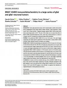
RESULTS In frontal sections of the medulla oblongata, the boundaries of the ION are easy to define, especially in the young rats in which the neurons are closely packed (Fig. 1).The MA0 is clearly separated from the surroundings but the limits between the other subnuclei are not so distinct (Fig. 1).As in the adults, the olivary cells in the newborn rats can easily be distinguished on the basis of their large, clear, somewhat eccentric nucleus and of their granular cytoplasmic bodies in the periphery of the cell (Fig. 1).In the youngest specimens, the nucleus of some cells contains two or several nucleoli. The proportion of these multinucleolated cells regularly decreases from 13.5 k 1.9% in newborn rats to 12.6 + 1.5 (day 31, 8.7 k 1.2 (day 5 ) , and 5 k 0.5 (day 7). A few degenerating cells are visible throughout the olivary complex in the young rats. The maximum number of pycnotic cells is 0.73 dead100 healthy cells on day 3, 0.77 dead100 healthy cells on day 4,and less than 0.02 d e a d 100 healthy cells on day 10. Table 1 presents the calculated mean length, the mean nucleolar diameter, the number of cell counts without correction and with correction for each developmental stage. The individual cell counts have been averaged for each stage investigated and the variation (difference between the extreme values expressed as a percentage of the highest value) from one result to another in each group is quite small, especially from 10 days onward (the lowest value being 80-94% of the highest). The number of cells actually counted represented 20-30% of the total estimated population.
POSTNATAL CELL DEATH IN RAT INFERIOR OLIVE
Fig. 1. Frontal sections in the olivary complex (photomontage). A. Adult rat. B. Three-day-old rat. Inserts show olivary neurons a t a higher magnification. dmcc, dorsomedial cell column; DAO, dorsal accessory olive; MAO, medial accessory olive; PO, principal olive.
301
N. DELHAYE-HOUCHAUI), H. GEOFFKOY, AND .J. MARIAN1
302
TABLE 1. Cell Counts in the Inferior Olivary Nucleus
Age’
Mean length of ION (prn)’
0 (8) 3 (9) 5 (8) 7 (6) 10 (6) 12 (41 15 (6) 20 (6) Adult (6)
1,210 i- 70 1,470 k 50 1,240 80 1,400 i 80 1,630 f 100 2,040 i 120 1,620 I 110 2,200 k 50 2,400 f 110
Mean nucleolar diameter (pmj’ .-
1.75 0.05 1.92 i 0.04 1.90 2 0.10 2.06 i 0.05 2.00 0.22 2.41 +_ 0.01 2.11 f 0.11 2.06 k 0.15 2.13 i 0.05
*
Uncorrected cell counts Left Right
31,542 30,577 22,576 27,186 29,438 30,218 31,105 35,183 32,122
31,899 32,030 24,920 25,646 29,816 30,600 29,373 34,365 32,723
Left
Right
Mean value per olive’
27,508 26,177 20,065 23,780 25,139 25,941 27,189 30,397 28,122
27,803 27,357 21,989 22,396 25.467 26,007 25,408 29,690 28,648
27,655 i 1,005 26,832 k 556 21,027 + 566 23,088 k 1,023 25,303 +_ 1,143 25,974 i 444 26,299 i 937 30,044 637 28,385 +_ 936
Corrected cell counts __---
*
‘Number of counted olives is In parenthesis in the first column. 2Mean values i standard error on the mean (SEMI.
1
b
0
3
5
7
10
12
AGE
(
15
20
Adult
days 1
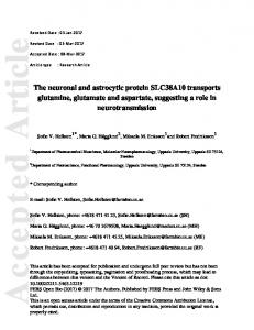
Fig. 2. Postnatal cvolution of inferior OIIVE cell population i n the rat. Each circle reprcwnts the mean valuc~per olive i standard error on the mean ISEMJ. Open circles, uncorrrvted counth: fillcd circlcx.i. corrected counts.
Examination of Table 1 and Figure 2 shows that the ION population size in the newborn rat does not differ significantly from that of the adult (27,655 k 1,005 as opposed to 28,385 +_ 936, P > ,901. The number of neurons decreases slightly but not significantly between birth and 3 days, then decreases markedly between 3 and 5 days (from 26,832 k 556 to 21,027 f 566), the difference being highly significant ( P < ,001); the cell loss during the first 5 days is approximately 25% of the newborn value. After this, there is a slow and regular increase and the cell population again reaches the adult value at 15 days ( P > .10 Student’s t test). The curve established with uncorrected values presents a very similar time course (Fig. 2). Counting the cell nuclei at different stages, according to Abercrombie’s method (’461, reveals a similar evolution of ION cell num-
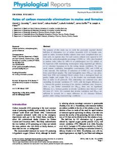
bers. No significant differences related to the sex 01 between left and right olives have been noted at any stage, as can be seen in Table 1.The variation between the two sides of a given animal is of the same order of magnitude as that observed between two animals of the same age. Moreover, summation of counts from both sides in a rat and comparison of the numbers obtained in rats at the same stage does not reduce the variation. The calculated length of the ION reveals a marked linear increase with age (Table l),and the mature size seems to be attained after the cell population has reached its adult value. As a result, the cell bodies are more closely packed in the immature animals, as illustrated in Figures 3 and 4, which represent the caudo-rostra1 distribution of the neurons within the four subnuclei in a 3-day-old rat and in a n adult. Each section in the young rat
POSTNATAL CELL DEATH IN RAT INFERIOR OLIVE
303
A---
MA0
\
A-
DAO
"'\
.........
po
\ \
dmcc
LENGTH ( m m )
caudal
rostral
Fig. 3. Caudo-rostra1 distribution of neurons within t h e four olivary subnuclei in a 3-day-old rat All counts have been corrected for fragmentation error.
A---
MA0
A-
DAO
.........
po dmcc
I
I I I I I
Fig. 4. Caudo-rostra1 distribution of neurons wtthin Lhe four olivary subnuclei in a n adult r a t
N. DELHAYE-ROUCHAUD, B. GEOFFROY, AND J. MARIAN1
304
rostra1
2 360
rost ral
1420
a dmcc
400
460
caudal
ADULT
RAT
3 DAY-OLD RAT
Fig. 5 . Camera lucida drawings ol' three corresponding frontal sections through the inferior olivary nucleus in an adult rat (left columnl and a Y~day~old r a t (right columnl. The numbers on each diagram indicate the distance in microns measured from the caudal pole of'the nucleus.
contains a larger number of neurons but the distribution of the cells along the extent of the nucleus is very similar in both animals, the M A 0 being the largest subnucleus, in terms of its size population and dimensions. Figure 5, which illustrates the aspect of the ION in transverse sections at three different levels in these animals, shows that the configuration of the olivary complex is very similar in corresponding sections. Examination of the cell counts versus age in the different subnuclei reveals a significant decrease of the M A 0 population in the first days so that at 5 days, the cell loss represents 32% of the population counted in the newborn ( P < ,001).Afterward, a slow increase occurs, and the adult value is attained ( P > .20) at 20 days. A comparison of the curve illustrating the evolution of the cell population in the M A 0 (Fig. 6) and of that illustrating the evolution of the ION "in toto" (Fig. 2) shows a similar time course. The cell loss in the other subnuclei is less pronounced (14% for the PO, 19%for the DAO) except for the dmcc (34%). Figure 7 and Table 2 show the distribution of cells in the four subnuclei expressed as a percentage of the whole population for each developmental stage. As in the adult animal, the M A 0 represents the most important subnucleus in the developing rats (42.8-48.3% of the whole population). The relative proportions of the subnuclei do not vary significantly with age ( P > .90 by x2 test) although the importance of the M A 0 decreases slightly with age (Fig. 7).
TABLE 2. Relative Proportions of the Four Main Subnuclei i n the Inferior Olivarv Nucleus Age' 0 (8) 3 (9) 5 (8) 7 (6) 10 (6) 12 (4) 15 (6) 20 (6) Adult (6)
MA02 48.3 44.7 44.2 42.8 43.0 46.0 44.8 44.1 45.8
f 1.6 0.7
+
k 0.3 f 1.5
f 1.2 5 0.7 0.6 0.6 1.6
+ + +
DAO~
PO2 20.8 21.6 23.8 22.1 23.9 23.7 23.7 23.1 23.1
k 0.7 f 0.3 f 1.2
k 0.6
+ 0.4 -
f 0.7 f 0.6 f 0.8 & 0.8
25.2 28.0 27.0 29.1 26.8 25.6 26.1 27.2 25.0
f 0.9
k 0.7
+ 0.7 k 0.9 + 0.7 5 0.9 + 0.7 + 0.3
+1
dmcc' 5.7 5.7 5.0 6.0 6.3 4.7 5.4 5.6 6.1
f 0.3
* 0.2
k 0.2
+ 0.4 k 0.6 f 0.2 f 0.1 0.4 k 0.5
'Number of counted olives is in parenthesis. 'Mean values f SEM.
DISCUSSION The present study was undertaken in developing rats with the aim of determining whether the elimination of redundant olivocerebellar synapses might be related to a reduction of the presynaptic cell population. The most salient result reported here is that cell loss does indeed occur in the ION of the rat after birth but that it takes place before the multiple innervation of Purkinje cells by olivary axons has reached its maximum, suggesting that neuronal death does not play a major role in the shaping of olivocer-
POSTNATAL CELL DEATH IN RAT INFERIOR OLIVE
305 -- M A 0
A-
DAO
A-
*. ....... PO 0 - - - -
drncc
T
I
!
0
3
5
7
10
12 AGE ( days)
20
15
Adult
Fig. 6. Postnatal evolution of the cell population in the four olivary subnuclci. Each symbol represents the mean corrected value per subnucleus 5 SEM.
...................................
A---
.................
OJ
,
0
.......................................
8'.'
3
5
7
10
12 AGE ( d a y s )
15
A---
MA0
A -
DAO
.........
PO
0-
drncc
~
~
~
......................................
----A
20
Adult
Fig. 7. Postnatal evolution of the relative proportions of the four olivary subnuclei
ebellar connections. Since quantitative studies have yielded controversial results in systems like the neuromuscular iunction (see above). the method will be discussed first.
eccentrically in a granular cytoplasm was used as a criterion for identifying ION neurons. The counts were performed everv third section in the voung animals because of the reducedsize of the ION, and* in eGery fifth section in Eva'uation Of the method used for counting the older rats. This sampling procedure has been used by In the present study, the presence of a visible nucleolus several investigators in the ION (Moatamed, '66; Escobar or fragment of nucleolus in a clear round nucleus lying et al., '68; Schild, '70;Mlonyeni, '73;Armstrong and Clarke,
306
N. DELHAYE-BOUCHAUU, R. GEOFFROY,AN11 J. MARIAN1
'79) and Mlonyeni reported that counting cells only every strong and Clarke, '79). The decline in the number of cells 20th section instead of every single section gives a maxi- occurs throughout the rostro-caudal extent of the nucleus mum deviation of 3.8%), which is even lower when the but the loss from the M A 0 is greater than that from the interval between the selected sections is reduced, as in the other nuclei. The rates of pycnotic cells observed in the ION present study. The outlines of the nucleus are distinct what- at 3 and 4 days, i.e., at the beginning of the drop in cell ever the age, so that the chance of including extra olivary number, are relatively low but in the same range as those reported in other parts of the brain (Finlay et al., '82; cells in the counts is very low. In adult and developing rats, a possible source of error is Janowsky and Finlay, '83). Insofar as the clearance of deintroduced by the splitting of the nucleolus during histolog- generating cells must be rapid in young animals, this is ical processing. However, since the nucleolus is the smallest likely to correspond to the observed loss in the ION. The available particle relative to section thickness, the chances presence of pycnotic profiles and the decrease in the counts of erroneous estimation due to double counting of split of healthy cells strengthen the argument that cell death neurons are low, compared to the methods based on the occurs postnatally in the ION and is for the most part counting of nuclei, the average size of which is large in responsible for the 25% decline in cell number between olivary cells, resulting in a higher proportion of nuclear days 3 and 5. In keeping with this, the percentage of pycfragments. Nevertheless, as splitting of the nucleolus in notic nuclei becomes very low at 10 days, after the drop in paraffin sections has been questioned (Konigsmark, '70) the cell number, and while the remodeling of the CF terminals uncorrected values have also been used (see Table 11, and it is not completed (Crepel et al., '81; Mariani and Changeux, appears that if no correction is applied, the same biphasic '81a). Nevertheless, a secondary outward migration of some evolution of the ION population is observed (see Results). neurons from the ION cannot be precluded as discussed in The results presented here give a n average number of other systems (see Clarke, '82). The apparent increase sub28,385 936 cells per olive in the adult rat. This is some- sequent to the cell decline occurs in all nuclei but more what higher than the estimation given by Schild ('70) in markedly in the MAO. No explanation is yet available for the same species, i.e.. 24,400 neurons, but it should be this apparent increase, which has also been observed in emphasized that Abercrombie's formula for correction, mice for the ION neurons during the same postnatal period which was used by Schild, leads to some underestimation (Caddy and Biscoe, '791, at later stages for the Purkinje of the population, compared to the Floderus formula used cells (Diglio and Herrup, '82),and in the locus coeruleus of in the present study. However, the variation from one re- the rat (Ruth and Goldsmith, '83).An explanation would be sult to another in the adult group is of the same magnitude that the whole stock of olivary neurons is already present in its adult location at birth, but some of them could not be as that reported by Schild i'70), the lowest value being 84'k of the highest (as opposed to 88%).Moreover, the relative recognized because of their immaturity, and the increase in proportions of the subnuclei given in Table 2 are in good cell numbers would simply reflect the maturation of these agreement with those already available on a more re- neurons. Alternatively, this increase could represent "newly stricted sample (Schild, '70). The MA0 is the largest olivary appeared" cells, the origin and nature of which are not 1.6% of the total number known. A local multiplication of the olivary neurons apsubnucleus, representing 45.8 0.8%)and 25 l%, pears unlikely as ION cells of the rat undergo their last icf. 49%), the DAO and the PO 23.1 respectively (cf. 25 and 26%). Since the dnicc was not distin- division on embryonic day (ED) 13 for the PO neurons and guished in the above report, possibly because it fuses with on ED 14 for the MA0 neurons (Altman and Bayer, '78). the ventral lamella of the PO in the upper two-thirds of the Residual migratory movements could be involved, espenucleus, and with the DAO at the rostra1 pole of the ION cially those giving rise to the MAO, which matures later iGwyn et al., '77), this probably accounts for the small than the other subnuclei (Altman and Bayer, '78). Whatever the origin of the apparent increase in the cell differences observed in the proportions of the subnuclei. Despite this slight discrepancy, the method used in the number, the evolution of the ION population as a function present study seems t o be accurate in the adult animals. of age could be explained by the combination of two proAs regards the immature rats, a source of error is the cesses: a phase of cell death occurring during the first few presence of multinucleolated cells. This is the reason why days, and a slow increase which becomes detectable when this material was also sectioned at 10 pm to reduce the cell death has slowed or stopped. proportion of nuclear fragments, all of which might contain Synapse elimination in the cerebellum and a nucleolus or a fragment of nucleolus. Although the possicell death in the ION bility of a small overestimation of the real number of cells between birth and 5 days cannot be discarded despite an In the rat cerebellum, multiple innervation of PC, by the appropriate correction, it should be noticed that our count- olivary axons can be detected on postnatal day 3, when 75% ing of the cell nuclei (Abercrombie, '46) confirmed the bi- of the PC, are innervated by a n average of 2.5 fibres (Crepel phasic evolution observed by the counting of nucleoli. et al., '81; Mariani and Changeux, '81a). The redundant contacts increase to a maximum on day 5 with a n average Evolution of the ION population versus age of 3.5 CF, for each PC. Therefore the period of increasing Examination of the curve illustrating the variation of multiple innervation of PC, parallels that of cell death ION population size versus age shows that the evolution is observed in the ION and the peak of multiple innervation biphasic. There is a n initial marked decrease of the cell coincides with the lowest value of the presynaptic neuron population between birth and 5 days, followed by a proges- population size. Moreover the period of apparent increase sive increase, so that the adult value is attained at 15 days. of the ION population (day 5-day 15) is coincident with the Between birth and 5 days, the number of neurons declines elimination of the redundant CF contacts in the cerebelto a value about 75% of that of the newborn. This 25% loss lum. Both facts rule out the possibility that cell death of of ION cells is close to the 30% loss of neurons found in the the olivary neurons could account entirelv for the loss of chick's ION during the 4 days preceding hatching (Arm- multiple innervation of PC, by CF,. It sgould be noticed
POSTNATAL CELL DEATH IN RAT INFERIOR OLIVE that in the chick ION in which the cell loss is exclusively prenatal (Armstrong and Clarke, '791, the cerebellum is much more mature at birth. Although contradictory results have been reported about synapse formation between CF, and PC, in this species, authors agree on the fact that it also begins prenatally (see review in Mugnaini, '69; Foelix and Oppenheim, '74). Unfortunately, no data are available about a possible polyneuronal innervation of the PC, by olivary axons in the chick. Our results and additional data obtained in adult rodents in which multiple innervation of PC, is maintained (Shojaeian et al, '85: companion paper) support the view that the remodeling of the CF terminals in the cerebellum results from a progressive reduction of' axonal branching such as occurs in the rabbit ciliary ganglion (Johnson and Purves, '81), in the avian cochlear nucleus (Jackson and Parks, '82), in the callosal system (Innocenti et al., "77; Innocenti, '81; O'Leary et al., '811,and in the pyramidal tract (Stanfield et al., '82).
307 Delhayc-Bonchaud, N., F. Crepel, and cJ. Mariani (1975) Mise en evidence d'une multi-innervation temporaire des cellules de Purkinje du cervelet par les fibres grimpantes a u cours d u ddveloppement chez le Rat. C.R. Acad. Sci. 281;909-912. Ilesclin, J.C. (19741 Histological evidence supporting the inferior olive a s the major source of cerebellar climbing fibers in the rat. Brain Res 77:365-384. Diglio, T.J., and K. Herrup (1982) A significant fraction of the adult number of mature cerebellar Purkinje cells first appears between postnatal days 16 and 30 in the mouse. Soc. Neurosci. Abstr. p. 636.
Escnbar, A , , E.D. Sampedro, and R.S. Dow (19661Quantitative data on the inferior olivary nucleus in man, cat and vampire h a t J. Comp. Ncxurol. 132:397-404. Finlay, B.L., A.T. Berg, and D.R. Sengelauh (19821 Cell death in the m a m ~ malian visual system during normal development. TI. Superior collicu~ lus. J. Comp. Neurol. 204:318-324. Floderus, S. (19441Ilntersuchungen uber den Bau der menschlichen hypo^ physe mit besonderer Berucksichtigung der qualitativen niikromorphol~ ogischc Verhaltnissc~.Acta Pathol. Microbiol. Scand 53isupp/.i:1-276. Forlix, R.F., and R. Oppenheim (1974) The development of synapses in the cerehellar cortex of the chick embryo. J. Neurocytol. 3.277-294. Frahet.. .J P. (19741A numerical study ofcervical and thoracic ventral nerve ACKNOW1,EDGMENTS roots. cJ. Anat. 118;127-142. The authors wish to thank Dr. P. Derer and Dr. H. Sho- Giordano, D.L., M. Murray, and T.J. Cunningham (1960) Naturally occurring neuron death in the optic layers of superior colliculus of the postjaeian for their help, Dr. C. Henderson for his critical renatal rat. J. Neurocytol. 9:603-614. view of the manuscript, and Drs. J.P. Changeux and P. Gwyn. D.G , G.P. Nicholson, a n d B.A. Flumerfelt (1977)The inferior olivary Laget for their helpful comments throughout the course of nucleus of the rat: A light and electron microscopic study. J. Comp. this study. This work was supported by grants from the Neurol. 174;489-520. College de France, the INSERM and the CNRS (ATP Or- Hamburger, 1'. (1975) Cell death in the development of the lateral motor column of the chick embryo. J. Comp. Neurol. 160.535-546. ganisation et fonctionnement du systeme nerveux). Hnttenlocher. P.R., C. de Courten, L.J. Carey, and H. Van Der 1,oos (1982) Synaptogenesis in human visual cortex. Evidence for synapse elimina~ LITERATIIRE CITED tion during normal development. Neurosci. Lett. 33247-252. Ahercromhie, M. (1946) Estimation of nuclear population from microtome Innocenti, G.M. (1961) Growth and reshaping of axons in the establishment of visual callosal connections. Science 212.824-827. sections. Anat. Rec 94:239-247 r 119781 Prenatal developmcnt of t h e cerrhellar Innocrnti, G.M , L Fiore, and R. Caniiniti (1977)Exuberant prqjectinn into the corpus callosum from the visual cortex of newborn rats. Neurosci. ytogenesis and histogenesis of the inferior olive, Lett. 4.231-242. pontine g r a y , and t h r preccrehellar reticular nuclei. J. Comp. Neurol. 179.49-76. Jackson, H. and T.N Parks (1982) Functional synapse elimination in the developing avian cochlear nucleus with simultaneous reduction in cochArmstrong, K.C., and P.G.H. Clarke (1979) Neuronal death and the devellear nerve axon branching. J. Neurosci 2:1736-1743. opment o f t h e pontinr nuclei and inferior olive in the chick. Neurosci~ ence 4:1635-1647. Janowsky, J.S., and B.L. Finlay 119831 Cell degeneration in early developBennett, M.R., P.A McGrath, D.F. Davey, and I. Hutchiiison 11983, Death ment of the forebrain and cerebellum. Anat. Emhryol. 167.439-447. of motorneurons during the postnatal loss of polyneuronal innervation Johnson, D.A., and D. Purves (19611 Postnatal reduction of neural unit size of rat niuscIc,s. J. Comp. Neurol. 218;351-:KX i n the rabbit ciliary ganglion. J. Physiol. iLond.1318:14:3-159. Brown, M.C., J.K.S. Janscm, and D. Van Essen (19761 Polyneuronal inner- Kdnigsmnrk, B.W. (1970) Methods for the counting of neurons. In W.J.H. vation of skeletal muscle in newborn rats and its elimination during Nauta and S.O.E. Ebbesson (eds): Contemporary Research Methods in maturation. 3.Physiol. (1,ond.I 2fi1.387-422. Neuroanatomy. Berlin: Springer-Verlag, pp. 315-380. Caddy, K.W.T.. a n d T.J. Biscoe (1979) Structural and quantitative studies Lance-Jones, C. (1982) Motoneuron cell death in the developing lumbar on the normal C3H and lurchrr mntant mouse. Philos. Trans. R. Soc. spinal cord o f t h e mouse. Dev. Brain Res. 4:473-479. Iniid. [Rial.] 287,167-201. Mariani. J. ( 1 9 8 3 Elimination of synapses during the development of the Cajal. S.R. (19111 IIistnlogic~du Systeme Nerveux dt, 1'Homme et des V e r ~ central nervous system. In J.P. Changeux, J. Glowinski, M. Imbert, and tebres. Paris: Maloine, 2:80-106. F.E. Bloom ieds): Molecular and Cellular Interactions Underlying Higher Camphell, N.C., and D.M. Armstrong (19831 Topographical localization in Brain Functions. Prog. Brain Res., Vol. 56. New York: Elsevier, pp. 383392. t h r olivocerebellar projection in the rat. An autoradiographic study. Brain Res. 275.235-249. and J.P. Changeux (1960) Etude par enregistrements intracelde I'innervation multiple des cellules de Purkinje par les fibres Chu-Wang, cJ.W.,and R.W. Oppenheim (1978, Cell death ofmotoneurons in grimpantes dans Ie cervelet du ral en developpement C.R. Acad. Sci. the chick embryo spinal cord. I. A light and electron microscopic study (Paris1 291;97-100. of naturally occurring and induced cell loss during development. J. Comp. Neurol. I77:33-58. Mariani, J.,and J.P. Changeux (1981a) Ontogenesis of olivocerebellar relationships. I. Studies by intracellular recordings of the multiple innerClarke, P G.H. (1982) Thc genuineness of isthnio~opticneuronal death in vation of Purkinje cells by climbing fibers in the developing r a t chick embryos. Anat. Embryol. 165389-404. cerebellum. J. Neurosci. 1:696-702. Clarke, P.G.H., L.A. Rogers, and W.M. Cowan (1976)The time of origin and the pattern of survival of neurons in the isthnio-optic nucleus of the Mariani, J., and J.P. Changeux (1981h) Ontogenesis of oli~ocerehellarrelationships. 11. Spontaneous activity of inferior olivary neurons and climbchick. J. Comp. Neurol. 167:125-142. ing fibei-f mediated activity of cerebellar Purkinje cells in developing Crepel, F. (19821 Regression of' functional synapses in the immature m a m ~ rats. J. Neurosci. 1.703-709. malian cerebellum. Tiends Neurosci. 5266-269. Crepel, F., N. Delhaye~Bouchaud,and J.1,. Dupont (1961) Fate of the multi- Mariani, J., N. Delhaye-Bouchaud, B. Geoffroy, a n d H. Shojaeian (1983) Cell counts in t h e inferior olivary nucleus a n d synapse elimination in the ple innervation of cerebellar Purkinje cells by climbing fibers in immadeveloping olivocerebellar system of rodents. Soc. Neurosci. Abstr. p. ture control, X-irradiated and hypothyroid rats. Dev. Brain Res. 1:59858. 71. Mlonyeni, M. (19731 The number of Purklnje cells and inferior olivary Crepel, F., J. Mariani, and N. Delhaye~Bouchaud(1976) Evidence for a neurones in the cat. J. Comp. Neurol. 147.1-10 multiple innervation of Purkinje cells by climbing fibers in the i m m a ~ Moatamed, F. (19661Cell frequencies in the human inferior olivary nuclear t u r e r a t cerebellum. J. Neurobiol. 7:567-578.
308
N. DELHAYE-BOUCHAUI), B. GEOFFROY, AND J. M A R I A N 1
complex. J. Comp. Neurol. 128:109-116. Mugnaini, E. (1969) Ultrastructural studies on the cerebellar histogenesis. 11. Maturation of nerve cell populations and establishment of synaptic connections in the cerebellar cortex of the chick. In R. Llinas (ed): Neurobiology of Cerebellar Evolution and Development. Chicago: Am. Med. Assoc., pp. 749-782. O’Leary, D.D.M., B.B. Stanfield, and W.M. Cowan (1981) Evidence that the early postnatal restriction of the cells of origin of the callosal projection is due to the elimination of axonal collaterals rather than to the death of neurons. Dev. Brain Res. 1:607-617. O’Leary, J.L., J. Inukai, and J.M. Smith (1971)Histogenesis of the cerebellar climbing fiber in the rat. J. Comp. Neurol. 142377-392. Oppenheim, R.W. (1981) Neuronal cell death and some related regressive phenomena during neurogenesis: A selective historical review and progress report. In W.M. Cowan, (ed): Studies in Developmental Neurobiology: Essays in Honor of Viktor Hamburger. New York: Oxford University Press, pp. 74-133. Palay, S.L., and V. Chan-Palay (1974) Cerebellar Cortex. Cytology and Organization. Berlin: Springer-Verlag. Purves, D., and J.W. Lichtman (1980)Elimination of synapses in the devcl-
oping nervous system. Science 210:153-157. Romanes, G.J. (1946) Motor localization and the effects of nerve injury on the ventral horn cells of the spinal cord. J. Anat. 80:117-131. Rootman, D.S., W.G. Tatton, and M. Hay (1981)Postnatal histogenetic death of rat forelimb motoneurons. J. Comp. Neurol. 199:17-27. Ruth, R.E., and S.K. Goldsmith (1983) Postnatal neuron death in the locus coeruleus of rats. Soc. Neurosci. Abstr. p. 857. Schild, R.F. (1970) On the inferior olive of the albino rat. J. Comp. Neurol. I40:255-260. Shojaeian, H., N. Delhaye-Bouchaud, and J. Mariani (1985) Neuronal death and synapse elimination in the olivocerebellar system: 11. Cell counts in the inferior olive of adult x-irradiated rats and zueauer and reeler mutant mice. J. Comp. Neurol. 232309-318. Stanfield, B.B., D.D.M. O’Leary, and C. Fricks (1982) Selective collateral elimination in early postnatal development restricts cortical distribution of rat pyramidal tract neurones. Nature 298:371-373. Tada, K., S. Ohshita, K. Yonenobu, K. Ono, K. Satoh, and N. Shimizu (1979) Development of spinal motoneurons innervation of the upper limb muscle in the rat. Exp. Brain Res. 35287-293.