6776 • The Journal of Neuroscience, July 28, 2004 • 24(30):6776 – 6784
Cellular/Molecular
Neuronal Nicotinic Synapse Assembly Requires the Adenomatous Polyposis Coli Tumor Suppressor Protein Murali Krishna Temburni,* Madelaine M. Rosenberg,* Narendra Pathak, Russell McConnell, and Michele H. Jacob Department of Neuroscience, Tufts University School of Medicine, Boston, Massachusetts 02111
Normal cognitive and autonomic functions require nicotinic synaptic signaling. Despite the physiological importance of these synapses, little is known about molecular mechanisms that direct their assembly during development. We show here that the tumor-suppressor protein adenomatous polyposis coli (APC) functions in localizing ␣3-nicotinic acetylcholine receptors (nAChRs) to neuronal postsynaptic sites. Our quantitative confocal microscopy studies indicate that APC is selectively enriched at cholinergic synapses; APC surface clusters are juxtaposed to synaptic vesicle clusters and colocalize with ␣3-nAChRs but not with the neighboring synaptic glycine receptors or perisynaptic ␣7-nAChRs on chick ciliary ganglion (CG) neurons. We identify PSD (postsynaptic density)-93, -catenin, and microtubule end binding protein EB1 as APC binding partners. PSD-93 and -catenin are also enriched at ␣3-nAChR postsynaptic sites. EB1 shows close proximity to and partial overlap with ␣3-nAChR and APC surface clusters. We tested the role of APC in neuronal nicotinic synapse assembly by using retroviral-mediated in vivo overexpression of an APC dominant-negative (APC-dn) peptide to block the interaction of endogenous APC with both EB1 and PSD-93 during synapse formation in CG neurons. The overexpressed APC-dn led to dramatic decreases in ␣3-nAChR surface levels and clusters. Effects were specific to ␣3-nAChR postsynaptic sites; synaptic glycine receptor and perisynaptic ␣7-nAChR clusters were not altered. In addition, APC-dn also reduced surface membrane-associated clusters of PSD-93 and EB1. The results show that APC plays a key role in organizing excitatory cholinergic postsynaptic specializations in CG neurons. We identify APC as the first nonreceptor protein to function in localizing nAChRs to neuronal synapses in vivo. Key words: nicotinic; cholinergic; nAChR; synapse formation; targeting; neuron; ciliary ganglion; adenomatous polyposis coli; APC
Introduction Nicotinic acetylcholine receptors (nAChRs) function at key interneuronal synapses. Their activation mediates excitatory transmission, reinforces nicotine addiction, and increases memory formation. Malfunction of cholinergic synapses has been implicated in Alzheimer’s disease, schizophrenia, nocturnal frontal lobe epilepsy, and autoimmune autonomic neuropathies. Despite the physiological importance of these synapses, little is known about molecular mechanisms that direct their assembly. Only two proteins have been identified that organize neuronal nicotinic synapses. First, one nAChR subunit, ␣3, is essential for targeting heteromeric ␣3-containing nAChRs to the postsynaptic membrane in autonomic neurons (Williams et al., 1998). Second, PSD (postsynaptic density)-93 regulates synaptic stability; ␣3nAChR clusters disassemble faster after denervation in PSD-93deficient versus wild-type autonomic ganglia (Parker et al., 2004). Received Oct. 15, 2003; revised June 11, 2004; accepted June 11, 2004. This work was supported by National Institutes of Health Grants NS21725 to M.H.J., DK34928 to the Gastroenterology Research on Absorptive and Secretory Processes Center, and P30 NS47243 to the Center for Neuroscience Research Cores. We thank Monica Giovanni and Howard Chen for expert technical assistance with quantitative immunofluorescence microscopy studies and yeast two-hybrid screens; Sean Sullivan for participating in preliminary studies of APC immunolocalization; and Kathleen Dunlap, Daniel Jay, and Angeles Ribera for insightful comments on this manuscript. *M.K.T. and M.M.R. contributed equally to this work. Correspondence should be addressed to Dr. Michele Jacob, Department of Neuroscience, Tufts University School of Medicine, 136 Harrison Avenue, Boston, MA 02111. E-mail:
[email protected]. DOI:10.1523/JNEUROSCI.1826-04.2004 Copyright © 2004 Society for Neuroscience 0270-6474/04/246776-09$15.00/0
In addition, PSD-93 and PSD-95 couple receptor activation to downstream signaling cascades that regulate plasticity at nicotinic and glutamatergic synapses (Migaud et al., 1998; Conroy et al., 2003). Because PSD-93 and PSD-95 organize both nicotinic and glutamatergic synapses, it is possible that a common core of proteins may organize diverse synaptic complexes. We propose that adenomatous polyposis coli (APC) may be one of these key organizers. APC is known best as a tumor-suppressor protein: lossof-function mutations cause colorectal cancer (Livingston, 2001). Although the function of APC in neurons has yet to be defined, several lines of evidence suggest that it may play a role in synaptic differentiation. Muscle-type nAChR clustering induced by agrin requires APC interactions in cultured myotubes (Wang et al., 2003). APC localizes to glutamatergic synapses in hippocampal neurons and binds PSD-95 (Matsumine et al., 1996; Yanai et al., 2000). Moreover, APC is expressed in multiple neuron types and binds -catenin, a widely expressed adhesion complex component of cadherin-mediated synapses, suggesting APC may have a broader synaptic distribution (Brakeman et al., 1999; Fearnhead et al., 2001). An essential role of APC in neurons is indicated by the correlation of APC gene deletions with human mental retardation (Raedle et al., 2001). We tested for a role of APC in neuronal synaptic assembly in vivo using the chick parasympathetic ciliary ganglion (CG). In these experimentally tractable neurons, excitatory cholinergic synapses lie adjacent to inhibitory glycinergic synapses (Tsen et
Krishna Temburni et al. • APC Is Essential for nAChRs Localizing at Synapses
al., 2000). Single CG neurons express two nAChR types, ␣3nAChRs and ␣7-nAChRs, and glycine receptors (GlyRs) that predominantly segregate to distinct synapse-associated surface membrane microregions. The ␣3-nAChRs are concentrated in postsynaptic membrane regions that oppose presynaptic terminal active zones (Jacob et al., 1986; Loring and Zigmond, 1987). Inhibitory GlyR clusters are present in separate but proximal postsynaptic membrane microregions, all under one presynaptic terminal (Tsen et al., 2000). In contrast, ␣7-nAChRs are excluded from the synapse and localize perisynaptically on somatic spines (Jacob and Berg, 1983; Shoop et al., 1999). The discrete localization of the different receptor types provides a valuable comparison for establishing the specificity of protein interactions responsible for orchestrating cholinergic synapse assembly. We show here that APC interactions are required for highdensity accumulations of ␣3-nAChRs, but not GlyRs, at postsynaptic sites in CG neurons in vivo. Our data identify APC as a key nicotinic cholinergic synapse-organizer and define a neural function for APC.
Materials and Methods Antibodies. Primary antibodies used for immunolabeling (1:100 dilution) were: rabbit C-20 to APC C terminus (Santa Cruz Biotechnology, Santa Cruz, CA), mouse ab58 to APC N terminus (Abcam, Cambridge, UK), anti-Chapsyn-110/PSD-93 antiserum (Alomone Labs, Jerusalem, Israel), anti-end binding protein 1 (EB1) monoclonal antibody (mAb) (Transduction Laboratories, Lexington, KY), rabbit anti-EB1 (H-70; Santa Cruz Biotechnology), anti--catenin mAb and anti-N-cadherin mAb (Zymed Laboratories, San Francisco, CA), rat mAb35 to ␣3nAChRs (Developmental Studies Hybridoma Bank, Iowa City, IA), biotinylated ␣-bungarotoxin (Molecular Probes, Eugene, OR) to detect ␣7nAChRs, mAb2b to GlyRs and mAb7a to gephyrin (Alexis Biochemicals, San Diego, CA), anti-synaptic vesicle 2 (SV2) mAb (Developmental Studies Hybridoma Bank), and pan-PSD-95 family mAb (clone K28/ 86.2; Upstate Biotechnology, Lake Placid, NY). Rat anti-hemagglutinin (HA) mAb (clone 3F10; Roche Diagnostics, Indianapolis, IN) and anti-HA Y-11 antisera (Santa Cruz Biotechnology) were used for detecting HA-tagged APC dominant-negative (APC-dn) protein. Secondary reagents used at 1:1000 –2000 dilution were as follows: Alexa 488- and 594-conjugated secondary antibodies raised in rabbit, rat, and mouse (affinity purified) (Molecular Probes), cyanine 3-conjugated streptavidin (Jackson ImmunoResearch, West Grove, PA), and FITC-conjugated avidin distinct cell sorter grade (Vector Laboratories, Burlingame, CA). Ciliary ganglion dissociation. White Leghorn embryonated chick eggs were obtained from Charles River Spafas (North Franklin, CT) and maintained at 37°C in a forced air-draft humidified incubator until use. Embryos were staged according to the classification scheme by Hamburger and Hamilton (1951), and the days of embryonic development refer to the stage (st) rather than the actual days of incubation. The developmental stages used in this study include the following: embryonic day 7 (E7) (st 31–32), E11–E13 (st 37–39), and E18 (st 44). CGs at E11–E13 and E18 were acutely dissociated in Ca 2⫹-free avian basal salt solution using collagenase (1 mg/ml; Type A; Roche) (Williams et al., 1998). Cells were plated onto poly-D-lysine and laminin-coated chamber slides (BD Labware, Bedford, MA) and allowed to attach to the substratum (30 min at 37°C) before immunolabeling. Quantitative confocal microscopy. Double-labeling immunofluorescence and confocal microscopy were done as described with minor modifications (Tsen et al., 2000). Surface nAChRs on freshly dissociated intact CG neurons were immunolabeled at 4°C for 1 hr to avoid staining the large internal biosynthetic pool. Next, cells were rinsed with cold basal salt solution, fixed (1% paraformaldehyde for 5 min), and permeabilized (0.1% saponin for 2 min) before labeling with other antibodies (to APC, PSD-93, -catenin, EB1, N-cadherin, gephyrin, or HA) followed by fluorescent-conjugated secondary reagents. Controls for specific binding in double-labeling studies included omitting the first or second primary antibody in separate tests; only background labeling was detected (data
J. Neurosci., July 28, 2004 • 24(30):6776 – 6784 • 6777
not shown). Cells were examined by confocal microscopy with a Leica (Heidelberg, Germany) TCS SP2 microscope equipped with HeNe (633 nm), Kr (568 nm), and Ar (488 nm) laser sources, using a 63⫻ 1.32 numerical aperture lens. Optical sections were acquired from the top to the bottom of each neuron at ⬃0.5 m step intervals. Laser intensity and photomultiplier tube gain were kept constant across experiments. Settings were chosen such that pixel intensities fell below saturation levels. In addition, the wavelengths of light collected in each detection channel were set such that no detectable bleed-through occurred between the different channels. Pixel intensity profiles along randomly chosen ⬃3 m segments of labeled neuronal surface membrane were assessed using Leica imaging software. Pearson’s correlation coefficients (r) were used to quantify the extent of overlap between two immunolabels and were calculated using Leica, Scion Image (Scion, Frederick, MD), and Microsoft Excel (Microsoft, Seattle, WA) software. Pearson’s correlation coefficient values represent the mean ⫾ SEM of calculations from two to three different labeled surface membrane areas per cell for four to six neurons for each immunolabel pair. Light microscopy. For some experiments, APC-dn infected and uninfected age-matched control CGs (E7, E11–E13, and E18) were processed in parallel for frozen sectioning and immunolabeling as described previously (Tsen et al., 2000). By E8, all CG neurons were functionally innervated. To detect surface ␣3-nAChRs, freshly dissected CGs were treated with collagenase (1 mg/ml; 10 min) to increase reagent access and incubated with mAb35 at 4°C. CGs were rinsed in cold PBS, fixed in 1% paraformaldehyde for 30 min, and processed for frozen sectioning (8 –10 m thick cryosections). Sections were labeled with other antibodies (to HA, gephyrin, APC, PSD-93, or N-cadherin), examined with a Zeiss Axioscope (Zeiss, Thornwood, NY) microscope, and photographed with a SPOT color CCD camera and software (Diagnostic Instruments, Sterling Heights, MI). For all experiments, images were acquired using identical settings for gain and exposure time such that pixel intensities were below saturation levels. To quantify changes in surface membraneassociated labeling in APC-dn versus uninfected control neurons, we assessed pixel intensity profiles along ⬃3 m length segments of the surface membrane using Scion Image. For each immunolabel, pixel intensities for three or more segments per neuron and a total of four to six neurons were evaluated for control and APC-dn expressing CGs. The pixel intensities of the sampled membrane segments were then binned into incremental groups of 10 pixel intensity steps (e.g., 0 –9, 10 –19. . . , up to 255 saturation). We then calculated the percentage of pixels that belonged to each pixel intensity category and plotted the relative frequency distributions of pixel intensities (data shown as graphs of the relative frequency polygons) (see Figs. 5i, 6). For HA with EB1, -catenin, and PSD-93, relative frequency distributions of pixel intensities were acquired using immunolabeled freshly dissociated neurons and confocal microscopy. In addition, we obtained similar results using either frozen sections or freshly dissociated neurons at both E11–E13 and E18. Cloning. An E15 chick CG cDNA library was constructed in Hybrizap vector (Stratagene, La Jolla, CA). Full-length human APC cDNA (gift from Dr. Bert Vogelstein, Johns Hopkins University, Baltimore, MD) was used as a probe to screen 5 ⫻ 10 8 clones by standard methods. Six positive clones were isolated, sequenced, and identified as overlapping cDNAs of APC. Yeast two-hybrid assay. Yeast two-hybrid assays were performed as described previously (Tsen et al., 2000). For the bait construct, chick APC cDNA encoding amino acids 2498 –2844 (numbering on the basis of homology to human and mouse sequences) was cloned into pBTM116 vector and expressed as a LexA DNA binding domain fusion in yeast L40 reporter strain. The target was an E15 chick CG cDNA library constructed in pAD-Gal4 vector (excised from Hybrizap). Coprecipitation assays. In vitro binding assays with recombinant fusion proteins were used to test whether HA-tagged APC dominant-negative peptide prevented the coprecipitation of APC with its targeted binding partners EB1 and PSD-93a. We generated glutathione S-transferase (GST) and maltose binding protein (MBP) fusions of chick APC, EB1, and PSD-93a proteins by cloning into pGEX4T-1 and pMalC2 vectors. The fusion proteins were expressed in Escherichia coli BL21-DE3 cells and purified using GST Sepharose (Sigma, St. Louis, MO) or amylose resin
6778 • J. Neurosci., July 28, 2004 • 24(30):6776 – 6784
Krishna Temburni et al. • APC Is Essential for nAChRs Localizing at Synapses
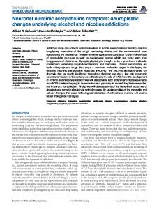
(New England Biolabs, Beverly, MA). The affinity-isolated recombinant fusion peptide was used to coprecipitate its binding partner from bacterial lysates. The coprecipitation assays were performed in the presence or absence of purified HA-tagged APC dominantnegative peptide. Bound protein complexes were eluted, separated by SDS-PAGE, transferred to nitrocellulose, and probed with antisera to the epitope tag. Specific binding was established by using GST alone (negative control) to precipitate the MBP-tagged protein, and using the MBP tag alone for binding to the recombinant GST protein. In vivo coimmunoprecipitation of APC with its binding partners -catenin and PSD-93 was performed on isolated postsynaptic densities (psds). The psds were isolated by subcellular fractionation using previously described methods (Ehlers, 2003; Parker et al., 2004). Briefly, two E18 chick brains (without the brain stem and cerebellum) were homogenized in homogenization buffer (HB) containing protease inhibitors (4 mM HEPES-NaOH, pH 7.4, 0.32 M sucrose, EDTA free protease inhibitor mixture Complete; Roche) and centrifuged at low speed to remove nuclei and blood cells. The supernatant was diluted with 2 ml of 10% Percoll-HB, layered on top of a discontinuous 10⬎20% Percoll-HB gradient, and centrifuged 33,000 ⫻ g in a Beckman L8 – 80 Ultracentrifuge (Beck- Figure 1. APC localization relative to synapses and the different neurotransmitter receptor types on the CG neuron surface. man Instruments, Fullerton, CA) using SW-28 Confocal micrographs of immunolabeled acutely dissociated E18 CG neurons. a, APC localized in patches along a portion of the rotor. The interface between the two Percoll neuronal surface membrane and small clusters throughout the cytoplasm and nucleus. The nuclear labeling was confirmed by layers was collected, diluted to 12 ml with HB, 4⬘,6⬘-diamidino-2-phenylindole staining (data not shown). b, Double-labeled neuron showing that APC surface clusters are and further centrifuged at 20,000 ⫻ g for 20 juxtaposed to synaptic vesicle clusters (SV2 labeling in red; presynaptic terminal). c, Double-labeled neuron showing strong min to pellet the psds. The pellet was resus- colocalization of APC and ␣3-nAChR surface clusters (predominance of yellow fluorescent patches). d, e, In contrast, APC and pended in psd buffer (40 mM HEPES-NaOH, ␣7-nAChRs ( d) and gephyrin (geph; e) (the GlyR directly associated protein) showed little overlap at the neuron surface (predompH 8.1) containing protease inhibitors plus 1 inance of distinct red or green patches). Insets, Threefold magnification views of the boxed regions. Bottom panels, Graphs show mM EDTA (Complete tablets; Roche), solubi- red and green fluorescence intensity profiles for the boxed regions. Staining intensities of surface clusters covaried and correlated lized with 1% Triton X-100, recentrifuged, and with each other for ␣3-nAChRs with APC but not for ␣7-nAChRs and GlyRs/gephyrin with APC (n ⫽ 2–3 surface areas per cell; the final pellet resuspended in 200 – 400 l of 4 – 6 neurons). f, g, Electron micrographs show that APC is localized at the postsynaptic density ( f) (APC detected by horseradish psd buffer. The isolated psds were stored at peroxidase); unlabeled control (ctl) synapse is included for comparison ( g). ⫺80°C and used in immunoprecipitation assays. The psds were incubated with 2 g of antact neurons from either two retroviral infected CGs or uninfected contibody to APC or -catenin, and the immune complexes were affinity trol CGs as we have done previously (Williams et al., 1998). precipitated with protein-G or protein-A Sepharose beads (for ms and rb Quantitative reverse transcription-PCR. To compare ␣3 subunit antibodies, respectively). The bound protein complexes were eluted, sepmRNA levels in individual APC-dn infected CGs versus uninfected conarated by SDS-PAGE, and analyzed by immunoblotting with the antitrol CGs at matched ages, total RNA was isolated, and quantitative rebody directed against the other protein of the candidate interacting pair verse transcription (RT)-PCR was performed using ␣3-specific primers (Ikonomov et al., 1998; Conroy et al., 2003). Controls for antibody bindand mutated internal standards as described previously (Levey et al., ing specificity were included. 1995). Retroviral vector-mediated gene transfer. APC-dn construct [amino acids 2500 –2844; corresponding to the C-terminus tail of APC that conResults tains the EB1 and PDZ (postsynaptic density-95/Discs large/zona APC localizes to nicotinic cholinergic, but not glycinergic, occludens-1) binding domains] (Fearnhead et al., 2001) was generated postsynaptic sites in chick CG neurons and coupled to the sequence encoding the hemaglutinin tag (YPYDVPOur immunofluorescent confocal microscopy studies showed DYA) at its 5⬘ end by PCR. The APC-dn cDNA was subcloned into the that APC is expressed in chick CG neurons. We used two different avian-specific retroviral vectors RCASBP of the A and B envelope subanti-APC antibodies that recognize distinct N-terminal and Cgroup types (Homburger and Fekete, 1996). Viral stocks were prepared terminal epitopes. APC was concentrated at or near the plasma in DF1 chicken fibroblast cells (American Type Culture Collection, Mamembrane in discrete patches that extended along a portion of nassas, VA). CGs were infected in ovo as described previously (Williams the neuron surface (Fig. 1a). APC was also localized in small et al., 1998) and sampled 1–2 weeks later. clusters throughout the cytoplasm and nucleus, as previously deElectron microscopy. E18 CGs were processed for ultrastructural localscribed for other cell types (Bienz, 2002). APC surface patches ization of APC relative to synapses using horseradish peroxidase for dewere juxtaposed to presynaptic terminals on the basis of their tection using previously described methods (Williams et al., 1998). partial overlap with synaptic vesicle clusters (Fig. 1b). APC Surface ␣3-nAChR binding assay. Surface levels of ␣3-nAChRs were strongly colocalized with ␣3-nAChR surface clusters, as indicated assayed by saturable specific binding of mAb35, followed by biotinylated by the predominance of yellow fluorescent patches on the neuanti-rat antibody and 125I-labeled strepavidin to freshly dissociated, in-
Krishna Temburni et al. • APC Is Essential for nAChRs Localizing at Synapses
rons (Fig. 1c). Fluorescence staining intensities of APC and ␣3nAChR surface clusters covaried and strongly correlated with each other [Pearson’s correlation coefficient (r) ⴝ 0.62 ⫾ 0.05; r can range from ⫺1.0 to ⫹1.0] (Fig. 1c, bottom panel). In contrast, little colocalization was observed for APC with either clusters of ␣7-nAChRs or GlyRs (detected by staining for the GlyR directly associated protein gephyrin), as indicated by the predominance of distinct red or green patches (Fig. 1d,e). Staining intensities of APC and ␣7-nAChR and GlyR/gephyrin surface clusters did not covary (r ⴝ 0.05 ⫾ 0.05 and 0.01 ⫾ 0.04, respectively) (Fig. 1d,e, bottom panels). In addition, we performed 10 separate double-labeling experiments and examined randomly selected neurons by confocal or epifluorescence microscopy. All 90 neurons examined for APC and ␣3-nAChR labeling showed strong colocalization at the surface membrane. Ultrastructural analysis demonstrated at high resolution that APC is concentrated at the postsynaptic density (Fig. 1f ). In contrast to the extensive overlap with ␣3-nAChRs, weak colocalization was observed between APC with ␣7-nAChRs and GlyRs: only 29% (23 of 80) and 15% (8 of 53) of neurons had any overlap, respectively (a few small punctae of yellow fluorescence). Given that perisynaptic ␣7-nAChRs and synaptic GlyRs are localized in close vicinity to ␣3-nAChR-rich postsynaptic membrane regions, some small amount of overlap with APC staining would be expected. Similar to our findings for APC, there is precedence for other proteins showing differential colocalization with the diverse receptor types despite their physical proximity to one another in the surface membrane. For example, PSD-93 and PSD-95 colocalize with ␣3-nAChRs but not ␣7-nAChRs, and gephyrin colocalizes with GlyRs but not ␣3-nAChRs on CG neurons as detected by immunolabeling with confocal microscopy (Tsen et al., 2000; Conroy et al., 2003). Overall, the colocalization studies showed that APC was selectively enriched at nicotinic cholinergic postsynaptic sites. Full-length APC and several binding partners are expressed in the CG and interact in vitro Microtubule end binding protein EB1, -catenin, and PSD-95 protein family members are known to interact with APC in noncholinergic cells (Fearnhead et al., 2001). We tested for the presence of these APC binding partners in chick CG neurons. Although a truncated APC isoform that lacks the EB1 and PSD-95 protein family binding domains has been reported (Nakagawa et al., 1998), we found that full-length APC is the dominant isoform expressed in the chick CG by library screening. All six isolated APC clones were overlapping cDNAs of a single isoform that contained the consensus EB1 and PDZ binding domains in the C terminus. Next, we identified PSD-93 as the PSD-95 protein family member that interacts with APC in the CG by yeast two-hybrid screens of our CG cDNA library using the chick APC C-terminal fragment (amino acids 2498 –2844) as bait. The screens identified EB1 and PSD-93 as APC binding partners in the CG. We obtained eight EB1 and three PSD-93 cDNA clones as positives and confirmed their interaction with the chick APC C-terminal fragment by coprecipitation in vitro using recombinant peptides (data not shown). The interacting PSD-93 protein is a short isoform (PSD93a) that lacks the Src homology 3 and guanylate kinase domains and has been shown previously to be expressed in the CG (Conroy et al., 2003). APC and its binding partners form a complex in the postsynaptic density in vivo We tested for APC interactions with its binding partners at neuronal synapses by coimmunoprecipitation from psds. The psds
J. Neurosci., July 28, 2004 • 24(30):6776 – 6784 • 6779
Figure 2. APC, -catenin (-Cat), and PSD-93 form a complex in the postsynaptic density in vivo. The psds were isolated from E18 chick brain (without the cerebellum and brainstem). The psd preparation was immunoprecipitated (IP) with anti-APC antibody, and the immunoprecipitate was probed with antibodies that recognize -catenin and PSD-93. In addition, the psds were immunoprecipitated with anti--catenin antibody and probed with antibodies that recognize APC and PSD-93. APC coprecipitated both -catenin and PSD-93, whereas -catenin coprecipitated APC, but not PSD-93, from the psds. In addition, -catenin is more abundant than APC in the coprecipitates, likely because of multiple -catenin molecules that bind to one APC molecule via the 15 amino acid and 20 amino acid repeat domains (Rubinfeld et al., 1995; Fearnhead et al., 2001).
were isolated from E18 chick brain by subcellular fractionation (Ehlers, 2003; Parker et al., 2004). We found that APC coprecipitates both -catenin and PSD-93, whereas -catenin coprecipitates APC, but not PSD-93, from the psds (Fig. 2). These findings confirm a previous report that -catenin does not bind directly to PSD-93 (Honjo et al., 2000). Thus, although cytoplasmic -catenin, which binds to APC, is targeted for rapid degradation in the Wnt pathway, our data indicate that APC, -catenin, and PSD-93 are sequestered and form a complex in the psd in vivo. APC binding partners localize to CG cholinergic synapses We determined by immunolabeling combined with confocal microscopy that PSD-93, -catenin, and N-cadherin (the cellular adhesion molecule that binds -catenin) were selectively enriched at cholinergic synapses on CG neurons (Fig. 3a–l ). They colocalized with ␣3-nAChRs and APC at the neuron surface (Fig. 3a,d,e,h,i,l ) and were juxtaposed to synaptic vesicle clusters (presynaptic terminals) (data not shown). Fluorescence staining intensities covaried for surface clusters of ␣3-nAChRs and APC with PSD-93, -catenin, and N-cadherin and strongly correlated with each other (Fig. 3, table). Thus, although APC and N-cadherin compete for interacting with the same domain of -catenin (Hulsken et al., 1994), the three proteins colocalized at nicotinic postsynaptic sites. In contrast, little colocalization was observed for either the neighboring synaptic GlyRs or perisynaptic ␣7-nAChRs. Staining intensities of surface clusters of ␣7-nAChR and GlyR/gephyrin and PSD-93, -catenin, and N-cadherin were not correlated with each other (Fig. 3, table). In addition, the APC binding partner EB1 showed close proximity to and partial colocalization with ␣3-nAChR and APC surface clusters but did not accumulate much at the surface membrane (Fig. 3m). As expected for a microtubule end binding protein, the bulk of EB1 staining was cytoplasmic. Altogether, our localization studies identify APC, PSD-93, -catenin, and N-cadherin as
6780 • J. Neurosci., July 28, 2004 • 24(30):6776 – 6784
Krishna Temburni et al. • APC Is Essential for nAChRs Localizing at Synapses
components of cholinergic, but not glycinergic, postsynaptic sites in CG neurons. Overexpressed APC dominant negative caused decreases in ␣3-nAChR surface levels and clusters in vivo We hypothesized that APC and its binding partners may function in nicotinic cholinergic synapse assembly on the basis of the role of APC surface clusters in capturing EB1-tagged microtubule plus ends at specific surface sites (cadherin/-cateninrich complexes in ectodermal cells) and the synapse organizing functions of PSD-93 and -catenin in glutamatergic neurons (Lee et al., 2000; Lu et al., 2001; Sheng and Sala, 2001; Malinow and Malenka, 2002; McGee and Bredt, 2003; Mimori-Kiyosue and Tsukita, 2003; Yu and Malenka, 2003). In particular, we speculate that the interactions of APC with EB1, -catenin, and PSD-93 make it a candidate for a role in directing ␣3-nAChR transport to and stabilization at postsynaptic sites. To test this hypothesis, we generated a dominantnegative APC C-terminal fragment (APCdn) to block the interaction of endogenous APC with both EB1 and PSD-93 during synapse formation in CG neurons in vivo. The APC-dn (amino acids 2500 – 2844) did not contain the -catenin binding domain to avoid disrupting the Wnt signaling pathway. In addition, the APC-dn was epitope tagged with HA to distinguish Figure 3. Localization of APC binding partners at nicotinic cholinergic postsynaptic sites. a, d, e, h, i, l, Confocal micrographs of infected cells from uninfected cells by double-labeled acutely dissociated E18 CG neurons showing the APC binding partners PSD-93 and -catenin (-Cat) and the HA immunolabeling. We used retroviral cellular adhesion molecule N-cadherin (N-Cad) predominantly colocalized with ␣3-nAChR (a, e, i) and APC (d, h, l ) surface clusters vector-mediated gene transfer to over- (overlap, yellow). b, c, f, g, j, k, In contrast, little colocalization was seen with ␣7-nAChRs (b, f, j) and gephyrin (geph) (c, g, k). m, In addition, the APC binding partner EB1 showed close proximity to and partial overlap with ␣3-nAChR surface clusters. However, express the APC-dn construct. EB1 did not accumulate much at the surface membrane. Instead, EB1 labeling was predominantly cytoplasmic. Insets, Twofold Before expression in CG neurons in magnification views of boxed regions. The table shows that the fluorescence staining intensities of surface clusters covaried and vivo, we tested the specificity and efficacy correlated with each other for PSD-93, -catenin, and N-cadherin with ␣3-nAChRs and APC but not with ␣7-nAChRs or GlyRs/ of the APC-dn construct. First, we estab- gephyrin (n ⫽ 2–3 surface areas per cell; 4 – 6 neurons). lished that HA-tagged APC-dn peptide specifically interacted in vitro with fullcaused no change in the amounts of APC and -catenin that length EB1 and PSD-93a and blocked the binding of APC recomcoprecipitated from lysates of retroviral infected versus uninbinant fusion protein to these two binding partners by using yeast fected control fibroblasts (data not shown). Based on the specitwo-hybrid directed interaction assays and coimmunoprecipitaficity and efficacy of the APC-dn construct in the in vitro binding tion assays (data not shown). Second, we tested the efficacy of the assays and DF1 fibroblasts, we overexpressed the blocking pepAPC-dn blocking peptide in chick DF1 fibroblasts in vitro. The tide in chick CG neurons during synapse formation in vivo. overexpressed APC-dn led to alterations in the localization of APC-dn led to specific and dramatic decreases in ␣3-nAChR endogenous APC and EB1 (Fig. 4). There were dramatic desurface clusters (Fig. 5a,b) relative to those on uninfected control creases in endogenous APC and EB1 surface membraneCG neurons (Fig. 5f ). In sharp contrast, neighboring ␣7-nAChR associated clusters in APC-dn infected fibroblasts compared with and GlyR clusters were not detectably altered (Fig. 5d,e). We uninfected control cells. The APC clusters were distributed in found a quantitative correlation between APC-dn expression levradial arrays throughout the cytoplasm of the APC-dn infected els (pixel intensity of HA immunolabeling) and the extent of cells (Fig. 4a). This staining pattern suggested localization of APC reductions in ␣3-nAChR clusters (Fig. 5h). In particular, the proalong microtubules, as expected on the basis of reports that APC portion of neurons per frozen ganglion cross-section that had binds to and moves along microtubules independent of its interlarge densely stained ␣3-nAChR surface clusters was sevenfold actions with EB1 (Barth et al., 2002; Jimbo et al., 2002). Unfortulower for heavily APC-dn infected neurons and twofold lower for nately, we were unable to test for changes in endogenous APC lightly APC-dn infected neurons relative to the values for control and EB1 coprecipitation levels, because the available anti-EB1 neurons [uninfected neurons within the same CG (internal conantibody did not work in the in vivo coimmunoprecipitation trol) and uninfected age-matched CGs]. In contrast, there were assays as reported previously (Su et al., 1995; Berrueta et al., no significant differences in the proportion of neurons with large densely stained GlyR and ␣7-nAChR surface clusters for APC-dn 1999). As an indication of specificity, the overexpressed APC-dn
Krishna Temburni et al. • APC Is Essential for nAChRs Localizing at Synapses
J. Neurosci., July 28, 2004 • 24(30):6776 – 6784 • 6781
tests for specificity, APC-dn overexpression caused no significant change in the following: intracellular ␣3-nAChR levels, representing the biosynthetic pool (Fig. 5c); ␣3 subunit mRNA levels (quantitative RT-PCR); and presynaptic terminal morphology (synaptic vesicle immunolabeling) (Fig. 5j). Additionally, there was no change in ␣3-nAChR surface clusters on retroviral-green fluorescent protein (GFP) infected CG neurons (as a negative control) (Fig. 5k). Overall, the overexpressed APC-dn caused specific changes in surface ␣3-nAChRs. To confirm the in vivo efficacy of the dominant negative construct and further define the synapse organizing functions of APC interactions, we looked for changes in surface levels of APC and its binding partners in infected versus uninfected control CG neurons. We found no change in APC or -catenin surface levels (frequency distribution of pixel intensities), whereas EB1 and PSD-93 surface membrane-associated labeling was reduced (Fig. 6). Similar to our results, APC interactions with EB1 are not required for APC localizing to specialized surface sites in polarized epithelial cells (Barth et al., 2002). Together, our findings suggest that the overexpressed APC-dn specifically blocked endogenous APC interactions with both EB1 and PSD-93 in CG neurons in vivo. Importantly, the specific decreases in PSD-93 and EB1 surface membrane-associated clusters further suggest that APC interactions are required to localize or selectively retain these two binding partners at nicotinic cholinergic postsynaptic sites. Figure 4. Efficacy of the overexpressed APC dominant-negative construct tested in chick fibroblasts in vitro. a–c, Epifluorescence micrographs of double-labeled chick DF1 fibroblasts showing that the overexpressed APC-dn ( a) led to drastic decreases in endogenous APC (green) and EB1 (red) surface membrane-associated clusters compared with uninfected control fibroblasts (b, c). The APC clusters were distributed in radial arrays throughout the cytoplasm of the APC-dn infected cells ( a). To distinguish endogenous APC from the exogenous APC-dn (APC C-terminal peptide fragment), we used the anti-APC antibody that recognizes the N-terminal epitope. Insets, Twofold magnification views of boxed regions in control uninfected fibroblasts (b, c).
infected neurons versus uninfected control neurons (Fig. 5h). In addition, we also measured the pixel intensities of ␣3-nAChR surface membrane labeling in heavily APC-dn infected versus uninfected control neurons (Fig. 5i). The relative frequency distribution of the pixel intensities shows that there is a substantial decrease in the high fluorescence intensity labeling in the APC-dn expressing neurons. In addition, decreases in ␣3-nAChR clusters were seen at both early (E7) (data not shown) and late (E13) stages of synapse formation and maturation, suggesting that the clusters never form (as opposed to their forming and not being retained). To test whether the decrease in ␣3-nAChR clusters reflected a diffuse distribution or reduced receptor numbers, we measured ␣3-nAChR surface levels by specific binding of anti-receptor antibody (mAb35), biotinylated secondary antibody, and 125Istrepavidin to freshly dissociated neurons from individual CGs (Williams et al., 1998). There were twofold reductions in the total number of surface ␣3-nAChRs per APC-dn infected CG relative to uninfected control CG values ( p ⬍ 0.01; Student’s t test; n ⫽ 35 APC-dn CGs and 38 control CGs from six separate experiments) (data not shown). This analysis underestimates the decrease in surface ␣3-nAChRs because not all neurons in the CG were infected (20 ⫾ 3% of the neurons per CG did not have detectable HA-APC-dn immunolabeling). The specific changes in ␣3-nAChRs, but not GlyRs and ␣7nAChRs, suggested that the effects of the overexpressed blocking peptide were restricted to the targeted interactions. As additional
Discussion The major finding reported here is that the tumor-suppressor protein APC is required for high-density accumulations of ␣3nAChRs at postsynaptic sites in CG neurons in vivo. This study identifies APC as the first nonreceptor protein to function in localizing nAChRs at neuronal synapses. We identify EB1, -catenin, and PSD-93 as APC binding partners in the CG. We show that APC, PSD-93, -catenin, and N-cadherin (the cell adhesion molecule that binds -catenin) are selectively enriched at cholinergic synapses; they colocalize with surface clusters of ␣3nAChRs and one another and are juxtaposed to synaptic vesicle clusters on CG neurons. In contrast, little colocalization was observed for either the neighboring synaptic GlyRs or perisynaptic ␣7-nAChRs. In addition, the microtubule end-binding protein EB1 shows close proximity to and partial overlap with surface clusters of ␣3-nAChRs and APC. Importantly, in vivo overexpression of an APC dominant-negative peptide causes dramatic decreases in ␣3-nAChR surface levels and clusters but not of GlyRs or ␣7-nAChRs. In addition, the overexpressed APC-dn also leads to reductions in PSD-93 and EB1 surface membraneassociated clusters. These results demonstrate that APC plays a key role in the assembly of excitatory cholinergic, but not inhibitory glycinergic, postsynaptic specializations in CG neurons. This work provides new insights into molecular interactions that direct the formation of neuronal nicotinic synapses in vivo and defines a neural function for APC. Blocking the interactions of APC with both EB1 and PSD-93 dramatically decreases ␣3-nAChR surface clusters. However, new studies indicate that PSD-93 is not required for ␣3-nAChR clustering at neuronal synapses (Conroy et al., 2003; Parker et al., 2004). Together, the data suggest that the interactions of APC with EB1, but not with PSD-93, target ␣3-nAChRs to postsynaptic sites in neurons. We speculate that the interactions of APC with EB1 direct ␣3-nAChR surface delivery (microtubule-mediated transport) or stabilization at postsynaptic sites by regulating the micro-
6782 • J. Neurosci., July 28, 2004 • 24(30):6776 – 6784
Krishna Temburni et al. • APC Is Essential for nAChRs Localizing at Synapses
tubule cytoskeleton. In our model, EB1 tags the plus ends of a subset of microtubules, and APC accumulates at synapses marked by -catenin/N-cadherin complexes. APC directs ␣3-nAChR delivery to the synapse by capturing EB1-tagged microtubule plus ends and thereby positioning a microtubulebased transport pathway in close proximity to -catenin/N-cadherin-marked postsynaptic regions. In addition, APC and EB1 interactions may be required for stabilizing ␣3-nAChRs at postsynaptic sites by anchoring microtubules and thereby regulating microtubule-cortical actin cytoskeletal interactions. Moreover, these two APC functions may not be mutually exclusive. Our model is based on the roles of APC and EB1 in polarized epithelial cells (Dikovskaya et al., 2001; Lu et al., 2001; Bienz, 2002). In these cells, APC accumulates at specific surface sites and directs the capture of EB1tagged microtubules (to the -catenin/ cadherin-rich adherens junctions in Drosophila ectodermal cells) (Lee et al., 2000; Lu Figure 5. In vivo overexpression of an APC dominant-negative peptide reduced surface clusters (surf) of ␣3-nAChRs, but not of et al., 2001; McCartney et al., 2001; Barth el ␣7-nAChRs or GlyRs, on CG neurons. Epifluorescence micrographs show double-labeled E11–E13 CG frozen sections. a–c, ␣3al., 2002; Mimori-Kiyosue and Tsukita, nAChR (a, b; red) surface clusters were dramatically reduced in neurons overexpressing the HA-tagged APC-dn (green) compared with those on nearby uninfected neurons [internal control (Ctl)], whereas intracellular ␣3-nAChR labeling (c; representing the 2003). Thus, APC and EB1 interactions anbiosynthetic pool) was not detectably altered. d, e, In sharp contrast to ␣3-nAChRs, GlyR ( d) and ␣7-nAChR ( e) surface clusters chor microtubule plus ends at precise posi- were not apparently altered in neurons overexpressing the APC-dn compared with those on nearby uninfected control neurons. tions of the cell surface. A key role for APC in Uninfected control CG age-matched and processed in parallel showed typical ␣3-nAChR surface clusters ( f) and internal staining tethering microtubules is also suggested by ( g). As additional indications of specificity, there were no detectable changes in presynaptic terminal morphology ( j) [synaptic studies that show heterozygous APC mutant vesicle (SV2) labeling; red] or ␣3-nAChR surface clusters on retroviral GFP-infected neurons (k; as a negative control). HA staining mice have substantially fewer microtubule (green) shows that retroviral infection was restricted to CG neurons and occasionally a few glial cells (small HA-positive cells arrays anchored to the specialized basal surrounding the neuronal somata). h, Histogram showing the proportion of the neuron population with large densely stained membrane of polarized epithelial support- neurotransmitter receptor surface clusters for APC-dn overexpressing CG neurons and uninfected control neurons. For infected ing cells (Mogensen et al., 2002). All to- CGs, individual neurons were classified into three groups on the basis of APC-dn expression levels as judged by the pixel intensity gether, the data suggest that the interactions of HA immunolabeling: heavy, light, or not detectable. Cells considered to be heavily labeled had pixel intensities ranging from 105 to 200, lightly labeled ranging from 50 to 105, and not detectable labeling had pixel intensity levels below 30 after subtracting of APC with EB1 may direct high-density background levels. Bars represent the mean (⫾SEM) from the analysis of ⬎427 randomly selected neurons (1125 APC-dn accumulations of ␣3-nAChRs at postsyn- infected neurons for ␣3-nAChR clusters) from three or more embryos. The asterisk indicates significant difference from controls aptic sites in vivo. ( p ⬍ 0.001; ANOVA). i, Graph showing substantial decreases in the pixel intensity of ␣3-nAChR surface labeling on heavily PSD-93 is a member of the PSD-95- APC-dn infected neurons versus uninfected control neurons at matched ages. Pixel intensities were measured along randomly related protein family and a well charac- chosen ⬃3 m segments of labeled neuronal surface membrane (n ⫽ 3 or more segments per neuron; 4 – 6 neurons). The values terized component of the glutamatergic were binned into incremental groups of 10 pixel intensity steps (from 0 to 9, 10 to 19. . . , up to saturation). We then calculated the postsynaptic complex. PSD-93 interacts percentage of pixels that belonged to each pixel intensity category and plotted the data as relative frequency polygons. with and links cytoskeletal, signaling, and overexpressed APC-dn led to decreases in PSD-93 surface clustransmembrane proteins (Brenman et al., 1996; Kim et al., 1996; ters in CG neurons, suggesting that APC interactions are required McGee et al., 2001; Sheng and Sala, 2001; Garner et al., 2002; to localize or selectively retain PSD-93 at nicotinic postsynaptic Malinow and Malenka, 2002; McGee and Bredt, 2003). Surprissites in vivo. ingly, we and others found that PSD-93 is also present at neuronal Similar to our findings in neurons in vivo, recent work in nicotinic cholinergic postsynaptic sites (Conroy et al., 2003; Parker et al., 2004; our study). However, studies of PSD-93 funcmuscle shows that APC localizes to the neuromuscular junction tion using knock-out mice or in vitro models have not yet eluciand is required for nAChR clustering induced by agrin on myodated the role of PSD-93 in cholinergic synapse formation. The tubes in vitro (Wang et al., 2003). Interestingly, APC binds distudies show that PSD-93 is not required for cholinergic synapses rectly to the 1-subunit of the muscle-type nAChRs. In contrast, and ␣3-nAChR clusters to form or for normal levels of functional we found that neither APC nor its binding partners EB1 and nAChRs on autonomic neurons (Conroy et al., 2003; Parker et PSD-93a directly bind to ␣3 (yeast two-hybrid-directed interacal., 2004). However, loss of synaptic PSD-93 and PSD-95 in vitro tion assays and in vitro coimmunoprecipitation assays; data not reduces spontaneous EPSC frequency (Conroy et al., 2003). The shown). We focused on ␣3 because our previous studies showed decreased synaptic activity suggests that PSD-93 may be required that this subunit is essential to target heteromeric nAChR chanto organize a complex that signals retrogradely to the presynaptic nels to synapses in CG neurons in vivo (Williams et al., 1998). terminal and thereby regulates synaptic function (Dean et al., However, there is no significant sequence identity between the region of 1 that binds to APC and any of the nAChR subunits 2003; Prange and El-Husseini, 2003). We identified PSD-93 as an expressed in CG neurons (␣3, ␣5, 2, 4, or ␣7). APC binding partner in the CG. Importantly, we show that the
Krishna Temburni et al. • APC Is Essential for nAChRs Localizing at Synapses
J. Neurosci., July 28, 2004 • 24(30):6776 – 6784 • 6783
Berrueta L, Tirnauer JS, Schuyler SC, Pellman D, Bierer BE (1999) The APC-associated protein EB1 associates with components of the dynactin complex and cytoplasmic dynein intermediate chain. Curr Biol 9:425– 428. Bienz M (2002) The subcellular destinations of APC proteins. Nat Rev Mol Cell Biol 3:328–338. Brakeman JS, Gu SH, Wang XB, Dolin G, Baraban JM (1999) Neuronal localization of the Adenomatous polyposis coli tumor suppressor protein. Neuroscience 91:661– 672. Brenman JE, Christopherson KS, Craven SE, McGee AW, Bredt DS (1996) Cloning and characterization of postsynaptic density 93, a nitric oxide synthase interacting protein. J Neurosci 16:7407–7415. Chen L, Chetkovich DM, Petralia RS, Sweeney NT, Kawasaki Y, Wenthold RJ, Bredt DS, Nicoll RA (2000) Stargazin regulates synaptic targeting of AMPA receptors by two distinct mechanisms. Nature 408:936 –943. Conroy W, Liu Z, Nai Q, Coggan J, Berg DK (2003) PDZ-containing proteins provide a functional postsynaptic scaffold for nicotinic Figure 6. In vivo overexpression of the APC dominant-negative peptide led to decreases in EB1 and PSD-93 surface membranereceptors in neurons. Neuron 38:759 –771. associated labeling in CG neurons. A–D, The graphs show decreases in the pixel intensity of EB1 ( B) and PSD-93 ( D) surface Dean C, Scholl FG, Choih J, DeMaria S, Berger J, membrane-associated labeling in APC-dn infected neurons versus uninfected control CG neurons. Inset, To enhance visualization Isacoff E, Scheiffele P (2003) Neurexin mediof the decrease in EB1 surface membrane-associated labeling, we separately calculated and graphed the relative frequency ates the assembly of presynaptic terminals. Nat distribution of pixel intensities in the higher range only (pixel intensities from 140 to 255). In contrast, APC ( A) and -catenin Neurosci 6:708 –716. (-cat) ( C) surface labeling was not detectably different in infected versus uninfected control CG neurons (n ⫽ 3 or more labeled Dikovskaya D, Zumbrunn J, Penman GA, Nathke IS (2001) The adenomatous polyposis coli surface membrane segments per neuron; 4 – 6 neurons for each immunolabel). protein: in the limelight out at the edge. Trends Cell Biol 9:378 –384. Consistent with our model that APC functions with other Ehlers MD (2003) Activity level controls postsynaptic composition and sigproteins to localize ␣3-nAChRs to synapses, ␣3-nAChRs and naling via the ubiquitin-proteasome system. Nat Neurosci 6:231–242. PSD-93 coprecipitate in a complex in vivo from mammalian and Fearnhead NS, Britton MP, Bodmer WF (2001) The ABC of APC. Hum Mol avian autonomic ganglia (Conroy et al., 2003; Parker et al., 2004). Genet 10:721–733. However, the proteins do not interact directly in the in vitro Garner CC, Zhai RG, Gundelfinger ED, Ziv NE (2002) Molecular mechabinding assays, similar to our findings (Conroy et al., 2003). nisms of CNS synaptogenesis. Trends Neurosci 25:243–251. None of the nAChR subunits have an apparent consensus PDZHomburger SA, Fekete DM (1996) High efficiency gene transfer into the embryonic chicken CNS using B-subgroup retroviruses. Dev Dyn binding motif. Thus, an adapter-like protein may be required to 206:112–120. link ␣3-nAChRs to PSD-93 or APC. Precedence exists for an Honjo Y, Nakagawa S, Takeichi M (2000) Blockade of cadherin-6B activity intermediate protein (Stargazin) linking neurotransmitter recepperturbs the distribution of PSD-95 family proteins in retinal neurones. tors (AMPA receptors) and PSD-95 (Chen et al., 2000; Schnell et Genes Cells 5:309 –318. al., 2002). Hulsken J, Birchmeier W, Behrens J (1994) E-cadherin and APC compete The importance of the role of APC in the vertebrate nervous for the interaction with beta-catenin and the cytoskeleton. J Cell Biol system is highlighted by the correlation of APC gene deletions 127:2061–2069. with various forms of human mental retardation (Raedle et al., Ikonomov O, Kulesa M, Shisheva A, Jacob MH (1998) Innervation and target tissue interactions induce Rab-GDP dissociation inhibitor (GDI) ex2001). However, the neural function of APC was previously unpression during peripheral synapse formation in developing chick ciliary defined. We show here that APC interactions with its binding ganglion neurons in situ. J Neurosci 18:6331– 6339. partners are required for high-density accumulations of ␣3Jacob MH, Berg DK (1983) The ultrastructural localization of ␣-bungaronAChRs at CG synapses in vivo and thereby define a neural functoxin binding sites in relation to synapses on chick ciliary ganglion tion for APC. In addition, APC is the first overlapping componeurons. J Neurosci 3:260 –271. nent in neurons and muscle identified to function in nicotinic Jacob MH, Lindstrom JM, Berg DK (1986) Surface and intracellular districholinergic synapse formation. Moreover, APC is also concenbution of a putative neuronal nicotinic acetylcholine receptor. J Cell Biol 103:205–214. trated at glutamatergic synapses in hippocampal neurons (MatJimbo T, Kawasaki Y, Koyama R, Sato R, Takada S, Haraguchi K, Akiyama T sumine et al., 1996). The APC binding partner PSD-93 has orga(2002) Identification of a link between the tumor suppressor APC and nizing functions at glutamatergic and nicotinic cholinergic the kinesin superfamily. Nat Cell Biol 4:323–327. synapses, whereas both proteins are excluded from inhibitory Kim E, Cho KO, Rothschild A, Sheng M (1996) Heteromultimerization and neuronal synapses (Sheng and Sala, 2001; Conroy et al., 2003; NMDA receptor clustering activity of Chapsyn-110, a member of the McGee and Bredt, 2003; Parker et al., 2004; our study). In sumPSD-95 family of proteins. Neuron 17:103–113. mary, accumulating evidence indicates that APC and its interactLee L, Tirnauer JS, Li J, Schuyler SC, Liu JY, Pellman D (2000) Positioning of ing proteins may be core organizers of excitatory, but not inhibthe mitotic spindle by a cortical-microtubule capture mechanism. Science 287:2260 –2262. itory, synapses in the vertebrate nervous system. Levey MS, Brumwell CL, Dryer SE, Jacob MH (1995) Innervation and target tissue interactions differentially regulate acetylcholine receptor subunit References transcript levels in developing neurons in situ. Neuron 14:153–162. Barth AI, Siemers KA, Nelson WJ (2002) Dissecting interactions between Livingston DM (2001) Cancer: chromosome defects in the colon. Nature EB1, microtubules and APC in cortical clusters at the plasma membrane. J Cell Sci 115:1583–1590. 410:536 –537.
6784 • J. Neurosci., July 28, 2004 • 24(30):6776 – 6784 Loring RH, Zigmond RE (1987) Ultrastructural distribution of 125I-toxin F binding sites on chick ciliary neurons: synaptic localization of a toxin that blocks ganglionic nicotinic receptors. J Neurosci 7:2153–2162. Lu B, Roegiers F, Jan LY, Jan YN (2001) Adherens junctions inhibit asymmetric division in the Drosophila epithelium. Nature 409:522–525. Malinow R, Malenka RC (2002) AMPA receptor trafficking and synaptic plasticity. Annu Rev Neurosci 25:103–126. Matsumine A, Ogai A, Senda T, Okumura N, Satoh K, Baeg GH, Kawahara T, Kobayashi S, Okada M, Toyoshima K, Akiyama T (1996) Binding of APC to the human homolog of the Drosophila discs large tumor suppressor protein. Science 272:1020 –1023. McCartney BM, McEwen DG, Grevengoed E, Maddox P, Bejsovec A, Peifer M (2001) Drosophila APC2 and Armadillo participate in tethering mitotic spindles to cortical actin. Nat Cell Biol 3:933–938. McGee AW, Bredt DS (2003) Assembly and plasticity of the glutamatergic postsynaptic specialization. Curr Opin Neurobiol 13:111–118. McGee AW, Topinka JR, Hashimoto K, Petralia RS, Kakizawa S, Kauer F, Aguilera-Moreno A, Wenthold RJ, Kano M, Bredt DS (2001) PSD-93 knock-out mice reveal that neuronal MAGUKs are not required for development or function of parallel fiber synapses in cerebellum. J Neurosci 21:3085–3091. Migaud M, Charlesworth P, Dempster M, Webster LC, Watabe AM, Makhinson M, He Y,Ramsay MF, Morris RG, Morrison JH, O’Dell TJ, Grant SG (1998) Enhanced long-term potentiation and impaired learning in mice with mutant postsynaptic density-95 protein. Nature 396:433– 439. Mimori-Kiyosue Y, Tsukita S (2003) “Search-and-capture” of microtubules through plus-end-binding proteins (⫹TIPs). J Biochem (Tokyo) 134:321–326. Mogensen MM, Tucker JB, Mackie JB, Prescott AR, Nathke IS (2002) The adenomatous polyposis coli protein unambiguously localizes to microtubule plus ends and is involved in establishing parallel arrays of microtubule bundles in highly polarized epithelial cells. J Cell Biol 157:1041–1048. Nakagawa H, Murata Y, Koyama K, Fujiyama A, Miyoshi Y, Monden M, Akiyama T, Nakamura Y (1998) Identification of a brain-specific APC homologue, APCL, and its interaction with beta-catenin. Cancer Res 58:5176 –5181.
Krishna Temburni et al. • APC Is Essential for nAChRs Localizing at Synapses Parker MJ, Zhao S, Bredt DS, Sanes JR, Feng G (2004) PSD93 regulates synaptic stability at neuronal cholinergic synapses. J Neurosci 24:378–388. Prange O, El-Husseini AE (2003) Instructive role for PSD-95 in neuroliginmediated synapse formation. Soc Neurosci Abstracts 783.1. Raedle J, Friedl W, Engels H, Koenig R, Trojan J, Zeuzem S (2001) A de novo deletion of chromosome 5q causing familial adenomatous polyposis, dysmorphic features, and mild mental retardation. Am J Gastroenterol 96:3016–3020. Rubinfeld B, Souza B, Albert I, Munemitsu S, Polakis P (1995) The APC protein and E-cadherin form similar but independent complexes with alpha-catenin, beta-catenin, and plakoglobin. J Biol Chem 270:5549–5555. Schnell E, Sizemore M, Karimzadegan S, Chen L, Bredt DS, Nicoll RA (2002) Direct interactions between PSD-95 and stargazin control synaptic AMPA receptor number. Proc Natl Acad Sci USA 99:13902–13907. Sheng M, Sala C (2001) PDZ domains and the organization of supramolecular complexes. Annu Rev Neurosci 24:1–29. Shoop RD, Martone ME, Yamada N, Ellisman MH, Berg DK (1999) Neuronal acetylcholine receptors with alpha7 subunits are concentrated on somatic spines for synaptic signaling in embryonic chick ciliary ganglia. J Neurosci 19:692–704. Su LK, Burrell M, Hill DE, Gyuris J, Brent R, Wiltshire R, Trent J, Vogelstein B, Kinzler KW (1995) APC binds to the novel protein EB1. Cancer Res 55:2972–2977. Tsen G, Williams B, Allaire P, Zhou YD, Ikonomov O, Kondova I, Jacob MH (2000) Receptors with opposing functions are in postsynaptic microdomains under one presynaptic terminal. Nat Neurosci 3:126 –132. Wang J, Jing Z, Zhang L, Zhou G, Braun J, Yao Y, Wang ZZ (2003) Regulation of acetylcholine receptor clustering by the tumor suppressor APC. Nat Neurosci 6:1017–1018. Williams B, Temburni MK, Levey M, Bertrand S, Bertrand D, Jacob MH (1998) The long internal loop of the ␣3 subunit targets nAChRs to subdomains within individual synapses on neurons in vivo. Nat Neurosci 1:557–562. Yanai H, Satoh K, Matsumine A, Akiyama T (2000) The colorectal tumour suppressor APC is present in the NMDA-receptor-PSD-95 complex in the brain. Genes Cells 5:815– 822. Yu X, Malenka RC (2003) Beta-catenin is critical for dendritic morphogenesis. Nat Neurosci 11:1169 –1177.